. 2025 Mar 5;20(1):27.
doi: 10.1186/s13024-025-00818-z.

Inactivation of NLRP3 inflammasome by dephosphorylation at Serine 658 alleviates glial inflammation in the mouse model of Parkinson's disease

Affiliations

Inactivation of NLRP3 inflammasome by dephosphorylation at Serine 658 alleviates glial inflammation in the mouse model of Parkinson's disease

Rong-Xin Zhu et al. Mol Neurodegener. .

Abstract

Background: Parkinson's disease (PD) is a leading neurodegenerative disorder characterized by the progressive loss of dopaminergic neurons, contributing to considerable disability worldwide. Current treatments offer only symptomatic relief, highlighting the need for novel therapeutic strategies targeting disease progression. Neuroinflammation plays a pivotal role in PD pathogenesis, with the NLRP3 inflammasome emerging as a key contributor.

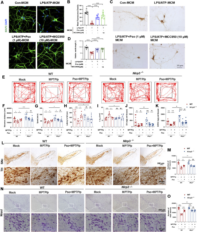
Methods: The virtual screening of a natural product library comprising 5,088 compounds was applied to identify five potential NLRP3 inhibitors through molecular docking scores. Then surface plasmon resonance assays were used to detect their binding affinities to the NLRP3 protein. Functional studies in macrophages and glial cells were used to demonstrate the effect of Psoralen on NLRP3 phosphorylation and inflammasome activation.

Results: Psoralen treatment improved PD-like symptoms and reduced dopaminergic neuronal death by targeting glial NLRP3 inflammasome activation in the MPTP/p mouse model. By performing 4D label-free quantitative phosphorylation proteomics and site mutation assays, we identified that Psoralen prevents NLRP3 phosphorylation at Serine 658 by binding to its NACHT and LRR domains.

Conclusions: These findings position Psoralen as a promising NLRP3 inflammasome inhibitor, offering a potential therapeutic avenue for PD and other NLRP3 inflammasome-related diseases. Additionally, this research highlights the innovative approach of targeting specific phosphorylation sites on the NLRP3 protein to reduce neuroinflammation.

Keywords: Inflammation; NLRP3 inflammasome; Parkinson’s disease; Phosphorylation; Serine 658.


Conflict of interest statement

Declarations. Conflict of interest: The authors declare no conflicts of interest.

Figures

Fig. 1
Fig. 1
Virtual screening and validation of potential NLRP3 inflammasome inhibitors in BMDMs. (A) The schematic model of screening NLRP3 inflammasome inhibitors from a nature compound library. (B) Five potential NLRP3 inflammasome inhibitors from 5,088 natural products were selected based on docking scores. (C) LPS (100 ng/mL) primed-BMDMs were treated with candidates and then stimulated with ATP (5 mmol/L). The levels of IL-1β in the supernatant were measured by ELISA, n = 6. MCC950 as a positive drug for inhibiting NLRP3 inflammasome activation. (D) The binding affinity between candidate compounds and purified human NLRP3 protein was examined by SPR assay. (E) Levels of IL-1β and caspase-1 in SN (Supernatant) and levels of pro-IL-1β/pro-caspase-1 in cell lysates (Lysates) were analyzed by Western blotting to examine the effects of candidate drugs on NLRP3 inflammasome activation. (F) The chemical structure of S4737 (Psoralen). Levels of IL-1β and caspase-1 in SN and levels of pro-IL-1β/pro-caspase-1 in Lysates were analyzed by immunoblotting in BMDMs pretreated with different concentrations of Psoralen (0.01, 0.1 and 1 µM) followed by stimulation with LPS/ATP (G) or LPS/Nigericin (I). Phos-tag SDS-PAGE and quantification of the phosphorylation levels of NLRP3 in BMDMs pretreated with Psoralen (0.01, 0.1 and 1 µM) followed by stimulation with LPS/ATP (H) or LPS/Nigericin (J). (K) Immunofluorescence staining for NLRP3 (red) and ASC (green) in the LPS/ATP treated BMDMs. DAPI stains the nucleus (blue). The scale bar represents 50 μm. Enlarge vision: 10 μm. (L) The percentage of cells containing an ASC speck was quantified. 100 BMDMs per group were analyzed. Data were analyzed by one-way ANOVA, followed by Tukey post-tests. *P < 0.05, **P < 0.01, and ***P < 0.001. ns: no significance
Fig. 2
Fig. 2
Psoralen prevents NLRP3 inflammasome activation in microglia and astrocytes. (A) LPS (100 ng/mL) primed microglia were treated with different concentrations (0.01, 0.1, and 1 µM) of Pso and then stimulated with ATP (5 mmol/L). The levels of IL-1β and caspase-1 in SN and levels of pro-IL-1β/pro-caspase-1 in Lysates were analyzed by immunoblotting. Quantification of relative expression of IL-1β (i), caspase-1(ii), pro-IL-1β (iii), and pro-caspase-1(iv) in microglia. All experiments were performed with three biological replicates. (B) Phos-tag SDS-PAGE and quantification of NLRP3 phosphorylation levels in microglia treated with LPS/ATP. Measurement of IL-1β (C), TNF-α (D), and IL-6 (E) levels in the supernatant of microglia, n = 3. (F) LPS (100 ng/mL) primed microglia were treated with different concentrations (0.01, 0.1, and 1 µM) of Pso and then stimulated with nigericin (5µmol/L). Quantification of relative expression of IL-1β (i), caspase-1(ii), pro-IL-1β (iii), and pro-caspase-1(iv) in microglia stimulated with LPS/nigericin. (G) Phos-tag SDS-PAGE and quantification on NLRP3 phosphorylation treated with LPS/nigericin in microglia. (H) LPS (100 ng/mL) primed astrocytes were treated with different concentrations (0.01, 0.1, and 1 µM) of Pso and then stimulated with ATP (5 mmol/L). Quantification of relative expression of IL-1β (i), caspase-1(ii), pro-IL-1β (iii), and pro-caspase-1(iv) in astrocytes stimulated with LPS/ATP. (I) Phos-tag SDS-PAGE and quantification on NLRP3 phosphorylation treated with LPS/ATP in astrocytes. Measurement of IL-1β (J), IL-6 (K), and TNF-α (L) levels in astrocytes, n = 3. Data were analyzed by one-way ANOVA, followed by Tukey post-tests. *P < 0.05, **P < 0.01, and ***P < 0.001. ns: no significance
Fig. 3
Fig. 3
Psoralen ameliorates PD-like motor symptoms and DA neuronal death in the presence of functional NLRP3. Primary neurons were incubated with conditioned medium (MCM) from microglia treated with LPS/ATP and Pso (1 µM). Representative immunostaining (A) and quantification (B) of MAP2 intensity (green). The scale bar represents 20 μm. (C-D) Effects of MCM on the morphology of DA neurons observed by immunohistochemical staining of TH. The scale bar represents 20 μm. (E-G) Travel path, movement distance and speed were recorded in the open field test, n = 6–10. (H) Latency to fall was recorded in the rotarod test, n = 6–8. (I) The time taken to descend a pole (T-TLA) was recorded in the pole test, n = 6–8. (J-K) The levels of dopamine and DOPAC in the striatum homogenate were detected by HPLC, n = 8–10. (L-M) Immunohistochemical staining and counting of TH+ neurons in the SNc. The scale bar represents 200 μm. Enlarge vision: 40 μm. (N-O) Staining and counting of Nissl+ neurons in the SNc. The scale bar represents 200 μm. Enlarge vision: 40 μm. Data were analyzed by one-way ANOVA, followed by Tukey post-tests. *P < 0.05, **P < 0.01, and ***P < 0.001. ns: no significance
Fig. 4
Fig. 4
Psoralen prevents glial NLRP3 inflammasome activation in the MPTP/p mouse model. Immunohistochemical staining and counting of IBA-1+ cells (A and C) and GFAP+ cells (B and D). The scale bar represents 200 μm. Enlarge vision: 40 μm. (E) Immunofluorescence staining for ASC (green) and microglia marker IBA1 (red) in the SNc of MPTP/p-treated WT and Nlrp3−/− mice. DAPI stains the nucleus (blue). The scale bar represents 100 μm. (F) Effects of Psoralen (1µM) on NLRP3 inflammasome activation in the LPS/ATP primed-BMDMs from WT and Nlrp3 KO mice. Data were analyzed by one-way ANOVA, followed by Tukey post-tests. *P < 0.05, **P < 0.01, and ***P < 0.001. ns: no significance
Fig. 5
Fig. 5
Biotinylated-Psoralen directly binds to the NACHT and LRR domains of NLRP3 protein. (A) Docking complex of human NLRP3 protein with Psoralen (Pso). (B) The chemical structure of Biotinylated-Psoralen. (C) Cell lysates of LPS-primed BMDMs were incubated with Pso or Bio-Pso for 2 h, which were then pulled down using streptavidin beads. The NLRP3 inflammasome components in the pull-down (PD) and total (Input) fractions were examined by Western blotting. Flag-tagged NLRP1, AIM2, NLRP3 or NLRC4 (D), NLRP3-LRR, NLPR3-NACHT or NLRP3-PYD (E) was expressed in HEK-293T cells. The HEK-293T cell lysates were incubated with Bio-Pso (10 µM) and then were pulled down using streptavidin beads for Western blotting assay. Bio-Oridonin (1 µM) as a positive drug targeting the NACHT domain of NLRP3 protein
Fig. 6
Fig. 6
S658 phosphorylation is required for NLRP3 inflammasome activation. (A) Heatmap demonstrated the differential phosphorylation peptides (fold change > 1) from 4D label-free quantitative phosphorylation proteomics. (B) Representative phosphorylation peptide Q3UZ39, annotated to NLRP3 protein. (C) The phosphorylation sites blocked by Psoralen across different species. BMDMs isolated from Nlrp3 KO mice were transfected with wtNLRP3, mutNLRP3-S658A plasmids, or mutNLRP3-S658D plasmids, then ELISA detected the production of IL-1β, IL-6, and TNF-α in the supernatant of BMDMs (D), Microglia (E), and Astrocytes (F). n = 6. (G) IL-1β and caspase-1 from SN and pro-IL-1β/pro-caspase-1 from Lysates were analyzed by immunoblotting. (H-I) Phos-tag SDS-PAGE and quantification of NLRP3 phosphorylation levels in Nlrp3 KO BMDMs transfected with wtNLRP3, mutNLRP3-S658A plasmids, or mutNLRP3-S658D plasmids. (J-K) Immunofluorescence staining and quantification of ASC (red) in Nlrp3 KO BMDMs transfected with wtNLRP3, mutNLRP3-S658A plasmids, or mutNLRP3-S658D plasmids. DAPI stains the nucleus (blue). The scale bar represents 5 μm. Data were analyzed by two-way ANOVA, followed by Tukey post-tests. *P < 0.05, **P < 0.01, and ***P < 0.001. ns: no significance
Fig. 7
Fig. 7
S658A mutation blocks the inhibitory effect of Psoralen on NLRP3 inflammasome activation. Effects of Psoralen treatment on the release of IL-1β (A), IL-6 (B), and TNF-α (C) from LPS + ATP-stimulated WT and S658A mutant BMDMs. (D) IL-1β and caspase-1 from SN and pro-IL-1β/pro-caspase-1 from Lysates were analyzed by immunoblotting after BMDMs were transfected with wtNLRP3 or mutNLRP3-S658A plasmids. (E) Immunoblots showed the binding of Pso and wtNLRP3 but not Pso and mutNLRP3-S658A in HEK-293T cells. (F-G) Phos-tag assay and quantification of NLRP3 phosphorylation in BMDMs pretreated with Pso followed by transfection with wtNLRP3 or mutNLRP3-S658A plasmids. (H-I) Immunostaining and quantification for ASC speck (red) in Nlrp3 KO BMDMs pretreated with Pso followed by transfection with wtNLRP3 or mutNLRP3-S658A plasmids. DAPI stains the nucleus (blue). The scale bar represents 5 μm. Data were analyzed by one-way ANOVA, followed by Tukey post-tests. *P < 0.05, **P < 0.01 and ***P < 0.001
Fig. 8
Fig. 8
Inhibition of NLRP3 phosphorylation at Serine 658 improves PD-like motor symptoms and pathology in the MPTP/p model. (A) Schematic illustration of AAVs carrying NLRP3 WT-EGFP (AAV-wtNLRP3) or NLRP3 S658A-EGFP (AAV-mutNLRP3). (B) Representative images showing co-label immunofluorescence of AAV(NLRP3-EGFP) with dopaminergic neuron marker (TH), astrocytes marker (GFAP), and microglia marker (IBA-1). Scale bar: 300 μm. Enlarge vision: 300 μm. Motor performance of Nlrp3 KO mice injected with AAV-wtNLRP3 or AAV-mutNLRP3 followed by treatment of MPTP/p and Psoralen in the open field test (C-D), the rotarod test (E), and the pole test (F), n = 6. (G-H) Representative immunohistochemical images and quantification of TH-positive neurons in the substantia nigra compacta. (I-J) Representative immunohistochemical images and quantification of Nissl+ neurons in the substantia nigra compacta. (K-L) Representative immunohistochemical images and quantification of IBA1+ cells in the substantia nigra compacta. (M-N) Representative immunohistochemical images and quantification of GFAP+ cells in the substantia nigra compacta, n = 6. Scale bars, 200 μm. Enlarge vision: 40 μm. (O) Immunofluorescence staining for NLRP3 (green), IBA1(red), and ASC (purple) in the substantia nigra compacta of Nlrp3 KO mice. The scale bar represents 50 μm. (P-Q) The protein levels of TH, IL-1β, and caspase-1 from the striatum were analyzed by immunoblotting, n = 3. Data were analyzed by two-way ANOVA, followed by Tukey post-tests. *P < 0.05, **P < 0.01 and ***P < 0.001. ns: no significance

References

    1. Ben-Shlomo Y, Darweesh S, Llibre-Guerra J, Marras C, San Luciano M, Tanner C. The epidemiology of Parkinson’s disease. Lancet. 2024;403:283–92. - PMC - PubMed
    1. Morris HR, Spillantini MG, Sue CM, Williams-Gray CH. The pathogenesis of Parkinson’s disease. Lancet. 2024;403:293–304. - PubMed
    1. Bloem BR, Okun MS, Klein C. Parkinson’s disease. Lancet. 2021;397:2284–303. - PubMed
    1. McGeer PL, Itagaki S, Boyes BE, McGeer EG. Reactive microglia are positive for HLA-DR in the substantia Nigra of Parkinson’s and Alzheimer’s disease brains. Neurology. 1988;38:1285–91. - PubMed
    1. Tansey MG, Wallings RL, Houser MC, Herrick MK, Keating CE, Joers V. Inflammation and immune dysfunction in Parkinson disease. Nat Rev Immunol. 2022;22:657–73. - PMC - PubMed

LinkOut - more resources