. 2025 Mar 4;18(1):84.
doi: 10.1186/s13071-025-06712-3.

Computational histology reveals that concomitant application of insect repellent with sunscreen impairs UV protection in an ex vivo human skin model

Affiliations

Computational histology reveals that concomitant application of insect repellent with sunscreen impairs UV protection in an ex vivo human skin model

Sophie Charrasse et al. Parasit Vectors. .

Abstract

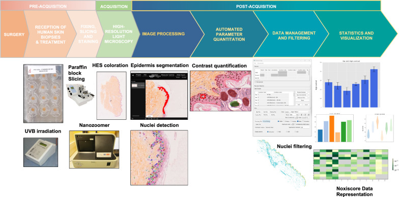
Background: Histological alterations such as nuclear abnormalities are sensitive biomarkers associated with diseases, tissue injury and environmental insults. While visual inspection and human interpretation of histology images are useful for initial characterization, such low-throughput procedures suffer from inherent limitations in terms of reliability, objectivity and reproducibility. Artificial intelligence and digital morphometry offer unprecedented opportunities to quickly and accurately assess nuclear morphotypes in relation to tissue damage including skin injury.

Methods: In this work, we designed NoxiScore, a pipeline providing an integrated, deep learning-based software solution for fully automated and quantitative analysis of nucleus-related features in histological sections of human skin biopsies. We used this pipeline to evaluate the efficacy and safety of three dermato-cosmetic products massively sold at the time of the study in the Montpellier area (South of France): a sunscreen containing UV filters, a mosquito repellent (with synthetic active ingredient IR3535) and a product combining a natural insect repellent plus a sunscreen. Hematoxylin and eosin or hematoxylin-eosin saffron staining was performed to assess skin structure before morphometric parameter computation.

Results: We report the identification of a specific nuclear feature based on variation in texture information that can be used to assess skin tissue damage after oxidative stress or UV exposure. Our data show that application of the commercial sun cream provided efficient protection against UV effects in our ex vivo skin model, whereas application of the mosquito repellent as a single product exerted no protective or toxic effect. Notably, we found that concurrent application of the insect repellent with the sunscreen significantly decreased the UVB protective effect of the sunscreen. Last, histometric analysis of human skin biopsies from multiple donors indicates that the sunscreen-insect repellent combo displayed variable levels of protection against UV irradiation.

Conclusions: To our knowledge, our study is the first to evaluate the potential toxicity of combining real-life sunscreen and insect repellent products using ex vivo human skin samples, which most closely imitate the cutaneous physiology. The NoxiScore wet-plus-dry methodology has the potential to provide information about the pharmaco-toxicological profile of topically applied formulations and may also be useful for diagnostic purposes and evaluation of the skin exposome including pesticide exposure, air pollution and water contaminants.

Keywords: Exposome; Histology; Image analysis; Insect repellent; Morphometry; Organelle biology; Sunscreen; Toxicology.


Conflict of interest statement

Declarations. Ethics approval and consent to participate: Not applicable. Consent for publication: Not applicable. Competing interests: The authors declare no competing interests.

Figures

Fig. 1
Fig. 1
NoxiScore image analysis pipeline for nuclei analysis in histological images of ex vivo skin samples. The figure describes a new methodology for quantitative characterization of nuclear appearance in large-scale whole-slide microscopic images using human skin explants as seed material. Schematic view of the system involving pre-acquisition (treatment and preparation of the biological material), acquisition (on a modern slide scanner producing high-resolution images within minutes), and post-acquisition steps (including segmentation and feature computation). Large-scale computerized slides are taken from hematoxylin and eosin (HE) [or hematoxylin-eosin saffron (HES)] stained permanent sections of formalin-fixed and paraffin-embedded tissues. Our in-house software (NoxiScore) then handles all fundamental operations from image segmentation to statistical analysis. NoxiScore software uses deep learning during the image segmentation phase and calculates about a dozen morphological descriptors (Additional file 3). A graphical interface facilitates data collection, handling and processing
Fig. 2
Fig. 2
Histometric analysis of human skin biopsies treated with hydrogen peroxide. A Histological characterization of skin explants (8 mm diameter) exposed to hydrogen peroxide (H2O2) 24 h during ex vivo culture; 3-µm skin cross-sections of fresh human skin fixed with 10% buffered formalin and embedded in paraffin wax. Hematoxylin-eosin saffron (HES) staining. Magnification is 20× (scale bar: 50 µM). All different layers of the epidermis were detected with the purple staining of nuclei in the pink cellular background. A bright line is visible at the top of the skin that corresponds to the stratum corneum. Dermis (collagen fibers) appears in orange. Inset details show the areas of interest with haloed nuclei indicated by arrows. B Histograms showing high contrast mean values (expressed in percent) between untreated and H2O2-treated human skin explants with a total of 80,914 analyzed nuclei. The number of nuclei analyzed per condition is given in Additional file 4. Results are from triplicate biopsies from the same donor. Standard Student’s t-test was used to measure significance between two groups
Fig. 3
Fig. 3
Histometric analysis of skin biopsies from a single donor treated with repellent and sunscreen formulations. Skin samples (from the same donor) were treated with (i) a sunscreen containing UV filters (panels Ad and Bd) and/or (ii) a mosquito repellent with synthetic active ingredient IR3535 (panels Ac and Bc) or (iii) a commercial preparation of a natural insect repellent plus sunscreen (‘combo’) (panels Af and Bf). The treated skin biopsies were exposed to outdoor sunlight (A) or to UVB irradiation in controlled conditions (B). Hematoxylin-eosin (HE) staining. Scale bar: 50 µM. Inset details show the areas of interest with haloed nuclei indicated by arrows. Separate treatment with the insect repellent (c) or the sunscreen (d) used as single products before sunlight exposure (2 h, UV index 8) (A) or UVB irradiation (B). Concurrent application of the insect repellent with the sunscreen. Treatment with a combination of sunscreen and insect repellent (in layers, first sunscreen and then insect repellent) (e). Treatment with a commercial preparation of a natural insect repellent plus sunscreen (‘combo’) (f). Histograms showing high contrast mean values between untreated and treated human skin explants exposed to solar radiation (C) or laboratory UVB irradiation (D). Results are from triplicate biopsies from a unique donor. Standard Student’s t-test was used to compare samples. A total of 1,185,854 nuclei were analyzed, and details of nucleus counting per condition is given in Additional file 4
Fig. 4
Fig. 4
Histometric analysis of skin biopsies from multiple donors treated with repellent and sunscreen formulations. Skin samples (from the same donor) were treated with (i) a sunscreen containing UV filters and/or (ii) a mosquito repellent with synthetic active ingredient IR3535 or (iii) a commercial preparation of a natural insect repellent plus sunscreen (‘combo’). The treated skin biopsies were exposed to outdoor sunlight (A) or to UVB irradiation in controlled conditions (B). Hematoxylin-eosin saffron (HES) staining. Scale bar: 50 µM. Inset details show the areas of interest with haloed nuclei indicated by arrows. Separate treatment with the insect repellent (c) or the sunscreen (d) used as single products before sunlight exposure (UV index 8) or UVB irradiation (300 mJ/cm2). Concurrent application (e) of the insect repellent with the sunscreen. Treatment with a combination of sunscreen and insect repellent (in layers, first sunscreen and then insect repellent) (e). Treatment with a commercial preparation of a natural insect repellent plus sunscreen (‘combo’) (f). Histograms showing high contrast mean values between untreated and treated human skin explants exposed to laboratory UVB irradiation for 24 h (C) or 48 h (D). Results are from triplicate biopsies from three different donors. Standard Student’s t-test was used to compare samples. A total of 725,397 nuclei were analyzed, and details of nuclei counting per condition are given in Additional file 4

References

    1. Brettmann EA, de Guzman SC. Recent evolution of the human skin barrier. Exp Dermatol. 2018;27:859–66. - PMC - PubMed
    1. Elias PM, Menon G, Wetzel BK, Williams JJW. Barrier requirements as the evolutionary ‘driver’ of epidermal pigmentation in humans. Am J Hum Biol. 2010;22:526–37. - PMC - PubMed
    1. Baker P, Huang C, Radi R, Moll SB, Jules E, Arbiser JL. Skin barrier function: the interplay of physical, chemical, and immunologic properties. Cells. 2023;12:2745. - PMC - PubMed
    1. Madison KC. Barrier function of the skin: ‘la raison d’être’ of the epidermis. J Invest Dermatol. 2003;121:231–41. - PubMed
    1. Matsui T, Amagai M. Dissecting the formation, structure and barrier function of the stratum corneum. Int Immunol. 2015;27:269–80. - PubMed

MeSH terms

LinkOut - more resources