. 2025 Feb 3;28(3):111947.
doi: 10.1016/j.isci.2025.111947. eCollection 2025 Mar 21.

Regeneration in planarians modifies behavioral switching

Affiliations

Regeneration in planarians modifies behavioral switching

Nayana S Tellakula et al. iScience. .

Abstract

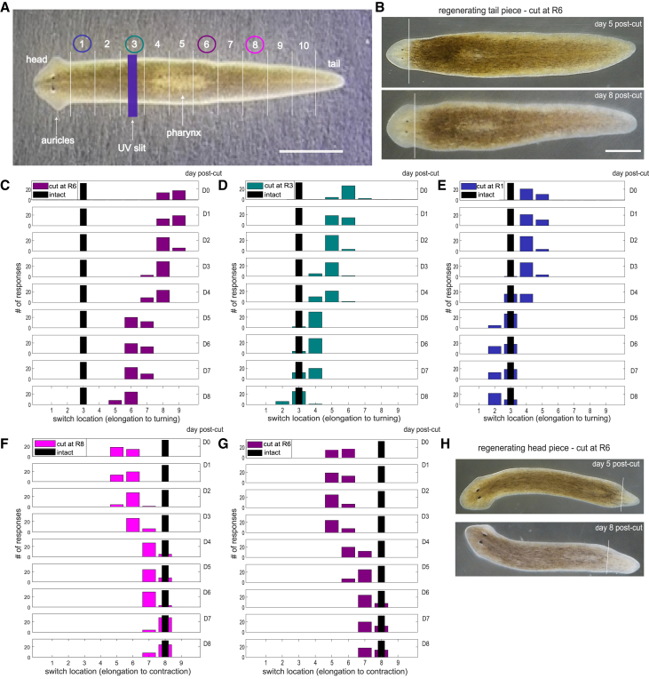
The planarian Dugesia japonica responds differently to localized stimuli: anterior regions turn, middle regions elongate, and posterior regions contract. If cut into several pieces, each piece immediately produces the same three responses. Over several days, each piece regenerates all transected body parts. This study tested how the pieces coordinate behavioral responses during regeneration. We first determined the locations of the turning/elongation and elongation/contraction behavioral switches. Immediately, all transections moved both switching sites away from the cut sites so that the worm pieces produced the same three responses as intact worms. During regeneration, the sites of behavioral switching moved progressively closer to the transection (now regeneration) sites. These results show that the immediate effects of transection (likely physiological) are coordinated with the addition of regenerating tissue (anatomical) to maintain as normal an animal as possible. Other animals that regenerate body parts, such as amphibians and reptiles, may use similar coordination mechanisms.

Keywords: Biological sciences; Developmental anatomy; Histology.


Conflict of interest statement

The authors declare no competing interests.

Figures

None
Graphical abstract
Figure 1
Figure 1
The locations of behavioral switching change during regeneration (A) Locations of the regions of behavioral switching, defining 12 body regions. Picture of an intact planarian with 12 regions indicated: head plus 11 body regions of equal length, one of which is the tail. The purple bar represents one location of a near-UV slit stimulus. Colored circles show the transection sites for the experiments corresponding to (C–G). Scale bar, 2 mm. (B) Pictures of a planarian tail piece resulting from transection at region 6, on day 5 (upper) and day 8 (lower) after transection. The tissue to the left of the white lines was regenerating tissue, indicated by the absence of dark pigmentation. The regions in the transected planarians retained the designations assigned to them in intact worms. For instance, the posterior end of worms transected at region 6 consisted of regions 6–10 and a tail. The regenerative tissue forming anterior to the cut site was designated region 5. Scale bar, 0.5 mm for both images. (C–E) Locating regions of switching in regenerating posterior pieces. Graphs show the region of behavioral switching from elongation to turning in the posterior pieces of planarians for transections at regions 6 (C), 3 (D), and 1 (E). Note that for a few cases on day 8 in the data of (D) and (E), the site of switching was in the regenerating tissue; accordingly, these were assigned to region 5 (D) and region 2 (E). (F and G) Locating regions of switching in regenerating anterior pieces. Graphs show the region of behavioral switching from elongation to contraction in the posterior pieces for transections at regions 8 (F) and 6 (G). In (F), many of the behavioral switches were observed in the regenerating tissue and therefore were assigned to regions 7 or 8, depending on how far it was away from the original cut site. Every graph used a different set of 16 planarians, each of which was stimulated twice on each of the 9 days for a total of 32 responses. Because our data were not normally distributed, we used mixed-effects ordinal logistic regression to account for the ordered values of the locations that behavioral switches occurred along the planarian, while accounting for random effects of individual planarian variability. For these measures, we used R version 4.4.2 with the “clmm” function from the “ordinal” package to model the ordered nature of the behavior response. We then used post hoc Tukey tests to compare differences between days within each cut location condition, using the “emmeans” package. We used location along the planarian as the ordinal response variable and the day of regeneration as a numeric predictor. Cut location was a categorical predictor. The method looked at the interaction between day and cut location to evaluate whether the effect of day post amputation varied across cut location conditions. There is a significant interaction between day number and cut location, so we did post hoc comparisons between days within each cut location condition. Since day was used as a numeric variable, we specified the numeric values at which the post hoc comparisons should be made, using day set to a value of 1, 2, 3, 4, 5, 6, 7, 8, and 9. Tukey comparisons of all different days against each other for each cut location condition were all significant with values of p < 0.0001. Hence, the switch location measured for each day after a given transection was strongly significantly different from the values for all other days. The black bars in each graph indicate the region of switching in intact planarians. (H) Pictures of a planarian head piece resulting from transection at region 6 on day 5 (upper) and day 8 (lower) after transection. The tissue to the right of the white lines was regenerating tissue, indicated by the absence of dark pigmentation. Scale bar shown in (B) (0.5 mm) applies to both of these images.

References

    1. Umesono Y., Tasaki J., Nishimura K., Inoue T., Agata K. Regeneration in an evolutionarily primitive brain--the planarian Dugesia japonica model. Eur. J. Neurosci. 2011;34:863–869. doi: 10.1111/j.1460-9568.2011.07819.x. - DOI - PubMed
    1. Reddien P.W., Sánchez Alvarado A. FUNDAMENTALS OF PLANARIAN REGENERATION. Annu. Rev. Cell Dev. Biol. 2004;20:725–757. doi: 10.1146/annurev.cellbio.20.010403.095114. - DOI - PubMed
    1. Le D., Sabry Z., Chandra A., Kristan W.B., Collins E.-M.S., Kristan W.B. Planarian fragments behave as whole animals. Curr. Biol. 2021;31:5111–5117.e4. doi: 10.1016/J.CUB.2021.09.056. - DOI - PubMed
    1. Inoue T., Kumamoto H., Okamoto K., Umesono Y., Sakai M., Sánchez Alvarado A., Agata K. Morphological and functional recovery of the planarian photosensing system during head regeneration. Zoolog. Sci. 2004;21:275–283. doi: 10.2108/zsj.21.275. - DOI - PubMed
    1. Deochand M.E., Birkholz T.R., Beane W.S. Temporal rgeulation of planarian eye regeneration. Regen (Oxford, England) 2016;3:209–221. doi: 10.1002/reg2.61. - DOI - PMC - PubMed

LinkOut - more resources