Review
. 2024 Nov 28;5(1):8-27.
doi: 10.1016/j.jncc.2024.11.001. eCollection 2025 Feb.

Unraveling molecular interconnections and identifying potential therapeutic targets of significance in obesity-cancer link

Affiliations
Review

Unraveling molecular interconnections and identifying potential therapeutic targets of significance in obesity-cancer link

Alanoud Abdulla et al. J Natl Cancer Cent. .

Abstract

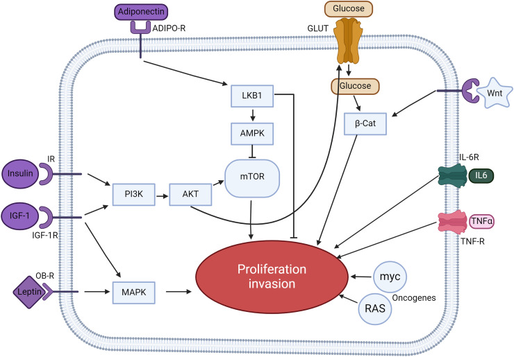
Obesity, a global health concern, is associated with severe health issues like type 2 diabetes, heart disease, and respiratory complications. It also increases the risk of various cancers, including melanoma, endometrial, prostate, pancreatic, esophageal adenocarcinoma, colorectal carcinoma, renal adenocarcinoma, and pre-and post-menopausal breast cancer. Obesity-induced cellular changes, such as impaired CD8+ T cell function, dyslipidemia, hypercholesterolemia, insulin resistance, mild hyperglycemia, and fluctuating levels of leptin, resistin, adiponectin, and IL-6, contribute to cancer development by promoting inflammation and creating a tumor-promoting microenvironment rich in adipocytes. Adipocytes release leptin, a pro-inflammatory substance that stimulates cancer cell proliferation, inflammation, and invasion, altering the tumor cell metabolic pathway. Adiponectin, an insulin-sensitizing adipokine, is typically downregulated in obese individuals. It has antiproliferative, proapoptotic, and antiangiogenic properties, making it a potential cancer treatment. This narrative review offers a comprehensive examination of the molecular interconnections between obesity and cancer, drawing on an extensive, though non-systematic, survey of the recent literature. This approach allows us to integrate and synthesize findings from various studies, offering a cohesive perspective on emerging themes and potential therapeutic targets. The review explores the metabolic disturbances, cellular alterations, inflammatory responses, and shifts in the tumor microenvironment that contribute to the obesity-cancer link. Finally, it discusses potential therapeutic strategies aimed at disrupting these connections, offering valuable insights into future research directions and the development of targeted interventions.

Keywords: Cancer risk; Gut microbiome; Inflammation; Obesity; Therapeutic interventions; Tumor microenvironment.


Conflict of interest statement

The authors declare that they have no known competing financial interests or personal relationships that could have appeared to influence the work reported in this paper.

Figures

Image, graphical abstract
Graphical abstract
Fig 1
Fig. 1
Overview of metabolic adaptations in cancer cells. Cancer cells exhibit distinct metabolic alterations to support their growth and survival. This illustration elucidates the key pathways and mechanisms involved. Cancer cells acquire FAs through de novo lipogenesis (where FAs are synthesized within the cell) and exogenous uptake (wherein FAs are absorbed from the tumor microenvironment, facilitated by transporters like CD36, FATPs, and FABPpm). Once inside, FAs are primarily stored in lipid droplets. These FAs can be metabolized to produce NADPH and acetyl-CoA via the β-oxidation process. Cancer cells metabolize glucose, glutamine, and acetate as primary substrates to produce citrate. Citrate is converted into palmitate through enzymatic reactions catalyzed by ACLY, ACC, and FASN. Subsequently, palmitate undergoes various modifications like desaturation and elongation, resulting in a diverse range of lipid species. Instead of the traditional pathway that leads to palmitoleate production, an alternative route exists where palmitate is desaturated to yield sapienate, primarily through the action of the FADS2 enzyme. ACC, acetyl-CoA carboxylase; ACSS2, acyl-CoA synthetase short-chain family member 2; ACLY, ATP–citrate lyase; CD36, cluster of differentiation 36; CDP, cytidine diphosphate; DAG, diacylglycerol; ELOVLs, elongation of very long-chain fatty acid protein; FAs, fatty acids; FABPpm, fatty acid-binding protein; FADS2, fatty acid desaturase 2; FASN, fatty acid synthase; FATPs, fatty acid transport proteins; GLS, glutaminase; GLUT1, glucose transporter 1; IDH, isocitrate dehydrogenase; LDLR, low density lipoprotein receptor; MCT, monocarboxylate transporter; MUFAs, monounsaturated fatty acids; NADPH, nicotinamide adenine dinucleotide phosphate; PA, phosphatidic acid; PE, phosphatidylethanolamine; PG, phosphatidylglycerol; PS, phosphatidylserine; PUFAs, polyunsaturated fatty acids; SCD, stearoyl-CoA desaturase-1; TAG, triacylglycerol.
Fig 2
Fig. 2
Inflammation-induced alterations in adipose tissue dynamics. This illustration captures the complex interplay between adipose tissue and immune cells during inflammation, particularly in the context of obesity. Obese-state adipocytes show elevated leptin expression, a hormone involved in appetite regulation and inflammation. Concurrently, these adipocytes also express II MHCII, which plays a role in immune cell recognition. MHCII-expressing adipocytes, in the presence of myeloid cells, facilitate the activation of TH1 cells. Upon TH1 activation, there's a subsequent secretion of the cytokine IFNγ. This cytokine is not just released by TH1 cells but also by NK and CD8+ T cells. The released IFNγ promotes further expression of adipocyte MHCII and encourages the polarization of macrophages towards the pro-inflammatory M1 phenotype. The aforementioned processes coincide with reduced abundance and function of Treg and TH2 cells. Both these cell types generally exert anti-inflammatory effects. Adipocytes, along with M1-polarized macrophages, release pro-inflammatory cytokines, specifically IL-1β, IL-6, and TNFα. These cytokines amplify the inflammatory cascade within the adipose tissue. Collectively, these mechanisms perpetuate inflammation in adipose tissue and are marked by reduced levels of Treg and TH2 cells, further compromising the tissue's anti-inflammatory response. IFNγ, interferon γ; IL, interleukin; ILC2, innate lymphoid type 2 cell; MHCII, class II major histocompatibility complex; NK, natural killer; NKT, natural killer T; TH1, T helper type 1; TH2, T helper type 2; TNFα, tumor necrosis factor α; Treg, regulatory T cell.
Fig 3
Fig. 3
The multi-faceted link between obesity and cancer development. This illustration delves into the intricate mechanisms by which expanded inflamed adipose tissue during obesity can accentuate cancer progression. Obesity is characterized by a significant increase in the size and inflammation of adipose tissue, contributing to a host of downstream effects. Inflamed adipose tissue releases adipokines, which are bioactive molecules. These adipokines have been implicated in enhancing tumor growth, metastasis and causing metabolic disturbances. Adipose tissue gives rise to CAAs, which in turn release FFAs and various inflammatory cytokines. These factors can directly influence cancer cell behavior and proliferation. ASCs, derived from adipose tissue, play a pivotal role in remodeling the tumor microenvironment. Their interaction with the surrounding matrix and cells can facilitate a niche conducive to tumor growth. Beyond direct interactions with tumors, adipose-derived proinflammatory factors have systemic effects. They can modify global metabolism and alter the behavior of tumor-supporting cells, fostering an environment favorable for tumor expansion. Collectively, these processes detail how obesity, through inflamed adipose tissue, can generate a milieu that is both directly and indirectly conducive to cancer development and progression. ASCs, adipose-derived stem cells; CAAs, cancer-associated adipocytes; FFAs, free fatty acids; IGF-1, insulin growth factor-1; IL, interleukin; MMP, matrix metalloproteinase; OPN, osteopontin; PAI-1, plasminogen activator inhibitor-1; SDF-1, stromal-derived factor-1; TGF-β, transforming growth factor β; TNFα, tumor necrosis factor α; VEGF, vascular endothelial growth factor.
Fig 4
Fig. 4
Intricate signaling pathways linking obesity to cancer progression. This figure provides a detailed account of the multiple signaling pathways implicated in connecting obesity to cancer. Insulin and IGF-1, when bound to their respective receptors (IR and IGF-1R), trigger the PI3K/AKT/mTOR signaling pathway. This cascade promotes tumor cell proliferation and invasion. Adiponectin, a hormone released predominantly by adipose tissue, interacts with ADIPO-R1 and ADIPO-R2. This binding initiates the LKB1/AMPK pathway which subsequently inhibits the mTOR pathway, acting as a counter-mechanism against tumor proliferation and metastasis. The insulin-driven PI3K/AKT pathway facilitates glucose uptake into cells via glucose transporters. Elevated glucose concentrations then amplify the Wnt/β-catenin signaling, further propelling tumor cell proliferation and invasion. Leptin, another hormone from adipose tissue, upon binding to its receptor OB-R, instigates the MAPK pathway. This cascade enhances cellular proliferation, augmenting tumor growth. Inflammatory cytokines IL-6 and TNFα, once bound to their receptors (IL-6R and TNF-R), activate the JAK/STAT/NF-kB signaling pathway. This cascade not only shields cells from apoptosis but also accentuates proliferation and metastasis. RAS and myc, being oncogenic proteins, modulate the expression of various metabolic enzymes. Their actions ramp up glycolysis, providing an energetic and metabolic advantage to tumor cells, leading to accelerated proliferation. ADIPO-R, adiponectin receptor; β-Cat, β-catenin; IGF-1, insulin growth factor; IGF-1R, insulin growth factor-1 receptor; IL6, interleukin 6; IL-6R, interleukin 6 receptor; IR, insulin reception; OB-R, leptin receptor; TNFα, tumor necrosis factor α; TNF-R, tumor necrosis factor receptor.
Fig 5
Fig. 5
Interconnections of gut microbiome and obesity-associated cancer. The interplay between diet, adipose tissue, gut microbiota dysbiosis, immune system and intestinal permeability leads to the development of obesity-induced gastrointestinal cancer. High-fat diet induces alterations in gut microbiota composition (gut dysbiosis) that increases the population of LPS-producing bacteria in the gut. This results in the downregulation of the expression of TJ proteins, leading to increased permeability of intestinal epithelial cells. Consequently, the defective intestinal barrier increasingly translocate LPS as well as bacteria and viruses. In circulation, LPS binds to macrophages, triggering immune activation via TLR signaling pathways and an upsurge in the release of proinflammatory cytokines such as IL-6, thus inducing inflammation. Additionally, gut dysbiosis induces hematopoietic dysfunction and metabolic alterations. Consumption of a high-fat diet promotes an increase in adipose tissue and fat mass as well as the risk of obesity, disrupting adipocyte function. This leads to increased release of leptin and resistin, which contribute to the amplification of inflammatory signaling pathways. All of these pathways contribute to the development of obesity-associated gastrointestinal cancers by creating an inflammatory environment conducive for tumorigenesis. Thus, this signifies the complex and interconnected relationship between diet, gut dysbiosis, obesity and GI cancers. IL-6, interleukin-6; LPS, liposaccharide; TJ, tight junctions; TLR, Toll-like receptor; TNF-α, tumor necrosis factor alpha.

References

    1. Swift D.L., McGee J.E., Earnest C.P., Carlisle E., Nygard M., Johannsen N.M. The effects of exercise and physical activity on weight loss and maintenance. Prog Cardiovasc Dis. 2018;61:206–213. - PubMed
    1. Apovian C.M. Obesity: definition, comorbidities, causes, and burden. Am J Manag Care. 2016;22:s176–s185. - PubMed
    1. WHO; Organization WH. Obesity; 2024.
    1. Fatima M.T., Ahmed I., Fakhro K.A., Akil A.S.A. Melanocortin-4 receptor complexity in energy homeostasis,obesity and drug development strategies. Diabetes Obes Metab. 2022;24:583–598. - PMC - PubMed
    1. Jung U.J., Choi M.S. Obesity and its metabolic complications: the role of adipokines and the relationship between obesity, inflammation, insulin resistance, dyslipidemia and nonalcoholic fatty liver disease. Int J Mol Sci. 2014;15:6184–6223. - PMC - PubMed

LinkOut - more resources