. 2025 Feb 13;11(4):e42691.
doi: 10.1016/j.heliyon.2025.e42691. eCollection 2025 Feb 28.

Macrophage expression of constitutively active TβRI alleviates hepatic injury in a mouse model of concanavalin A-induced autoimmune hepatitis

Affiliations

Macrophage expression of constitutively active TβRI alleviates hepatic injury in a mouse model of concanavalin A-induced autoimmune hepatitis

Arnon Pudgerd et al. Heliyon. .

Abstract

Transforming growth factor-β (Tgf-β) contributes to the development of liver diseases through its regulation of various cell types. While Tgf-β signaling to hepatic stellate cells (HSCs) and hepatocytes was shown to mediate hepatic damage, the effect of Tgf-β on other cells in liver is yet to be clearly defined. Herein we identified a regulatory function of macrophage Tgf-β signaling in liver injury. We found that transgenic mice expressing constitutively active Tgf-β receptor type I (TβRI CA ) under the control of Fsp1-Cre (TβRI CA /Fsp1-Cre mice) were less susceptible to concanavalin A (conA)-induced autoimmune hepatitis. Liver tissue examination showed a decrease of necrotic area in conA-treated TβRI CA /Fsp1-Cre liver compared to those of wild-type mice. Blood test revealed that serum aminotransferases were significantly reduced in conA-treated TβRI CA /Fsp1-Cre mice as compared to those of wild-type mice. Immunohistochemistry for CD3 and myeloperoxidase demonstrated that there was a decreased accumulation of T cells and neutrophils, respectively, whereas ELISA showed that IL-4, IL-5, IL-10, IL-12 and IFN-γ was increased in livers of conA-treated TβRI CA /Fsp1-Cre mice. Alternatively activated macrophage (M2) polarization was significantly elevated in livers of conA-treated TβRI CA /Fsp1-Cre mice as indicated by enhanced hepatic expression of CCR2 and CD206 as well as increased numbers of liver macrophages expressing M2 subtype marker, CD163. qPCR analysis indicated an increased expression of TβRI CA , Arg1, Ym1, CD206, Snail1, Foxo1 and IRF4 as well as a decreased expression of MHC class II and CD1d in liver macrophages that were isolated from TβRI CA /Fsp1-Cre mice. Moreover, flow cytometry analysis showed a lower number of NKT cells in livers of conA-treated TβRI CA /Fsp1-Cre mice when compared to those of wild-type mice. In conclusion, Fsp1-Cre-mediated expression of TβRI CA lead to a decreased conA-induced liver injury that was associated with enhanced M2 macrophage polarization and reduced NKT cell recruitment.


Conflict of interest statement

The authors declare that they have no known competing financial interests or personal relationships that could have appeared to influence the work reported in this paper.

Figures

Fig. 1
Fig. 1
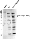
Expression analysis of TβRICAtransgenes in livers of TβRICA/Fsp1-Cre mice. (A) Detection of TβRICA transcripts by qPCR performed on total RNA from livers of wild-type and TβRICA/Fsp1-Cre mice. (B) Expression of Tgf-β, Snail1, Twist and PAI-1 in livers of wild-type and TβRICA/Fsp1-Cre mice (n = 6). (C) Phosphorylated form of Smad2 and 3 (pSmad2/3) level was analyzed by Western blot analysis. β-actin proteins served as a control. Non-adjusted images of pSmad2/3 and β-actin Western blots can be found in supplemental materials. (D) Hematoxylin/Eosin and Sirius Red/Fast green staining of liver sections obtained from control and TβRICA/Fsp1-Cre mice. The mRNA expression of TβRICA (E) and Snail1 (F) in macrophages isolated from livers of wild-type and TβRICA/Fsp1-Cre mice (n = 6). Data are shown as means ± SD. ∗P < 0.05, ∗∗P < 0.01, ∗∗∗∗P < 0.0001 between the two indicated groups.
Fig. 2
Fig. 2
Fsp1-Cre-mediated expression of TβRICAis protective against conA-induced liver injury. (A–B) Serum level of aspartate transaminase (AST) and alanine transaminase (ALT) in control and TβRICA/Fsp1-Cre mice treated with PBS or conA for 6 h. AST (A) and ALT (B) level was lower in TβRICA/Fsp1-Cre mice compared to those of control mice. Data are means ± SD of n = 10 per experimental group. (C) Survival experiments were performed in wild-type and TβRICA/Fsp1-Cre mice treated with conA 20 mg/kg body weight (n = 11–12 per group). ∗∗∗P < 0.001, ∗∗∗∗P < 0.0001 between the two indicated groups.
Fig. 3
Fig. 3
Assessment of liver histopathology following conA treatment. (A) Paraffin sections of liver from conA-treated wild-type and TβRICA/Fsp1-Cre mice. The fraction of necrotic area was enclosed by dash lines and percentage necrotic area was calculated by examining 10 high-power fields/livers (B). Data are shown as means ± SD. ∗∗∗∗P < 0.0001 between the two indicated groups.
Fig. 4
Fig. 4
Hepatic immune infiltration induced by conA. Hematoxylin and Eosin-stained liver sections obtained from conA-treated wild-type (A) and TβRICA/Fsp1-Cre mice (B). Myeloperoxidase immunohistochemical staining of liver sections from conA-treated wild-type (C) and TβRICA/Fsp1-Cre mice (D). CD3 immunohistochemical staining of liver sections from conA-treated wild-type (E) and TβRICA/Fsp1-Cre mice (F). Quantification of myeloperoxidase-(G), and CD3-(F) positive cells in livers from PBS-treated and conA-treated mice (n = 6). Flow cytometry analysis of CD4+ cells (I), CD4+ IFN-γ+ cells (J) and CD4+ IL-4+ cells (K) in livers from conA-treated wild-type and TβRICA/Fsp1-Cre mice (n = 6). (L) Representative results of TCR-β and NK1.1 positivity by flow cytometry. (M) Quantitative analysis of TCR-β- and NK1.1-positive NKT cells by flow cytometry (n = 6). Data are shown as means ± SD. ∗P < 0.05, ∗∗∗∗P < 0.0001 between the two indicated groups.
Fig. 5
Fig. 5
Hepatic expression of inflammatory cytokines after conA treatment. Relative mRNA expression of IL-4 (A), IL-5 (B), IL-12 (C), IFN-γ (D), TNF-α (E), and IL-10 (F) was determined quantitative PCR. Expression level was normalized to GAPDH. Six mice from each experimental group were used for the analysis. Data are shown as means ± SD. ∗P < 0.05, ∗∗P < 0.01 between the two indicated groups.
Fig. 6
Fig. 6
M2 macrophage polarization was enhanced in livers of conA-treated TβRICA/Fsp1-Cre mice. (A) Representative results of F4/80 and CD163 positivity in liver macrophages by flow cytometry. (B) Quantitative analysis of F4/80- and CD163-positive liver macrophages by flow cytometry (n = 6). Expression of CD206 (C) and CCR2 (D) in livers of PBS- and conA-treated wild-type and TβRICA/Fsp1-Cre mice (n = 6). Data are shown as means ± SD. ∗P < 0.05, ∗∗P < 0.01 between the two indicated groups.
Fig. 7
Fig. 7
Gene expression analysis of liver macrophages from conA-treated TβRICA/Fsp1-Cre mice. (A) Detection of Arg1, Ym1, and CD206 transcripts by qPCR performed on total RNA from liver macrophages of conA-treated mice. (B) Expression of H2-Aa and H2-k1 in liver macrophages from conA-treated wild-type and TβRICA/Fsp1-Cre mice. (C) Expression of FOXO1 and IRF1 in liver macrophages from conA-treated wild-type and TβRICA/Fsp1-Cre mice. (D) Quantification of CD1d mRNA expression levels in liver macrophages from conA-treated wild-type and TβRICA/Fsp1-Cre mice. Data are shown as means ± SD of n = 5 per experimental group. ∗P < 0.05, ∗∗P < 0.001, ∗∗∗P < 0.001 between the two indicated groups.
figs1
figs1
figs2
figs2

Similar articles

References

    1. Fabregat I., Moreno-Caceres J., Sanchez A., Dooley S., Dewidar B., Giannelli G., et al. TGF-beta signalling and liver disease. FEBS J. 2016;283(12):2219–2232. doi: 10.1111/febs.13665. - DOI - PubMed
    1. Dewidar B., Meyer C., Dooley S., Meindl-Beinker A.N. TGF-Beta in hepatic stellate cell activation and liver fibrogenesis-updated 2019. Cells. 2019;8(11) doi: 10.3390/cells8111419. - DOI - PMC - PubMed
    1. Li Y., Fan W., Link F., Wang S., Dooley S. Transforming growth factor beta latency: a mechanism of cytokine storage and signalling regulation in liver homeostasis and disease. JHEP Rep. 2022;4(2) doi: 10.1016/j.jhepr.2021.100397. - DOI - PMC - PubMed
    1. Massague J., Sheppard D. TGF-beta signaling in health and disease. Cell. 2023;186(19):4007–4037. doi: 10.1016/j.cell.2023.07.036. - DOI - PMC - PubMed
    1. Kongphat W., Pudgerd A., Sridurongrit S. Hepatocyte-specific expression of constitutively active Alk5 exacerbates thioacetamide-induced liver injury in mice. Heliyon. 2017;3(5) doi: 10.1016/j.heliyon.2017.e00305. - DOI - PMC - PubMed

LinkOut - more resources