Case Reports
. 2025 Feb 28:16:66.
doi: 10.25259/SNI_889_2024. eCollection 2025.

High-grade astrocytoma with piloid features: A case report and review of literature

Affiliations
Case Reports

High-grade astrocytoma with piloid features: A case report and review of literature

Zainab Haider Ejaz et al. Surg Neurol Int. .

Abstract

Background: High-grade astrocytoma with piloid features (HGAP) is a rare, newly recognized brain tumor, typically seen in middle aged to elderly patients, often associated with neurofibromatosis type 1.

Case description: We report the first documented case of HGAP in Pakistan in a 57-year-old woman with tremors, vertigo, and cerebellar signs. Magnetic resonance imaging showed a cerebellar lesion, and after resection, initial pathology suggested a pilocytic astrocytoma. Molecular testing confirmed HGAP with a CDKN2A/B deletion. Despite treatment, including a second surgery, the disease progressed.

Conclusion: This case highlights the diagnostic challenges of HGAP and underscores the importance of advanced molecular testing for accurate diagnosis. Given the poor prognosis and limited treatment options, further research is needed to understand this rare tumor entity better and improve patient outcomes.

Keywords: Brain tumors; High-grade astrocytoma; High-grade astrocytoma with piloid features; Neurofibromatosis type 1; Piloid features; South Asia.


Conflict of interest statement

There are no conflicts of interest.

Figures

Figure 1:
Figure 1:
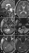
Magnetic resonance imaging findings in a 57-year-old woman with a posterior fossa lesion. (a). Axial T2-weighted image shows a partly defined lesion in the right paravermian location (arrow) with surrounding edema and mass effect over the fourth ventricle. (b). Coronal fluid-attenuated inversion recovery image demonstrates the same lesion (arrow)with similar features. (c). Axial T1-weighted image shows a hypointense lesion (arrow) with a subtle hyperintense rim. (d). Coronal T1 postcontrast image reveals intense nodular enhancement of the lesion (arrow) after gadolinium injection (arrow). (e). Axial T1 postcontrast image also shows intense nodular enhancement of the lesion (arrow). (f). Sagittal T1 postcontrast image further illustrates the intense nodular enhancement of the lesion (arrow) after gadolinium injection.
Figure 2:
Figure 2:
High-grade astrocytoma with piloid features. Hematoxylin and eosin (H&E) image (a) shows tumor cells with bipolar cytoplasmic processes with round to elongated hyperchromatic nuclei, focally centered around vessels with prominent myxoid background and occasional Rosenthal fibers. H&E image (b) shows a tumor exhibiting high cellularity with a prominent gemistocytic population and foci of microvascular proliferation. Magnification: × 40.
Figure 3:
Figure 3:
Immunohistochemical profile of high-grade astrocytoma with piloid features. IDH1: Isocitrate Dehydrogenase 1, ATRX: Alpha-Thalassemia/Mental Retardation Syndrome X-Linked, H3K27M: Histone H3 Lysine 27 Mutant, BRAF: B-Raf Proto-Oncogene, Serine/Threonine Kinase, P53: Tumor Protein p53, KI67: Antigen Ki-67.
Figure 4:
Figure 4:
High-grade astrocytoma with piloid features. The DNA copy number profile derived from the Illumina array data shows several alterations, including characteristic CDKN2A/B deletion. Green: Gains/amplifications represent positive deviation from the baseline. Red/pink: chromosome losses/deletions negative deviations from the baseline.
Figure 5:
Figure 5:
Postoperative magnetic resonance imaging findings after the first surgery. (a). Sagittal T2-weighted image reveals a resection cavity (arrow). (b). Axial T2-weighted image shows the resection cavity (arrow). (c). Axial T2-weighted image displays a rounded hyperintense lesion with a hypointense rim in the right cerebellar hemisphere paravermian location (arrow).( d). Axial T1-weighted image shows the lesion appearing hypointense (arrow). (e). Sagittal T1 postcontrast image demonstrates nodular peripheral enhancement of the lesion (arrow). (f). Axial T1 postcontrast image also shows nodular peripheral enhancement of the same lesion (arrow), suggestive of residual disease.
Figure 6:
Figure 6:
Postoperative magnetic resonance imaging findings after the second surgery. (a). Sagittal T2-weighted image reveals a resection cavity (arrow). (b). Axial T2-weighted image shows a ventricular drain in place (arrow). (c). Axial T2-weighted image also reveals a resection cavity (arrow). (d). Susceptibility-weighted image (SWI) shows marginal susceptibility and pneumocephalus (arrow). (e). Axial T1-weighted image shows marginal hyperintensity, indicating hemorrhage (arrow). (f). Axial T1 postcontrast image shows subtle reactive enhancement along the resection cavity (arrow), representing postsurgical changes. No discrete residual disease was appreciated on imaging.

References

    1. Bender K, Perez E, Chirica M, Onken J, Kahn J, Brenner W, et al. High-grade astrocytoma with piloid features (HGAP): The charité experience with a new central nervous system tumor entity. J Neurooncol. 2021;153:109–20. - PMC - PubMed
    1. Biczok A, Strübing FL, Eder JM, Egensperger R, Schnell O, Zausinger S, et al. Molecular diagnostics helps to identify distinct subgroups of spinal astrocytomas. Acta Neuropathol Commun. 2021;9:119. - PMC - PubMed
    1. Cielo B. QLTI-01. High-grade astrocytoma with piloid features response to treatment with carboplatin and vincristine. Neuro Oncol. 2023;25(Suppl 5):v244–5.
    1. Cimino PJ, Ketchum C, Turakulov R, Singh O, Abdullaev Z, Giannini C, et al. Expanded analysis of high-grade astrocytoma with piloid features identifies an epigenetically and clinically distinct subtype associated with neurofibromatosis type 1. Acta Neuropathol. 2023;145:71–82. - PMC - PubMed
    1. Louis DN, Perry A, Wesseling P, Brat DJ, Cree IA, FigarellaBranger D, et al. The 2021 WHO classification of tumors of the central nervous system: A summary. Neuro Oncol. 2021;23:1231–5. - PMC - PubMed

Publication types

LinkOut - more resources