. 2024 Sep 13;14(2):103230.
doi: 10.1016/j.eats.2024.103230. eCollection 2025 Feb.

Minimally Invasive 2-Incision Patellar Tendon Autograft Anterior Cruciate Ligament Reconstruction Using Retrograde Reamer Guide to Prevent Graft-Construct Mismatch

Affiliations

Minimally Invasive 2-Incision Patellar Tendon Autograft Anterior Cruciate Ligament Reconstruction Using Retrograde Reamer Guide to Prevent Graft-Construct Mismatch

Dylan S Koolmees et al. Arthrosc Tech. .

Abstract

Graft-tunnel mismatch (GTM) is a known technical challenge that can occur with anterior cruciate ligament reconstruction when using a patellar tendon autograft. Two-incision anterior cruciate ligament reconstruction is a well-established technique with excellent outcomes and can serve as an excellent tool to prevent GTM. Traditionally, 2-incision femoral tunnel drilling has been performed using an over-the-top guide through a lateral incision, but more modern retrograde reamer guides can allow this to be done percutaneously. We detail how a minimally invasive 2-incision femoral tunnel drilling technique can be used in patients with patellar tendon lengths that are longer than average to avoid GTM.


Conflict of interest statement

The authors declare the following financial interests/personal relationships which may be considered as potential competing interests: D.L.B. reports funding grants from the 10.13039/100011549American Orthopaedic Society for Sports Medicine. A.D.N. reports financial support from Campbell Clinic. F.M.A. reports administrative support from and employment with Campbell Clinic. J.D.L. reports editorial board for Arthroscopy. All other authors (D.S.K., J.D.L., T.J.C.) declare that they have no known competing financial interests or personal relationships that could have appeared to influence the work reported in this paper.

Figures

Fig 1
Fig 1
Anteroposterior radiographs of a left knee from 2 different patients, with the left radiograph demonstrating screw insertion retrograde through an anteromedial portal and the right demonstrating screw insertion antegrade through a 2-incision technique.
Fig 2
Fig 2
Clinical photographs of a left knee demonstrating the optimal setup of leg holder and positioning of patient to ensure knee hyperflexion can be achieved. The leg holder is brought all the way to the end of the bed and angled superiorly 30°.
Fig 3
Fig 3
Clinical photograph of a left knee demonstrating standard incision for patellar tendon autograft incision. This incision runs from the inferior aspect of the patella to the medial side of the tibial tubercle.
Fig 4
Fig 4
Clinical photograph demonstrating incision through the paratenon to expose the patellar tendon. It is important to carefully dissect this layer to allow for repair of the paratenon at the end of the case.
Fig 5
Fig 5
Clinical photograph demonstrating harvest of the soft tissue portion of the patellar tendon autograft measured directly with a ruler. A 10-mm width graft is harvested from the central one-third of the patellar tendon.
Fig 6
Fig 6
Clinical photograph demonstrating harvest of the patellar bone plug. To achieve minimal bone harvested on the patellar side, the saw blade is angled approximately 45° toward the opposite side so as to create a triangular shaped plug, which is later reshaped to trapezoidal.
Fig 7
Fig 7
Clinical photograph demonstrating creation of an anterolateral portal just inferior to the level of inferior pole of the patella and just off of the lateral border of the patellar tendon. The portal incision is made within the central skin incision as well as inside of the paratenon flap that was created.
Fig 8
Fig 8
Arthroscopic photograph of a left knee after ACL tear demonstrating the use of a bone-cutting shaver to debride remnant anterior cruciate ligament tissue.
Fig 9
Fig 9
Arthroscopic photograph of a left knee demonstrating the use of a bone-cutting shaver to perform a notchplasty addressing the lateral intercondylar ridge. The arthroscope is located within the anterolateral portal with the shaver in the anteromedial portal.
Fig 10
Fig 10
Arthroscopic photograph demonstrating the placement of the retrograde guide at the anatomic location of anterior cruciate ligament femoral attachment with guide inserted through anterolateral portal as the arthroscope is located in the anteromedial portal.
Fig 11
Fig 11
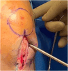
Clinical photograph demonstrating retrograde drill guide in place with marked incision for 2-incision technique. The dot immediately to the left of the marked incision is the lateral epicondyle of the femur.
Fig 12
Fig 12
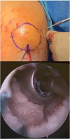
Clinical photograph on the right demonstrating drilling of a 2.4-mm guide pin for planned 2-incision femoral tunnel. Arthroscopic photograph on the left demonstrating 2.4-mm guide pin drill exiting through the anatomic anterior cruciate ligament femoral footprint, centered within the retrograde drill guide.
Fig 13
Fig 13
Arthroscopic photograph demonstrating retroreamer exiting through the anatomic anterior cruciate ligament femoral footprint with the drill blade deployed to allow for creation of a socket.
Fig 14
Fig 14
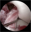
Arthroscopic photograph demonstrating a completed tunnel/socket on the lateral wall of the femoral notch, over the anterior cruciate ligament femoral footprint. There is a passing stitch noted within the tunnel. As we cannot see the end of the tunnel/socket, it cannot be determined whether this photograph represents standard or 2-incision type of femoral drilling.
Fig 15
Fig 15
Arthroscopic photograph demonstrating a Kocher clamp being inserted through the anteromedial portal and clamped onto the end of the 2.4-mm guide pin drill to protect from overpenetration of 10-mm reamer. The arthroscope is in the anterolateral portal.
Fig 16
Fig 16
Clinical photograph demonstrating 10-mm reamer being used to over-ream the 2.4-mm guide pin to create a complete tunnel on the femoral side using 2-incision technique.
Fig 17
Fig 17
Arthroscopic photograph demonstrating placement of a 2.4-mm guide pin for drilling of the ACL tibial bundle. The pin is located in the midbundle position and in line with the posterior aspect of the anterior horn of the lateral meniscus. LM represents the anterior horn of the lateral meniscus.
Fig 18
Fig 18
Arthroscopic photograph demonstrating use of a hemostat to guide the femoral-sided patellar graft bone block into the femoral tunnel. The arthroscope is inserted in the anterolateral portal with the hemostat clamp being used through the anteromedial portal.
Fig 19
Fig 19
Clinical photograph demonstrating use of index finger into the small lateral-sided incision to palpate when the bone plug is pulled flush with the lateral aperture of the tunnel.
Fig 20
Fig 20
Clinical photograph on the left demonstrates use of arthroscope into the small lateral-sided incision to visualize location of bone block relative to tunnel aperture to confirm what is expected from digital palpation. The arthroscopic photograph on the right demonstrates the bone plug flush with the aperture of the tunnel.
Fig 21
Fig 21
Top, insertion of the screw through lateral incision. Bottom, placement of the screw flush with the bone plug and tunnel aperture, with excellent compression of the bone plug achieved.
Fig 22
Fig 22
Clinical photograph demonstrates insertion of the tibial screw over a guidewire with tension held on graft and knee in full extension while a reverse Lachman maneuver is applied to the knee.
Fig 23
Fig 23
Arthroscopic photograph demonstrates a well-tensioned patellar tendon anterior cruciate ligament reconstruction graft, viewed with arthroscope in anterolateral portal and probe placed in anteromedial portal.
Fig 24
Fig 24
Anteroposterior and lateral radiographs demonstrating the fixation of a bone–patellar tendon–bone graft using titanium interference screws by way of a 2-incision drilling technique and antegrade screw at the far lateral cortex.

References

    1. Graf R.M., Dart S.E., MacLean I.S., et al. The “N+10 rule” to avoid graft-tunnel mismatch in bone–patellar tendon–bone ACL reconstruction using independent femoral tunnel drilling. Orthop J Sports Med. 2023;11 - PMC - PubMed
    1. Black K.P., Saunders M.M., Stube K.C., Moulton M.J., Jacobs C.R. Effects of interference fit screw length on tibial tunnel fixation for anterior cruciate ligament reconstruction. Am J Sports Med. 2000;28:846–849. - PubMed
    1. Mayr R., Heinrichs C.H., Eichinger M., Coppola C., Schmoelz W., Attal R. Biomechanical comparison of 2 anterior cruciate ligament graft preparation techniques for tibial fixation: Adjustable-length loop cortical button or interference screw. Am J Sports Med. 2015;43:1380–1385. - PubMed
    1. Gill T.J., Steadman J.R. Anterior cruciate ligament reconstruction the two-incision technique. Orthop Clin North Am. 2002;33:727–735. vii. - PubMed
    1. Hettrich C.M., Dunn W.R., Reinke E.K., MOON Group, Spindler K.P. The rate of subsequent surgery and predictors after anterior cruciate ligament reconstruction: Two- and 6-year follow-up results from a multicenter cohort. Am J Sports Med. 2013;41:1534–1540. - PMC - PubMed

LinkOut - more resources