. 2025 Jan;15(1):224-238.
doi: 10.1016/j.apsb.2024.12.011. Epub 2024 Dec 16.

Free fatty acid receptor-4 regulates T-cell-mediated allogeneic reaction through activating an aryl hydrocarbon receptor pathway

Affiliations

Free fatty acid receptor-4 regulates T-cell-mediated allogeneic reaction through activating an aryl hydrocarbon receptor pathway

Maxwell Duah et al. Acta Pharm Sin B. 2025 Jan.

Abstract

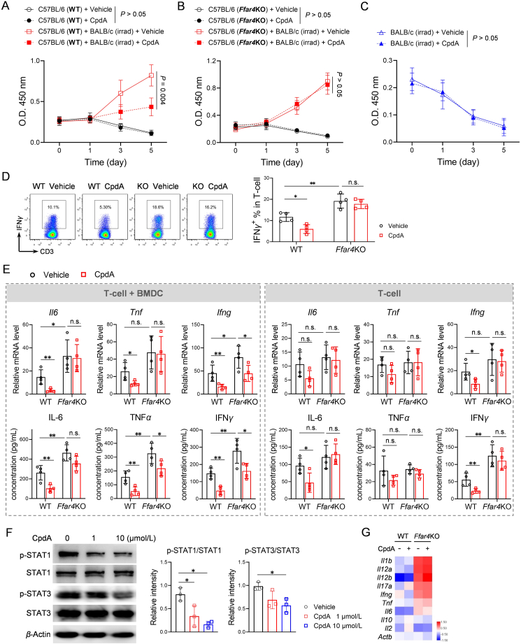
Targeting T-cell is a strategy to control allogeneic response disorders, such as acute graft-versus-host disease (GVHD) which is an important cause of therapy-failure after allogeneic hematopoietic cell transplants. Free fatty acid receptor-4 (FFAR4) is a regulator of obesity but its role in T-cell and allogeneic reactions is unknown. Here, we found knockout of Ffar4 in donor T-cells in a mouse allograft model increased acute GVHD whereas the natural FFAR4 ligands and the synthetic FFAR4 agonists decreased it. FFAR4 agonist-mediated anti-acute GVHD effects depended on FFAR4-expression in donor T-cells. The FFAR4 agonist CpdA suppressed donor T-cell-mediated alloreaction by activating an aryl hydrocarbon receptor (AhR) pathway. CpdA recruited β-Arrestin2 to FFAR4 which facilitated nuclear translocation of AhR and upregulation of IL-22. The CpdA-mediated anti-acute GVHD effect was absent in mice receiving Ahr-knockout or Il22-knockout T-cells. Recipient-expressing Ffar4 was also important for the anti-acute GVHD effect of CpdA which inhibited activation of antigen presenting cells. Importantly, CpdA decreased acute GVHD in obese mice, an effect also depended on Ffar4-expression in donor T-cells and recipients. Our study shows the immunoregulatory effect of FFAR4 in T-cell, and targeting FFAR4 might be a relative option for controlling allogeneic reactions in obese patients.

Keywords: Allogeneic reaction; Aryl hydrocarbon receptor; CpdA; Free fatty acid receptor-4; Graft-versus-host disease; Interleukin-22; Obesity; T-cell.


Conflict of interest statement

The authors declare no conflicts of interest.

Figures

Image 1
Graphical abstract
Figure 1
Figure 1
CpdA decreased acute GVHD (aGVHD) in murine allograft models. (A) In the B6 (donor) → BALB/c (recipient) model, recipient mice were transplanted with TCD-BM cells and spleen T-cells from donor mice. Recipient mice were injected with vehicle or different doses of CpdA thrice weekly for 3 weeks posttransplant. Survival and aGVHD score of recipient mice (n = 18 in each group). Recipient mice transplanted with donor TCD-BM cells alone were used as aGVHD-negative controls (n = 8). (B) In the B6 → BALB/c model, recipient mice were transplanted with wild type (WT) donor TCD-BM cells together with WT T-cells or Ffar4KO T-cells. CpdA (30 mg/kg) injection was performed as described above. Survival and aGVHD score (n = 13 in each group). (C, D) On Day 21, colon and liver tissues were collected from recipient mice described in Fig. 1A and B. H&E staining and histological score, TUNEL assay used for staining apoptotic cells in colon tissues (n = 4). Scale bar = 500 μm. GVHD features were magnified. Black arrows indicated inflammatory cells in portal tracts of liver tissues and crypt apoptotic bodies of colon tissues. (E) On Day 21, spleen cells were isolated from recipient mice described in Fig. 1B. Flow cytometry was used to analyze IFNγ-positive spleen T-cells (n = 5). (F) In the BALB/c → B6 model, recipient mice were transplanted with TCD-BM cells and spleen T-cells from donor mice. CpdA injection was performed as described above. Survival and aGVHD score (n = 8 in TCD-BM group, n = 18 in other groups) were recorded. Survival was compared using log-rank test. Data were from two or three independent experiments. Data are mean ± SD, compared using one-way ANOVA test or Student's t test. ∗P < 0.05; ∗∗P < 0.01; ∗∗∗P < 0.001; n.s., not significant.
Figure 2
Figure 2
CpdA altered T-cell related inflammatory profiles. (A–C) Mixed lymphocyte reaction was performed by co-culturing B6 spleen lymphocytes with irradiated BALB/c spleen lymphocytes. Non-mixed lymphocytes (B6 lymphocytes or irradiated BALB/c lymphocytes cultured alone) were used as controls. Cell viability was measured by CCK-8 assay on Days 0, 1, 3 and 5 (n = 4). (D) Cells from mixed lymphocyte reaction were analyzed by flow cytometry for detecting IFNγ-positive T-cells (n = 4). (E) T-cells from WT or Ffar4KO B6 mice were cultured alone (stimulated by CD3/CD28 Dynabeads) or co-cultured with BALB/c BMDCs (stimulated by LPS) for 24 h in the presence of vehicle or 10 μmol/L CpdA. mRNA of cytokine was measured by qPCR (n = 4). mRNA levels were relative to T cells not stimulated by CD3/CD28 Dynabeads. Supernatants of cell culture were used for measuring concentrations of cytokines (cytometric beads array for IL-6, TNFα and IFNγ, ELISA for IL-22) (n = 4). (F, G) CD3/CD28 Dynabeads-stimulated T-cells were treated by CpdA for 24 h. (F) Whole cell proteins were detected by Western blotting with indicated antibodies (n = 3). ImageJ was used to analyze band intensity. (G) RNA-seq analysis of T-cells treated by 10 μmol/L CpdA for 24 h. Figure shows the average of three independent values. Data are mean ± SD, compared using one-way ANOVA test or Student's t test. ∗P < 0.05; ∗∗P < 0.01; n.s., not significant.
Figure 3
Figure 3
CpdA activated AhR/IL-22 in T-cells. (A) On Day 21, flow cytometry was used to analyze IL-22-positive spleen T-cells of recipient mice described in Fig. 1A (n = 5). (B) T-cells from WT or Ffar4KO B6 mice were stimulated by CD3/CD28 Dynabeads for 24 h in the presence of vehicle or 10 μmol/L CpdA. IL-22 mRNA levels were measured by qPCR as described in Fig. 2E. (C) RNA-seq analysis of WT T-cells as described Fig. 2. All three replicates are shown. (D) In the B6 → BALB/c model, recipient mice were transplanted with WT or Il22KO T-cells. CpdA (30 mg/kg) injection was performed as described above. Survival and aGVHD score (n = 13 in each group). (E) In the B6 → BALB/c model, recipient mice were transplanted with WT or Ffar4KO T-cells. Recipient mice were injected with 10 μg/kg recombinant murine IL-22 twice weekly for 3 weeks. Survival and aGVHD score (n = 10 in each group). (F) CD3/CD28 Dynabeads-stimulated T-cells and LPS-primed BMDCs were treated by 10 μmol/L CpdA for 24 h. Cells were detected by immunofluorescence. ImageJ was used to analyze colocalization of AhR (red signal) and DAPI (blue signal). Figure shows one representative photo of three independent experiments. (G, H) CD3/CD28 Dynabeads-stimulated T-cells were treated by CpdA for 24 h. (G) Western blot analysis of AhR. Figure shows one representative band of three independent experiments. (H) Cyp1a1 mRNA level was measured by qPCR (n = 4). The AhR agonist FICZ was used as a positive control. (I) In the B6 → BALB/c model, recipient mice were transplanted with Lck-cre;Ahrfl/fl T-cells. CpdA (30 mg/kg) injection was performed as described above. Survival and aGVHD score (n = 15 in each group). Survival was compared using log-rank test. Data were from three independent experiments. Data are mean ± SD, compared using one-way ANOVA test. ∗P < 0.05; ∗∗P < 0.01; ∗∗∗P < 0.001; n.s., not significant.
Figure 4
Figure 4
β-Arrestin2 was involved in activation of AhR/IL-22 pathway. (A) UHPLC–MS/MS analysis for tryptophan metabolites of fecal samples collected on Day 21 from recipient mice in the B6 → BALB/c model. Normal indicates mice not receiving an allotransplant (n = 6). Other mice all received an allotransplant and treated by vehicle (n = 4), CpdA (n = 5), GW9508 (n = 4) or TUG891 (n = 4). Concentrations of 5-hydroxytryptophan, kynurenine and tryptophan are shown. (B) RNA-seq analysis was described in Fig. 2. Figure shows the average of three independent values. (C) T-cells were transfected with 300 nmol/L siRNAs followed by stimulation with CD3/CD28 Dynabeads (48 h) and treatment with 10 μmol/L CpdA (24 h). mRNA levels were measured by qPCR (n = 4). (D) CD3/CD28 Dynabeads-stimulated T-cells were treated by 10 μmol/L CpdA for 24 h with/without a PKC inhibitor (1 μmol/L) and a PKA inhibitor (0.1 μmol/L). mRNA levels were measured by qPCR (n = 4). Data are mean ± SD, compared using one-way ANOVA test or Student's t test. ∗P < 0.05; ∗∗P < 0.01; ∗∗∗P < 0.001; n.s., not significant.
Figure 5
Figure 5
CpdA increased nuclear translocation of AhR. Immunoprecipitation was performed with whole cell proteins (A, B), cytoplastic proteins (C) or nuclear proteins (D) extracted from Jurkat cells. Anti-β-Arrestin2 (Arrb2), anti-AhR and anti-ARNT were used for pull-down assay. Western blot was used to detect proteins in precipitate. (E) Jurkat cells were treated by CpdA (10 μmol/L) or/and FICZ (0.1 μmol/L) for 24 h. Western blot was used to detect AhR in cytoplastic and nuclear proteins. (F) Proposed actions of CpdA in T-cell. (G, H) T-cells were transfected with siRNAs and stimulated by CD3/CD28 Dynabeads. (G) Cells were detected by immunofluorescence and analyzed as described in Fig. 3F. (H) Western blot was used to detect AhR in cytoplastic and nuclear proteins. Figures show representative result from three (A–C, G) or two (D, E, H) independent experiments. Data are mean ± SD, compared using Student's t test. ∗P < 0.05.
Figure 6
Figure 6
Recipient-expressing FFAR4 contributed to FFAR4 agonism-mediated anti-aGVHD effect. (A, B) In the BALB/c → B6 model, WT and Ffar4KO B6 mice were used as recipients. CpdA (30 mg/kg) injection was performed as described above. (A) Survival and aGVHD score (n = 12 in each group). (B) H&E staining and histological score (n = 4). (C, D) In the BALB/c → B6 model, Il22KO (C) and CD11c-cre;Ahrfl/fl (D) B6 mice were used as recipients. CpdA (30 mg/kg) injection was performed as described above. Survival and aGVHD score (Il22KO, n = 13; CD11c-cre;Ahrfl/fl, n = 10). (E) LPS-primed BMDCs were treated by 10 μmol/L CpdA for 24 h mRNA levels were measured by qPCR (n = 4). (F) BMDCs treated or not treated by LPS and CpdA. Western blot analysis of proteins as indicated (n = 3). Survival was compared using log-rank test. Data were from three independent experiments. Data are mean ± SD, compared using one-way ANOVA test or Student's t test. ∗P < 0.05; n.s., not significant.
Figure 7
Figure 7
FFAR4 agonism alleviated aGVHD in obese mice. In the BALB/c → B6 model, Lepob obesity B6 mice were used as recipients. CpdA (30 mg/kg) injection was performed as described. (A) Survival and aGVHD score (n = 10 in each group). (B) H&E staining and histological score (n = 4). Normal group indicated the obesity mice were not treated by irradiation or transplant. (C) Plasma samples were used for measuring concentrations of cytokines as described (n = 5). (D) In the B6 → BALB/c model, diet-induced obesity (DIO) BALB/c mice were recipients. Recipient mice received WT or Ffar4KO T-cells (n = 13 in each group). (E) In the BALB/c → B6 model, DIO mice derived from WT or Ffar4KO B6 mice were used as recipients (n = 12 in each group). CpdA (30 mg/kg) injection was performed as described. Survival and aGVHD score were recorded. Survival was compared using log-rank test. Data were from two independent experiments. Data are mean ± SD, compared using Student's t test. ∗P < 0.05; ∗∗P < 0.01; n.s., not significant.

References

    1. Zeiser R., Blazar B.R. Acute graft-versus-host disease–biologic process, prevention, and therapy. N Engl J Med. 2017;377:2167–2179. - PMC - PubMed
    1. Penack O., Marchetti M., Ruutu T., Aljurf M., Bacigalupo A., Bonifazi F., et al. Prophylaxis and management of graft versus host disease after stem-cell transplantation for haematological malignancies: updated consensus recommendations of the European Society for Blood and Marrow Transplantation. Lancet Haematol. 2020;7:157–167. - PubMed
    1. Tan J.K., McKenzie C., Marino E., Macia L., Mackay C.R. Metabolite-sensing g protein-coupled receptors-facilitators of diet-related immune regulation. Annu Rev Immunol. 2017;35:371–402. - PubMed
    1. Duah M., Zhang K., Liang Y., Ayarick V.A., Xu K., Pan B. Immune regulation of poly unsaturated fatty acids and free fatty acid receptor 4. J Nutr Biochem. 2023;112 - PubMed
    1. Oh H., Loberiza F.R., Jr., Zhang M.J., Ringden O., Akiyama H., Asai T., et al. Comparison of graft-versus-host-disease and survival after HLA-identical sibling bone marrow transplantation in ethnic populations. Blood. 2005;105:1408–1416. - PubMed

LinkOut - more resources