. 2025 Jan;15(1):349-370.
doi: 10.1016/j.apsb.2024.11.019. Epub 2024 Nov 26.

The PGAM5-NEK7 interaction is a therapeutic target for NLRP3 inflammasome activation in colitis

Affiliations

The PGAM5-NEK7 interaction is a therapeutic target for NLRP3 inflammasome activation in colitis

Cheng-Long Gao et al. Acta Pharm Sin B. 2025 Jan.

Abstract

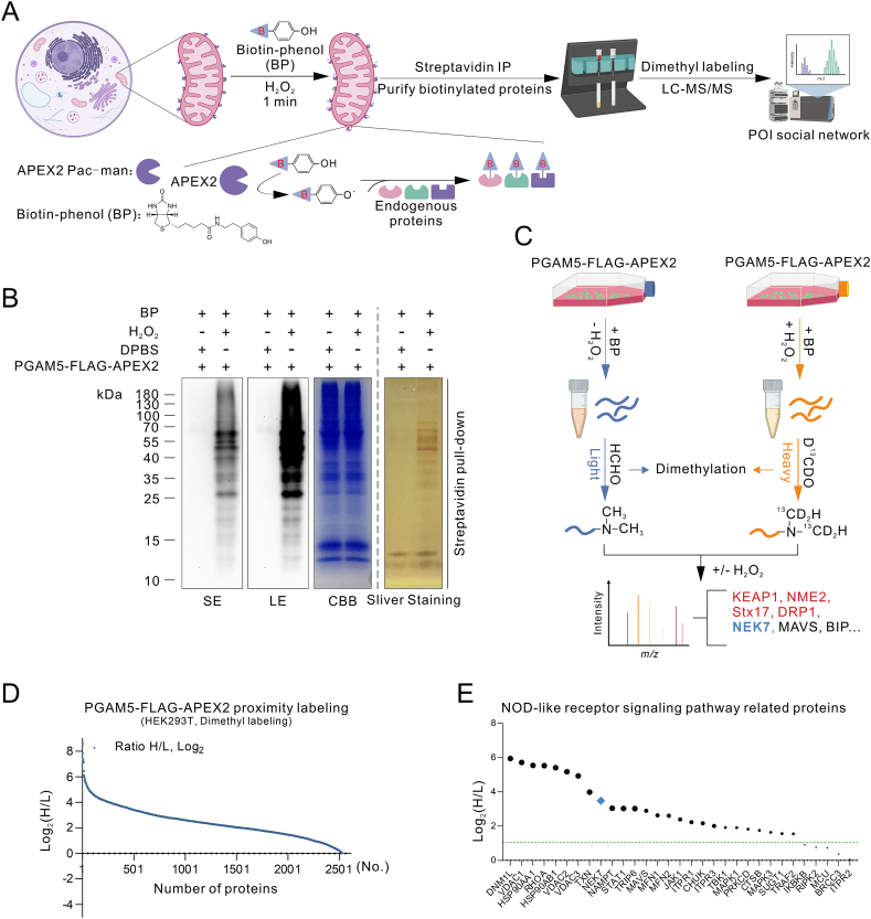
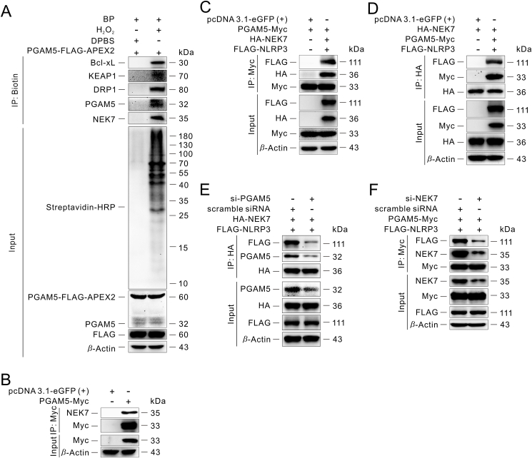
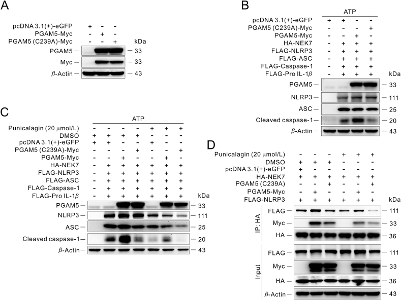
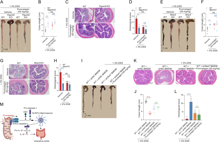
The innate immune sensor NLRP3 inflammasome overactivation is involved in the pathogenesis of ulcerative colitis. PGAM5 is a mitochondrial phosphatase involved in NLRP3 inflammasome activation in macrophages. However, the role of PGAM5 in ulcerative colitis and the mechanisms underlying PGAM5 regulating NLRP3 activity remain unknown. Here, we show that PGAM5 deficiency ameliorates dextran sodium sulfate (DSS)-induced colitis in mice via suppressing NLRP3 inflammasome activation. By combining APEX2-based proximity labeling focused on PGAM5 with quantitative proteomics, we identify NEK7 as the new binding partner of PGAM5 to promote NLRP3 inflammasome assembly and activation in a PGAM5 phosphatase activity-independent manner upon inflammasome induction. Interfering with PGAM5-NEK7 interaction by punicalagin inhibits the activation of the NLRP3 inflammasome in macrophages and ameliorates DSS-induced colitis in mice. Altogether, our data demonstrate the PGAM5-NEK7 interaction in macrophages for NLRP3 inflammasome activation and further provide a promising therapeutic strategy for ulcerative colitis by blocking the PGAM5-NEK7 interaction.

Keywords: APEX2 proximity labeling; Colitis; Macrophage; NEK7; NLRP3 inflammasome; PGAM5; Protein–protein interaction; Punicalagin.


Conflict of interest statement

The authors declare no competing interests.

Figures

Image 1
Graphical abstract
Figure 1
Figure 1
PGAM5 deficiency attenuates the activation of NLRP3 inflammasome in vivo and protects against DSS-induced colitis. (A) Schematic representation of evaluation of the biological activity of PGAM5 deficiency on DSS-induced ulcerative colitis. (B, C) The change of body weight and disease activity index (DAI) of WT and Pgam5 deficiency mice in (A) (n = 8). The DAI value was calculated as described in Methods. (D, E) Colon length in WT and Pgam5 deficiency mice was measured after being treated with DSS for 7 days (n = 8). (F, G) H&E staining and histological score of colons from DSS-induced WT and Pgam5 deficiency mice at day 7 (n = 8). (H) Immunoblot analysis and statistical analysis of PGAM5, NEK7, NLRP3 and cleaved caspase-1 protein expression of mouse colon from WT and Pgam5 deficiency mice treated with DSS for 7 days (n = 8). (I, J) ELISA analysis of IL-1β protein in serum and colons from WT and Pgam5 deficiency mice treated with DSS for 7 days (n = 8). All data are represented as mean ± SEM in (B, C, E, G–J). Statistical analysis was carried out using unpaired t-test for (B, C, E, G–J). ∗P < 0.05, ∗∗P < 0.01, and ∗∗∗P < 0.001.
Figure 2
Figure 2
Proteomics analysis of PGAM5-interacting proteins using APEX2-based proximity labeling. (A) Schematic overview of PGAM5-FLAG-APEX2 catalyzed biotinylation in the mitochondria. (B) Streptavidin blot and streptavidin pull-down analysis of biotinylated proteins from PGAM5-FLAG-APEX2 proximity labeling, and the right shows CBB staining of the same samples. HEK293T cells were transfected with PGAM5-FLAG-APEX2 fusion construct for 24 h, and then biotin phenol (0.5 mmol/L, 30 min, 37 °C) was added, followed by with or without H2O2 (1 mmol/L, 1 min, RT). Then, quickly aspirate the labeling solution and wash cells three times with quencher buffer. The biotinylated proteins were enriched using streptavidin beads and then analyzed by silver staining (streptavidin pull-down) and streptavidin blotting analysis (Input). Negative control is shown with omission of H2O2 treatment. (C) Illustration depicting the strategy to identify PGAM5 interaction partners by dimethyl labeling and quantitative LC–MS/MS-based proteomics. The control group was labeled with light isotope-labeled HCHO and the PGAM5-FLAG-APEX2 group was labeled with heavy isotope-labeled D13CDO. (D) The rank plot of streptavidin magnetic beads enriched proteins, according to the ranking of H/L ratios. (E) The rank plot of enriched proteins related with Nod-like receptor signaling pathway based on KEGG analysis. Similar results were obtained from three independent experiments.
Figure 3
Figure 3
NEK7 is a new binding partner of PGAM5. (A) Verification of the PGAM5-NEK7 interaction by streptavidin pull-down assay in HEK293T. The Bcl-xL, KEAP1 and DRP1 proteins serve as positive controls for PGAM5-FLAG-APEX2 proximity labeling. (B–D) Immunoprecipitation of the interaction between PGAM5 and NEK7 in the absence or presence of NLRP3 in HEK293T. (E) Immunoprecipitation analysis of the effect of knockdown of PGAM5 on the interaction between PGAM5 and NEK7, similarly between NEK7 and NLRP3 in HEK293T. (F) Immunoprecipitation analysis of the effect of knockdown of NEK7 on the interaction between PGAM5 and NEK7, similarly between PGAM5 and NLRP3 in HEK293T. Similar results were obtained from three independent experiments (A–F).
Figure 4
Figure 4
PGAM5 phosphatase activity-independent interaction of PGAM5 and NEK7 is responsible for NLRP3 inflammasome activation. (A) Immunoblot analysis of the effect of knockdown of PGAM5 on the activation of NLRP3 inflammasome in HEK293T. (B) Immunoblot analysis of the effect of PGAM5 overexpression on the activation of NLRP3 inflammasome in HEK293T. (C) Immunoblot analysis of the effect of the phosphatase catalysis activity of PGAM5 on the activation of NLRP3 inflammasome in HEK293T. (D) Immunoprecipitation analysis of the effect of the phosphatase catalysis activity of PGAM5 on the interaction between PGAM5 and NEK7, similarly between NEK7 and NLRP3 in HEK293T. Similar results were obtained from three independent experiments (A–D).
Figure 5
Figure 5
Natural product punicalagin blocks PGAM5–NEK7 interaction and inhibits NLRP3 inflammasome activation. (A) Immunoblot analysis of the effect of punicalagin treatment on the activation of NLRP3 inflammasome in BMDMs. (B) ELISA analysis of IL-1β secretion in supernatants of LPS-primed and ATP-activated BMDMs with punicalagin treatment. (C) Cell viability analysis of BMDMs subjected to LPS and ATP stimulation followed by punicalagin treatment. (D) Immunoblot analysis of the effect of punicalagin treatment at the level of post-translational modifications on the activation of NLRP3 inflammasome in HEK293T cells. (E, F) Immunoprecipitation analysis of the effect of punicalagin treatment or PGAM5 deficiency on the interaction between PGAM5 and NEK7, similarly between NEK7 and NLRP3 in BMDMs subjected to LPS and ATP stimulation. (G, H) SPR analysis of the binding between punicalagin and PGAM5. (I–K) Immunoblot analysis of the binding between punicalagin and PGAM5 in BMDMs via DARTS (I), CETSA (J) and SIP (K). Similar results were obtained from three independent experiments (A, D–K). All data are represented as mean ± SEM in (B, C). Statistical analysis was carried out using One-way ANOVA followed by Tukey's post hoc test for (B, C). ∗∗∗P < 0.001; ##P < 0.01, and ###P < 0.001.
Figure 6
Figure 6
Cysteine 239 residue of PGAM5 is responsible for punicalagin activity and is necessary for the interaction between PGAM5 and NEK7. (A) Immunoblot analysis of overexpression of PGAM5 (C239A)–Myc compared with PGAM5–Myc in HEK293T. (B and C) Immunoblot analysis of the effect of PGAM5 (C239A)-Myc or punicalagin treatment compared with or without PGAM5–Myc on the activation of NLRP3 inflammasome at the level of post-translational modifications in HEK293T. (D) Immunoprecipitation analysis of the effect of PGAM5 (C239A)–Myc or punicalagin treatment compared with or without PGAM5–Myc on the interaction between PGAM5 and NEK7, similarly between NEK7 and NLRP3 in HEK293T. Similar results were obtained from three independent experiments (A–D).
Figure 7
Figure 7
Punicalagin inhibits the activation of NLRP3 inflammasome in vivo and protects against DSS-induced colitis. (A, B) The change of body weight and DAI of mice administrated with or without punicalagin in DSS-induced ulcerative colitis. The DAI value was calculated as described in the Methods (n = 6). (C, D) Colon length of mice administrated with or without punicalagin was measured after induced by DSS for 7 days (n = 6). (E, F) H&E staining and histological score of colons from DSS-induced mice administrated with or without punicalagin on Day 7 (n = 6 for E and n = 3 for F). (G, H) Immunoblot analysis and statistical analysis of PGAM5, NEK7, NLRP3 and Cleaved caspase-1 protein expression of mouse colon from mice administrated with or without punicalagin and subjected to DSS for 7 days (n = 6). (I, J) ELISA analysis of IL-1β protein in serum and colon from DSS-induced mice administrated with or without punicalagin for 7 days (n = 6). (K) Immunoprecipitation analysis of the effect of punicalagin on the interaction between PGAM5 and NEK7, similarly between NEK7 and NLRP3 in colon of mice subjected to DSS. All data are represented as mean ± SEM in (D, F and H–J). Statistical analysis was carried out using One-way ANOVA followed by Tukey's post hoc test for (D, F and H–J). ∗∗P < 0.01, and ∗∗∗P < 0.001; #P < 0.05, ##P < 0.01, and ###P < 0.001; &&P < 0.01, and &&&P < 0.001; $$$P < 0.001.
Figure 8
Figure 8
Punicalagin improves DSS-induced ulcerative colitis depending on PGAM5–NEK7–NLRP3 complex. (A, B) Colon length of Pgam5 knockout or WT mice administrated with or without punicalagin was measured after induced by DSS for 7 days (n = 6). (C, D) H&E staining and histological score of colons from DSS-induced Pgam5 knockout or WT mice administrated with or without punicalagin on Day 7 (n = 6). (E, F) Colon length of Nlrp3 knockout or WT mice administrated with or without punicalagin was measured after induced by DSS for 7 days (n = 6–8). (G, H) H&E staining and histological score of colons from DSS-induced Nlrp3 knockout or WT mice administrated with or without punicalagin on Day 7 (n = 6–8). (I, J) Colon length of mice reconstituted of Nek7 knockdown macrophages in the presence or absence of punicalagin treatment was measured after DSS administration for 7 days (n = 5–6). (K, L) H&E staining and histological score of colons from mice reconstituted of Nek7 knockdown macrophages in the presence or absence of punicalagin treatment (n = 5–6). (M) Schematic diagram for the PGAM5-NEK7 interaction playing a critical role for the NLRP3 inflammasome activation in colitis. All data are represented as mean ± SEM in (B, D, F, H, J and L). Statistical analysis was carried out using Two-way ANOVA followed by Bonferroni's post hoc test for (B, D, F and H). Statistical analysis was carried out using unpaired t-test between mice reconstituted of normal control macrophages group and mice reconstituted of Nek7 knockdown macrophages group, or mice reconstituted of Nek7 knockdown macrophages group and mice reconstituted of Nek7 knockdown macrophages followed by treatment with punicalagin (50 mg/kg) group, respectively for (J, L). ∗P < 0.05, ∗∗P < 0.01, and ∗∗∗P < 0.001; ns, no significance.

References

    1. Broz P., Dixit V.M. Inflammasomes: mechanism of assembly, regulation and signalling. Nat Rev Immunol. 2016;16:407–420. - PubMed
    1. Gong T., Liu L., Jiang W., Zhou R. DAMP-sensing receptors in sterile inflammation and inflammatory diseases. Nat Rev Immunol. 2020;20:95–112. - PubMed
    1. Swanson K.V., Deng M., Ting J.P. The NLRP3 inflammasome: molecular activation and regulation to therapeutics. Nat Rev Immunol. 2019;19:477–489. - PMC - PubMed
    1. Mangan M.S.J., Olhava E.J., Roush W.R., Seidel H.M., Glick G.D., Latz E. Targeting the NLRP3 inflammasome in inflammatory diseases. Nat Rev Drug Discov. 2018;17:588–606. - PubMed
    1. Zindel J., Kubes P. DAMPs, PAMPs, and LAMPs in immunity and sterile inflammation. Annu Rev Pathol. 2020;15:493–518. - PubMed

LinkOut - more resources