. 2025 Apr 29;121(3):507-521.
doi: 10.1093/cvr/cvaf016.

Vasoconstriction-inhibiting factor: an endogenous inhibitor of vascular calcification as a calcimimetic of calcium-sensing receptor

Affiliations

Vasoconstriction-inhibiting factor: an endogenous inhibitor of vascular calcification as a calcimimetic of calcium-sensing receptor

Sofía de la Puente-Secades et al. Cardiovasc Res. .

Abstract

Aims: Patients with chronic kidney disease (CKD) show a high risk of cardiovascular diseases, predominantly caused by accelerated vascular calcification. Vascular calcification is a highly regulated process with no current treatment. The vasoconstriction-inhibiting factor (VIF) peptide was recently discovered with vasoregulatory properties, but no information regarding calcification has been described.

Methods and results: In the present work, the inhibitory calcification effect of the VIF peptide was analysed in vitro in vascular smooth muscle cells (VSMCs), ex vivo in rat aortic rings, as well as in vivo in rats treated with vitamin D and nicotine (VDN). The VIF peptide inhibits vascular calcification by acting as a calcimimetic for the calcium-sensing receptor, increasing carboxylated matrix Gla protein production and blocking the activation of calcification pathways. The VIF peptide decreased calcium influx, the production of reactive oxygen species, and the activation of multiple kinases in VSMCs. Furthermore, calcium deposition in the aortas of patients with CKD negatively correlates with the VIF peptide concentration. Moreover, we show the cleavage of the VIF peptide from chromogranin-A by 'proprotein convertase subtilisin/kexin type 2' and 'carboxypeptidase E' enzymes. In addition, 'cathepsin K' degrades the VIF peptide. The active site of the native 35 amino acid-sequence long VIF peptide was identified with seven amino acids, constituting a promising drug candidate with promise for clinical translation.

Conclusion: The elucidation of the underlying mechanism by which the VIF peptide inhibits vascular calcification, as well as the active sequence and the cleavage and degradation enzymes, forms the basis for developing preventive and therapeutic measures to counteract vascular calcification.

Keywords: CKD; CaSR; Vascular Calcification; inhibitor; smooth muscle cells.


Conflict of interest statement

Conflict of interest: A.A. has received payment or honoraria from Astra Zeneca. L.S. received research funding from Gnosis by Lesaffre, Bayer, Boehringer Ingelheim, not related to this work, and is a stockholder in Immuno diagnostic systems. T.S. has received a research grant from Else Kröner Fresenius. E.P.C.v.d.V., H.N. and J.J. are founding shareholders of AMICARE Development GmbH. R.V. is adviser to AstraZeneca, Glaxo Smith Kline, Fresenius Kabi, Novartis, Kibow, Baxter, Nipro, Fresenius Medical Care and Nextkidney. All other authors have no conflicting interests to declare.

Figures

Graphical Abstract
Graphical Abstract
Created in BioRender. Jankowski, J. (2023) BioRender.com/h88z261
Figure 1
Figure 1
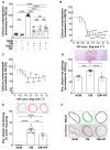
The vasoconstriction-inhibiting factor (VIF) peptide does not affect the angiotensin II inhibitory calcification effect in human aortic smooth muscle cells (hAoSMCs) and inhibits vascular calcification in in vitro and ex vivo. (A) Relative quantification of Ca2+ in hAoSMCs incubated in non-calcifying medium (NCM; circle dots) and calcifying medium (CM) in the absence (square dots) or presence of Ang II (100 nmol L−1; CM + Ang II; inverse triangle dots); Ang II and the VIF peptide (each 100 nmol L−1; CM + Ang II + VIF; diamond dots); or the VIF peptide (100 nmol L−1; NM + VIF; black and white circles dots and CM + VIF; triangle dots). Data are compared with NM, CM, or CM + Ang II (N = 4 independent experiments with 3–4 pseudoreplicates per group). Dose–response effect of the VIF peptide on Ca2+ content of (B) cultivated human aortic smooth muscle cells (N = 6 independent experiments with 3 pseudoreplicates per group) and (C) isolated thoracic rat aortic rings (N = 3 independent experiments with 3 pseudoreplicates per group). Representative images of alizarin red staining in (D) hAoSMCs and (E) thoracic aortic rat rings incubated in NCM (circle dots) and in a CM in the absence (square dots) or presence of VIF (100 nmol L−1; CM + VIF; triangle dots; magnification: 4×; scale bar: 1000 μm for hAoSMCs; and magnification: 2.5×; scale bar: 200 μm for the rings) and the corresponding quantification (N = 4 independent experiments with 3 pseudoreplicates per group). (F) Representative MALDI images of calcium phosphate crystals in the thoracic aortic rings incubated in a NCM and calcifying medium in the absence (CM) or presence of the VIF peptide (100 nmol L−1; CM + VIF) and the corresponding von Kossa staining (magnification: 2.5×; scale bar: 200 μm; N = 3).
Figure 2
Figure 2
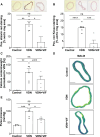
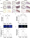
The vasoconstriction-inhibiting factor (VIF) peptide inhibits vascular calcification and influences pulse pressure in vivo. Representative images of (A) alizarin red-stained and (B) von Kossa-stained thoracic aortic rings of Wistar rats (control; circle dots) and VDN Wistar rats treated with a vehicle (VDN; square dots) or VIF peptide infusion (31 µg kg−1 per day for 4 weeks; VDN + VIF; triangle dots) via an osmotic pump (magnification: 2.5×; scale bar: 200 μm) and the corresponding quantification of the stained area (percentage of total aortic area section; N = 4–6 per group). (C) Calcium content of thoracic aorta of Wistar rats (control; circle dots) and VDN Wistar rats treated with a vehicle (VDN; square dots) or VIF peptide infusion (31 µg kg−1 per day for 4 weeks; VDN + VIF; triangle dots; N = 4–6 per group). (D) Representative images of calcium phosphate crystals in the thoracic aortic rings from Wistar rats (control) and VDN Wistar rats treated with a vehicle (VDN) or VIF peptide infusion (31 µg kg−1 per day for 4 weeks; VDN + VIF; magnification: 2.5×; scale bar: 200 μm). (E) Quantification of carotid arterial pulse pressure of Wistar rats (control; circle dots) and VDN Wistar rats treated with a vehicle (VDN; square dots) or VIF infusion (31 µg kg−1 per day for 4 weeks; VDN + VIF; triangle dots; N = 4–6 per group).
Figure 3
Figure 3
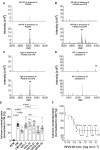
Identification of synthesizing and degrading enzymes and active amino acid sequence causing the inhibitory effect of vasoconstriction-inhibiting factor (VIF) peptide. (A) MALDI mass-spectrometric analyses of ‘20 + VIF’ peptide incubated in the absence (upper panel) and presence (lower panel) of ‘proprotein convertase subtilisin/kexin type 2’ (PCSK2). The molecular mass (m/z) of 3908.7 [M + H]+ corresponds to VIF. (B) MALDI mass-spectrometric analyses of ‘VIF + 20’ peptide incubated in the absence (upper panel) and presence (lower panel) of PCSK2 and ‘carboxypeptidase E’ (CPE). The molecular mass (m/z) of 3908.7 [M + H]+ corresponds to the VIF peptide. (C) MALDI mass-spectrometric analyses of CgA incubated in the absence (upper panel) and presence (lower panel) of PCSK2 and CPE. The molecular mass (m/z) of 3908.7 [M + H]+ corresponds to the VIF peptide. (D) MALDI mass-spectrometric analyses of the VIF peptide incubated in the absence (upper panel) and presence (lower panel) of cathepsin K. The molecular mass (m/z) of 3908.7 [M + H]+ corresponds to the VIF peptide. (E) Quantification of Ca2+ content of cultivated human aortic smooth muscle cell (hAoSMC) incubated in non-calcifying medium (NCM; circle dots) or calcifying medium in the absence (CM; square dots) or presence of VIF (CM + VIF; triangle dots) or VIF fragments (white triangle dots; see Supplementary material online, Figure S2; each 100 nmol L−1; N = 7 independent experiments with 3 pseudoreplicates per group). (F) Dose–response effect of VIF peptide fragment VIF22–28 on Ca2+ content of cultivated hAoSMC (N = 10 per group independent experiments with 3 pseudoreplicates per group).
Figure 4
Figure 4
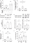
The vasoconstriction-inhibiting factor (VIF) peptide inhibits calcium influx and reactive oxygen species (ROS) production in human aortic smooth muscle cells (hAoSMCs) and reduces the phosphate-induced activity and phosphorylation of P38 and ERK1/2, and expression and secretion of IL-6. (A) Relative quantification of calcium influx in cultivated hAoSMCs incubated in non-calcifying medium (NCM; circle dots) or in calcifying medium (CM) in the absence (square dots) or presence of the VIF peptide (100 nmol L−1; CM + VIF; triangle dots), and NCM in the presence of the VIF peptide and ionomycin (Iono; NCM + VIF + ionomycin; white circle dots) and CM in the presence of the VIF peptide and ionomycin (CM + VIF + ionomycin; white triangle dots; N = 3–4 independent experiments with 3 pseudoreplicates per group). (B) Relative quantification of ROS activity in cultivated hAoSMCs incubated in NCM (circle dots) or in calcifying medium in the absence (CM; square dots) or presence of the VIF peptide (100 nmol L−1; CM + VIF; triangle dots) for 24 h. Fold change compared with NCM at time point zero (N = 7 per group). (C–D) Western blot and quantitative analysis of P38− (C) and ERK1/2- (D) phosphorylation in hAoSMCs incubated in a NCM (circle dots) or calcifying medium in the absence (CM; square dots) or presence of the VIF peptide (100 nmol L−1; CM + VIF; triangle dots) after 72 h (N = 3 independent experiments with 2–3 pseudoreplicates per group). (E–F) Relative quantification of interleukin-6 (IL-6) gene expression (E) or secretion (F) in hAoSMCs incubated in a NCM (circle dots) or CM in the absence (square dots) or presence of the VIF peptide (100 nmol L−1; NM + VIF; black and white circle dots and CM + VIF; triangle dots) after 48 h using RT-qPCR analyses and ELISA (N = 3–4 independent experiments with 3 pseudoreplicates per group).
Figure 5
Figure 5
The vasoconstriction-inhibiting factor (VIF) peptide reduces the expression of genes involved in the development of vascular calcification and increases cMGP production after binding to CaSR. (A) Relative quantification of ’bone morphogenetic protein 2’ (BMP2) gene expression in human aortic smooth muscle cell (hAoSMC) incubated for 5 days in a non-calcifying medium (NCM; circle dots) or calcifying medium (CM) in the absence (square dots) or presence of the VIF peptide (100 nmol L−1; CM + VIF; triangle dots) using RT-qPCR analyses (N = 4–5 independent experiments with 3 pseudoreplicates per group). (B) Relative quantification of ’α-smooth muscle actin’ (α-SMA), ’msh homeobox 2’ (MSX2) and ’SRY-box transcription factor 9’ (SOX9), ’osteocalcin’ (OCN) and ’sodium-dependent phosphate cotransporter-1’ (PIT-1) gene expression in vitro by RT-qPCR analyses in hAoSMCs incubated in a NCM (circle dots) or calcifying medium (CM) in the absence (square dots) or presence of the VIF peptide (100 nmol L−1; CM + VIF; triangle dots; N = 4–5 independent experiments with 3 pseudoreplicates per group). (C) Relative quantification of ‘matrix Gla protein’ (MGP) gene expression in vitro by RT-qPCR analyses in hAoSMC incubated 2 or 5 days in a NCM (circle dots) or calcifying medium in the absence (CM; square dots) or presence of the VIF peptide (100 nmol L−1; CM + VIF; triangle dots; N = 3 independent experiments with 2–3 pseudoreplicates per group). (D) Western blot and quantitative analysis of cMGP production in hAoSMCs incubated 5 days in a NCM (circle dots) or calcifying medium in the absence (CM; square dots) or presence of the VIF peptide (100 nmol L−1; CM + VIF; triangle dots; N = 3 independent experiments with 2–3 pseudoreplicates per group). (E) Effect of the VIF peptide (300 nmol L−1; Ca + VIF; square dots) on CaSR-mediated Ca2 +  i mobilisation upon stimulation with 2.5 mmol l−1 calcium (Ca) (bf: before addition of VIF; circle dots and af: after addition of VIF peptide, triangle dots). (F) Representative MALDI image of the co-localization (yellow) of calcium-sensing receptor (red) and VIF (green) in thoracic aortic rings from VDN Wistar rats treated with VIF peptide (31 µg kg−1 per day for 4 weeks; 2.5x; scale bar: 200 μm; N = 4).
Figure 6
Figure 6
The vasoconstriction-inhibiting factor (VIF) peptide increases cMGP production and reduces reactive oxygen species (ROS) and apoptosis in the VDN rat model. Representative images of (A) cMGP, ucMGP, and von Kossa staining in the thoracic aortic rings from control, VDN, or VDN + VIF rats (magnification: 2.5×; scale bar: 200 μm and 40×; scale bar 50 μm) (B–C) and the corresponding ratio of cMGP/ucMGP and vice versa (N = 4–6 per group; arrows pointing to positive staining). (D) Percentage of positive 8OdGH staining (red) in the nuclei of SMCs (blue) in rat aortic rings, elastin fibers are shown in green (autoflurescence; magnification: 100×; scale bar: 100 μm) and its representative pictures. (E) Percentage of positive TUNEL staining (red) in rat aortic rings (percentage of total aortic area section), nuclei of SMCs (blue), elastin fibers are shown in green (autofluorescence; magnification: 5×; scale bar: 500 μm) and its representative pictures of Wistar rats (control; circle dots) and VDN Wistar rats treated with a vehicle (VDN; square dots) or VIF peptide infusion (31 µg kg−1 per day for 4 weeks; VDN + VIF; triangle dots) via an osmotic pump (N = 4–6 per group).
Figure 7
Figure 7
The vasoconstriction-inhibiting factor (VIF) peptide decreases apoptosis of human aortic smooth muscle cells (hAoSMC) culture in calcifying medium (CM) and correlates negatively with CAC volume in patients with chronic kidney disease (CKD). (A) Representative flow cytometry histograms of hAoSMCs with the population of FITC-annexin V-positive cells. (B) Relative quantification of FITC-annexin V-positive cells in hAoSMC incubated in a non-calcifying medium (NCM; circle dots) or calcifying medium in the absence (CM; square dots) or presence of the VIF peptide (100 nmol L−1; CM + VIF; triangle dots). (C) Correlation between the VIF peptide concentration analysed by ELISA in plasma and the coronary artery calcium of patients with CKD under haemodialysis (r = −0.5025; P-value = 0.0398; N = 17). (D) Correlation between the VIF peptide concentration analysed by ELISA in plasma and the coronary artery calcium of patients with CKD under haemodialysis with (triangle dots; r = −0.3480; P-value = 0.3588; N = 9) or without (circle dots; r = −0.5978; P-value = 0.1175; N = 8) statin treatment.

References

    1. Muhire G, Iulita MF, Vallerand D, Youwakim J, Gratuze M, Petry FR, Planel E, Ferland G, Girouard H. Arterial stiffness due to carotid calcification disrupts cerebral blood flow regulation and leads to cognitive deficits. J Am Heart Assoc 2019;8:e011630. - PMC - PubMed
    1. Nemcsik J, Kiss I, Tisler A. Arterial stiffness, vascular calcification and bone metabolism in chronic kidney disease. World J Nephrol 2012;1:25–34. - PMC - PubMed
    1. Alexopoulos N, Raggi P. Calcification in atherosclerosis. Nat Rev Cardiol 2009;6:681–688. - PubMed
    1. Chertow GM, Correa-Rotter R, Block GA, Drueke TB, Floege J, Goodman WG, Herzog CA, Kubo Y, London GM, Mahaffey KW, Mix TC, Moe SM, Wheeler DC, Parfrey PS. Baseline characteristics of subjects enrolled in the Evaluation of Cinacalcet HCl Therapy to Lower Cardiovascular Events (EVOLVE) trial. Nephrol Dial Transplant 2012;27:2872–2879. - PubMed
    1. Chen J, Budoff MJ, Reilly MP, Yang W, Rosas SE, Rahman M, Zhang X, Roy JA, Lustigova E, Nessel L, Ford V, Raj D, Porter AC, Soliman EZ, Wright J Jr, Wolf M, He J, Investigators C. Coronary artery calcification and risk of cardiovascular disease and death among patients with chronic kidney disease. JAMA Cardiol 2017;2:635–643. - PMC - PubMed

MeSH terms

Substances

LinkOut - more resources