. 2025 Mar 25;10(3):e0098324.
doi: 10.1128/msphere.00983-24. Epub 2025 Mar 5.

The role of l-serine and l-threonine in the energy metabolism and nutritional stress response of Trypanosoma cruzi

Affiliations

The role of l-serine and l-threonine in the energy metabolism and nutritional stress response of Trypanosoma cruzi

Mayke Bezerra Alencar et al. mSphere. .

Abstract

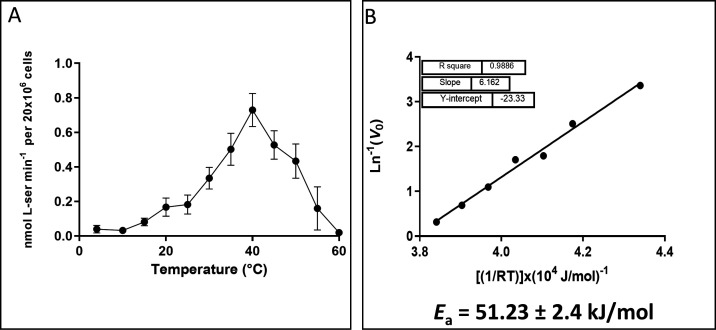
l-Serine and l-threonine have versatile roles in metabolism. In addition to their use in protein synthesis, these amino acids participate in the biosynthesis pathways of other amino acids and even phospholipids. Furthermore, l-serine and l-threonine can be substrates for a serine/threonine dehydratase (Ser/ThrDH), resulting in pyruvate and 2-oxobutyrate, respectively, thus being amino acids with anaplerotic potential. Trypanosoma cruzi, the etiological agent of Chagas disease, uses amino acids in several biological processes: metacyclogenesis, infection, resistance to nutritional and oxidative stress, osmotic control, etc. This study investigated the import and metabolism of l-serine, l-threonine, and glycine in T. cruzi. Our results demonstrate that these amino acids are transported from the extracellular environment into T. cruzi cells through a saturable transport system that fits the Michaelis-Menten model. Our results show that l-serine and l-threonine can sustain epimastigote cell viability under nutritional stress conditions and stimulate oxygen consumption, maintaining intracellular ATP levels. Additionally, our findings indicate that serine plays a role in establishing the mitochondrial membrane potential in T. cruzi. Serine is also involved in energy metabolism via the serine-pyruvate pathway, which stimulates the production and subsequent excretion of acetate and alanine. Our results demonstrate the importance of l-serine and l-threonine in the energy metabolism of T. cruzi and provide new insights into the metabolic adaptations of this parasite during its life cycle.IMPORTANCETrypanosoma cruzi, the parasite responsible for Chagas disease, impacts 5-6 million individuals in the Americas and is rapidly spreading globally due to significant human migration. This parasitic organism undergoes a complex life cycle involving triatomine insects and mammalian hosts, thriving in diverse environments, such as various regions within the insect's digestive tract and mammalian cell cytoplasm. Crucially, its transmission hinges on its adaptive capabilities to varying environments. One of the most challenging environments is the insect's digestive tract, marked by nutrient scarcity between blood meals, redox imbalance, and osmotic stresses induced by the triatomine's metabolism. To endure these conditions, T. cruzi has developed a remarkably versatile metabolic network enabling it to metabolize sugars, lipids, and amino acids efficiently. However, the full extent of metabolites this parasite can thrive on remains incompletely understood. This study reveals that, beyond conventional carbon and energy sources (glucose, palmitic acids, proline, histidine, glutamine, and alanine), three additional metabolites (serine, threonine, and glycine) play vital roles in the parasite's survival during starvation. Remarkably, serine and threonine directly contribute to ATP production through a serine/threonine dehydratase enzyme not previously described in T. cruzi. The significance of this metabolic pathway for the parasite's survival sheds light on how metabolic networks aid in its endurance under extreme conditions and its ability to thrive in diverse metabolic settings.

Keywords: Trypanosoma cruzi; amino acid metabolism; bioenergetics; nutritional stress; transport.


Conflict of interest statement

The authors declare no conflict of interest.

Figures

Fig 1
Fig 1
Uptake of l-Ser, l-Thr, and Gly in T. cruzi epimastigote as a function of time and concentration. (A–C) Time-course transport of 5 mM l-Ser, l-Thr, and Gly, respectively. (D–F) Uptake of l-Ser, l-Thr, and Gly, respectively, as a function of their concentration. The insets represent the adjustment of incorporation as a function of time to a linear function. The text below the graphs gives the values of the kinetic parameters calculated by the Michaelis-Menten model. r2 values of 0.96, 0.95, and 0.94 for l-Ser, l-Thr, and Gly, respectively. All data were shown as mean  ±  SD (n = 3). All experiments were replicated three times or more in three biological replicates.
Fig 2
Fig 2
Effect of oligomycin A (Oly) and CCCP on l-Ser uptake: the dependence of l-Ser transport on intracellular ATP levels (Oly 30′) and the H+ gradient (CCCP) were assessed. CCCP can rapidly trigger ATP hydrolysis by mitochondrial ATPase to reestablish the H+ gradient, leading to ATP depletion in the cell. To distinguish between these phenomena, the parasites were incubated with CCCP in the presence and absence of Oly (CCCP + Oly). To control for non-specific (off-target) inhibition of the transport system by oligomycin A, we added it without pre-incubation (Oly T0). All data were shown as mean  ±  SD (n = 3). All experiments were replicated three times or more in three biological replicates. Statistical analysis was performed using one-way ANOVA with Tukey’s post-test (****P < 0.0001; **P: 0.002).
Fig 3
Fig 3
Effect of temperature on l-Ser uptake. (A) The V0 was measured at saturating concentrations of l-Ser after 3 min of uptake at specific temperatures. (B) An Arrhenius plot was created by performing a linear fit between the V0 values measured at temperatures ranging from 4°C to 40°C. All data were shown as mean ± SD (n = 3). All experiments were replicated three times or more in three biological replicates.
Fig 4
Fig 4
Transport of Ser in different life-cycle stages of T. cruzi. (A) Transport of l-Ser. (B) Transport of l-Ser (normalized). All data were shown as mean  ±  SD (n = 3). All experiments were replicated three times or more in three biological replicates. The panel on the right represents the normalized transport rates, considering the transport measured in epimastigotes as 100%.
Fig 5
Fig 5
(A) Viability assay of T. cruzi epimastigotes as a function of nutritional stress: viability was measured by the irreversible reduction of resazurin to resorufin. l-His (5 mM) was used as a positive control, and no exogenous carbon source (PBS) was used as a negative control. (B) Recovery assay of T. cruzi epimastigotes (as described in Materials and Methods) after NS conditions: the proliferation profile was evaluated after 72 h of NS in the presence or absence (only PBS) of exogenous carbon sources, 5 mM His (positive control) and 5 mM Gly, l-Ser, and l-Thr. Calibration curves were performed using known parasite densities. As a negative control, PBS without exogenous carbon sources was used. All data were shown as mean ± SD (n = 3). All data were compared with the PBS control and were replicated three times or more in three biological replicates. Two-way ANOVA with Tukey’s post-test was used for statistical analysis: **P < 0.0029; ***P < 0.0007; and ****P < 0.0001.
Fig 6
Fig 6
Production of 14CO2 by breaking down l-Ser, l-Thr, and Gly in T. cruzi epimastigote. To trap the produced 14CO2, pieces of Whatman filter soaked in 2 M KOH were placed on the top of the tubes where the parasites were incubated. The filters were recovered and mixed with a scintillation cocktail, and the K214CO3 trapped on the paper was measured by using a scintillation counter (as described in Materials and Methods). The data were shown as mean ± SD (n = 3). All data were compared with the values obtained for Gly, and the experiments were replicated three times or more in three biological replicates. Two-way ANOVA with Tukey’s post-test was used for statistical analysis: ****P < 0.0001.
Fig 7
Fig 7
High-resolution respirometry in T. cruzi epimastigotes. Respiration rates were measured after 16 h of NS when the parasites were recovered for 3 h in the presence of a substrate. (A–C) Respiration rates after 3 h of incubation with 5 mM l-Ser, l-Thr, and Gly, respectively; (D and E) respiration rates after 3 h of incubation with 5 mM l-His and without exogenous carbon sources, respectively (MCR, mitochondrial cell respiration buffer). (F) Bar plot analysis of all data collected from respiration assay. (G) Free routine activity in epimastigotes recovered from NS with Gly, l-Ser, l-Thr, and l-His. The free routine activity was obtained by subtracting the respiratory rates measured after adding oligomycin A. The graph is representative of this subtraction, using the average of the slopes obtained with the amino acids and after inhibition of Fo-ATP synthase with oligomycin A. (H) The parasites were recovered for 3 h in the presence (or not, negative control—only PBS) of 5 mM l-His (positive control) Gly, l-Ser, or l-Thr. ATP levels were measured by detecting luminescence in a coupled luciferase reaction. All data were shown as mean ± SD (n = 3). All experiments were replicated three times or more in three biological replicates. Two-way ANOVA with Tukey post-test was used for statistical analysis. ****P < 0.0001 and ***P < 0.001.
Fig 8
Fig 8
Quantification of ΔΨm in the presence of l-Ser and l-Pro. (A) Representative trace of the ΔΨm quantification experiment by fluorometry, using safranine O in the presence of l-Ser, UK-5099 (UK), and Pro (positive control). Inset: calibration curve performed with K+ in the presence of 5 nM valinomycin (V). (B) Quantification of ΔΨm under the previously mentioned conditions using the Nernst equation (see Materials and Methods). All data were shown as mean ± SD (n = 3). All experiments were replicated three times or more in three biological replicates. An unpaired t test was used. **P = 0.0011. *P = 0.0225.
Fig 9
Fig 9
Excretion profile of metabolites from T. cruzi epimastigotes from l-Ser metabolism. (A) Proton resonance profile of metabolites excreted from parasites incubated for 6 h with PBS or (B) PBS supplemented with 5 mM l-Ser. (C–F) Quantification of identified metabolites. ICS, inner carbon sources. All data were shown as mean ± SD (n = 3). All experiments were replicated three times or more in three biological replicates. A two-tailed unpaired t test was used for statistical analysis. P < 0.05 was considered statistically significant.
Fig 10
Fig 10
SerDH and ThrDH activities were measured by monitoring the decrease in absorbance at 340 nm (formation of NAD+) by coupling their reactions with recombinant lactate dehydrogenase (LDH). Briefly, l-SerDH produces pyruvate from l-Ser, which, through LDH, produces lactate and oxidizes NADH. For ThrDH activity, 2-oxobutyrate is produced, which through LDH produces 2-hydroxybutyrate, also oxidizing NADH. (A) Activity using l-Ser as a substrate in soluble extract of epimastigotes. (B) Activity using l-Thr as a substrate in soluble extract of epimastigotes. (C) NAD+ produced in the reaction at different concentrations of soluble parasite extract. (D) SerDH activity of the purified recombinant enzyme. All data were shown as mean ± SD (n = 3). All experiments were replicated three times or more in three biological replicates. Two-way ANOVA with Tukey post-test was used for statistical analysis. ****P < 0.0001.
Fig 11
Fig 11
Metabolism of l-Ser and l-Thr in Trypanosoma cruzi. Metabolic steps are represented with different colors according to the origin of data: green-labeled steps correspond to data obtained in this work; yellow-labeled steps correspond to data obtained from the literature; blue-labeled steps correspond to inferred reactions according to annotations of the T. cruzi genome. In the light blue box, the same transport system for the three amino acids; in the gray box, plasma-membrane H+-ATPase (75); pyruvate dehydrogenase (PDH) (76); mitochondrial electron-transfer complexes (CI-CIV) (40, 77, 78); NADH-fumarate reductase (mFR) (79–81) (39, 76); F1Fo-ATP synthase (40, 77); adenine nucleotide translocase (ANT) (TcCLB.511249.10) (82); SPT (TcCLB.506405.50) (9, 83); serine acetyltransferase and cysteine synthase (SAT/CS) (84, 85); SHMT (22); branched-chain alpha-keto acid dehydrogenase complex (BCAKDH) (Tc001047053506295.160; Tc001047053506853.50; Tc001047053507601.70; and Tc001047053507757.70);TDH (73); 2-amino-3-ketobutyrate coenzyme A ligase (AKL) (TcCLB.511899.10); ASCT (TcCLB.504153.360); SCS (α: TcCLB.508479.340 and β: TcCLB.507681.20).

References

    1. Cazzulo JJ. 1992. Aerobic fermentation of glucose by trypanosomatids. FASEB J 6:3153–3161. doi:10.1096/fasebj.6.13.1397837 - DOI - PubMed
    1. Cazzulo JJ. 1994. Intermediate metabolism in Trypanosoma cruzi. J Bioenerg Biomembr 26:157–165. doi:10.1007/BF00763064 - DOI - PubMed
    1. Cazzulo JJ, de Cazzulo BM, Higa AI, Segura EL. 1979. NAD-linked glutamate dehydrogenase in Trypanosoma cruzi. Comp Biochem Physiol B 64:129–131. doi:10.1016/0305-0491(79)90197-4 - DOI - PubMed
    1. Cazzulo JJ, Franke de Cazzulo BM, Engel JC, Cannata JJ. 1985. End products and enzyme levels of aerobic glucose fermentation in trypanosomatids. Mol Biochem Parasitol 16:329–343. doi:10.1016/0166-6851(85)90074-x - DOI - PubMed
    1. Cazzulo JJ, Juan SM, Segura EL. 1977. Glutamate dehydrogenase and aspartate aminotransferase in Trypanosoma cruzi. Comp Biochem Physiol B Biochem Mol Biol 56:301–303. doi:10.1016/0305-0491(77)90020-7 - DOI - PubMed

LinkOut - more resources