. 2025 Mar 11;122(10):e2419244122.
doi: 10.1073/pnas.2419244122. Epub 2025 Mar 5.

BIN1 reduction ameliorates DNM2-related Charcot-Marie-Tooth neuropathy

Affiliations

BIN1 reduction ameliorates DNM2-related Charcot-Marie-Tooth neuropathy

Marie Goret et al. Proc Natl Acad Sci U S A. .

Abstract

Charcot-Marie-Tooth (CMT) disease, the most common inherited neuromuscular disorder, manifests as progressive muscle weakness and peripheral nerve defects. Dominant mutations in DNM2, encoding the large GTPase dynamin 2, result in CMT without any suggested therapeutic strategy. Different dominant mutations in DNM2 also cause centronuclear myopathy (CNM), and increasing BIN1 (amphiphysin 2), an endogenous modulator of DNM2, rescued CNM in mice. Here, we found that increasing BIN1 level exacerbated the phenotypes of the Dnm2K562E/+ mouse carrying the most common DNM2-CMT mutation. Conversely, whole-body reduction of Bin1 expression level, through the generation of Dnm2K562E/+ mice with heterozygous loss of BIN1, restored motor performance and ameliorated muscle organization and structural defects of peripheral nerves. The rescue of motor defects was maintained at least up to 1 y of age. BIN1 inhibited the GTPase activity of DNM2, and the rescue was driven by an increased activity of the K562E DNM2-CMT mutant, and a normalization of integrin localization in muscle. Overall, this study highlights BIN1 as a modifier of DNM2-CMT, and its reduction as a potential therapeutic strategy. It also revealed an opposite pathological mechanism and inverse therapeutic concepts for DNM2-CMT peripheral neuropathy versus DNM2-CNM myopathy.

Keywords: Charcot–Marie–Tooth neuropathy; amphiphysin; dynamin; gene modulation; hereditary motor and sensory neuropathy.


Conflict of interest statement

Competing interests statement:Jocelyn Laporte is inventor on a patent describing BIN1 overexpression to cure centronuclear myopathies.

Figures
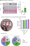
Fig. 1.
Fig. 1.
BIN1 overexpression in the Dnm2K562E/+ mouse leads to perinatal lethality. (A) Representative western blot and quantification of BIN1 protein level in WT and TgBIN1 TA at 16 wk, normalized to Ponceau S staining (5 ≤ n ≤ 7). (B) Genotypes obtained at E18.5, assessment of fetuses’ viability and weight (n = 8). (C) Births ratio expected and obtained at P8 for the four groups in percentage and n number (n = 33). Each dot represents a mouse. Values are represented as mean ± SD, ***P < 0.001. (A) Unpaired-t test. (C) Chi-square test.
Fig. 2.
Fig. 2.
BIN1 reduction restores Dnm2K562E/+ whole-body motor performance. (A) Genotypes analyzed and nomenclature used. (B) Body length measured during the gait analysis at 8 wk (15 ≤ n ≤ 25). (C) Hanging test performance at 8 wk. Maximum hanging time = 60 s (17 ≤ n ≤ 29). (D) Total locomotor activity during the night. One locomotor count corresponds to one back-and-forth movement in the cage (8 ≤ n ≤ 14). (E) Total rear activity during the night (8 ≤ n ≤ 15). Each dot represents a mouse. Values are represented as mean ± SD, *P < 0.05, **P < 0.01, ***P < 0.001, ****P < 0.0001. (BD) Kruskal–Wallis test. (E) ANOVA test.
Fig. 3.
Fig. 3.
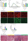
BIN1 reduction improves Dnm2K562E/+ muscle organization. (A) TA mass normalized to body weight at 8 wk (7 ≤ n ≤ 14). (B) TA fibers distribution based on their MinFeret diameter (7 ≤ n ≤ 8). (C) Proportion of small fibers (MinFeret<35 µm) in TA sections (7 ≤ n ≤ 8). (D) Immunolabeling of desmin (Upper panel) and β1-integrin (Lower panel) in transversal TA sections. Arrows indicate examples of fibers presenting an abnormal central accumulation of the staining. (Scale bar, 50 µm.) (E and F) Proportion of fibers presenting an abnormal localization of (E) desmin, and (F) β1-integrin in TA sections at 8 wk (5 ≤ n ≤ 7). (G) Soleus transversal sections stained with Masson Trichrome, arrows indicate fibrosis. (Scale bar, 100 µm.) Each dot represents a mouse. Values are represented as mean ± SD, *P < 0.05, **P < 0.01, ***P < 0.001, ****P < 0.0001. (A, E, and F) ANOVA test. (C) Kruskal–Wallis test.
Fig. 4.
Fig. 4.
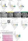
BIN1 reduction improves Dnm2K562E/+ peripheral nerve defects. (AC) RT-qPCR analysis of (A) Chrna1, (B) Chrng, and (C) Chrne expression in TA at 8 wk (4 ≤ n ≤ 6). (D) Electron microscopy images of sciatic nerve transversal sections. (Scale bar, 10 µm.) (EG) Violin plot showing (E) (g=axon diameteraxon + myelin diameter), (F) axon diameter, and (G) myelin thickness of sciatic nerve myelinated fibers (633 ≤ n = nerves ≤ 1,000, n = 3 mice). (H) Representative western blot and quantification of EGR2 protein in sciatic nerves, normalized to Ponceau S staining (n = 7). (AC and H) Each dot represents a mouse. (EG) Each fiber is plotted. Values are represented as mean ± SD, *P < 0.05, **P < 0.01, ***P < 0.001, ****P < 0.0001. (AC) ANOVA test. (EH) Kruskal–Wallis test.
Fig. 5.
Fig. 5.
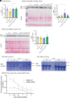
The BIN1-mediated rescue is driven by the modulation of DNM2 activity. (A) RT-qPCR analysis of Dnm2 expression in TA at 8 wk (5 ≤ n ≤ 7). (B and C) Representative western blot and quantification of DNM2 protein in TA (n = 7), and (C) sciatic nerve (n = 7), normalized to Ponceau S staining. (D and E) Coomassie Blue-stained gels of pull-down assays of recombinant DNM2-WT and DNM2-K562E using (D) GST-ubiquitous uBIN1 or (E) GST-muscle-specific mBIN1 columns. (F) Lipid (PS, phosphatidylserine)-induced GTPase activity of recombinant DNM2-WT and DNM2-K562E with increasing BIN1 concentrations. The red dashed line at Y = 0.25 highlights different BIN1/DNM2 ratios to reach the same GTPase activity for DNM2-WT and DNM2-K562E (6 ≤ n ≤ 9 technical replicates). (AC) Each dot represents a mouse. Values are represented as mean ± SD, *P < 0.05, **P < 0.01, ***P < 0.001, ****P < 0.0001. (AC) ANOVA test. (F) Multiple unpaired t tests.
Fig. 6.
Fig. 6.
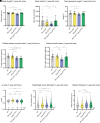
The BIN1-mediated rescue of the Dnm2K562E/+ muscular phenotypes is maintained over time. (A) Body length measured during the gait analysis at 1 y (12 ≤ n ≤ 17). (B) Total rear activity during the night (12 ≤ n ≤ 16). (C) Angle of feet between paw and body line when walking, measured during gait analysis (12 ≤ n ≤ 17). (D) TA mass normalized to body weight (12 ≤ n ≤ 17). (E) Soleus mass normalized to body weight (12 ≤ n ≤ 17). Each dot represents a mouse. (FH) Violin plots showing (F) (g=axon diameteraxon + myelin diameter), (G) axon diameter, and (H) myelin thickness of sciatic nerve myelinated fibers (874 ≤ n = nerves ≤ 1,022, n = 3 mice). Each fiber is plotted. Values are represented as mean ± SD, *P < 0.05, **P < 0.01, ***P < 0.001, ****P < 0.0001. (AE) ANOVA test. (FH) Kruskal–Wallis test.

References

    1. Estévez-Arias B., et al. , Genetic approaches and pathogenic pathways in the clinical management of Charcot-Marie-Tooth disease. J. Transl. Genet. Genom. 6, 333–352 (2022).
    1. Laurá M., Pipis M., Rossor A. M., Reilly M. M., Charcot–Marie–Tooth disease and related disorders: An evolving landscape. Curr. Opin. Neurol. 32, 641–650 (2019). - PubMed
    1. Bolino A., D’Antonio M., Recent advances in the treatment of Charcot-Marie-Tooth neuropathies. J. Peripher. Nerv. Syst. 28, 134–149 (2023). - PubMed
    1. Pisciotta C., Saveri P., Pareyson D., Challenges in treating Charcot-Marie-Tooth disease and related neuropathies: Current management and future perspectives. Brain Sci. 11, 1447 (2021). - PMC - PubMed
    1. Züchner S., et al. , Mutations in the pleckstrin homology domain of dynamin 2 cause dominant intermediate Charcot-Marie-Tooth disease. Nat. Genet. 37, 289–294 (2005). - PubMed

MeSH terms

LinkOut - more resources