. 2025 Mar 24;6(2):zqaf014.
doi: 10.1093/function/zqaf014.

High-Salt Diet Increases Suprachiasmatic Neuronal Excitability Through Endothelin Receptor Type B Signaling

Affiliations

High-Salt Diet Increases Suprachiasmatic Neuronal Excitability Through Endothelin Receptor Type B Signaling

Jodi R Paul et al. Function (Oxf). .

Abstract

Circadian rhythms are 24-h oscillations in behavioral and biological processes such as blood pressure and sodium excretion. Endothelin B (ETB) receptor has been connected to the molecular clock in peripheral tissues and plays a key role in the regulation of sodium excretion, especially in response to a high-salt diet. However, little is known about the role of ETB in the primary circadian pacemaker in the brain, the suprachiasmatic nucleus (SCN), despite recent reports showing its enrichment in SCN astrocytes. In this study, we tested the hypothesis that high-salt diet (4.0% NaCl) impacts the circadian system via the ETB receptor at the behavioral, molecular, and physiological levels in C57BL/6 mice. Two weeks of high-salt diet feeding changed the organization of nighttime wheel-running activity, as well as increased the SCN expression of ETB mRNA determined by fluorescence in situ hybridization at night. Neuronal excitability determined using loose-patch electrophysiology was also elevated at night. This high-salt diet-induced increase in SCN activity was ameliorated by ex vivo bath application of an ETB antagonist and could be mimicked with acute treatment of endothelin-3. Finally, we found that the excitatory effects of endothelin-3 were blocked with co-application of an N-methyl-D-aspartate (NMDA) receptor antagonist, suggesting that glutamate mediates endothelin-induced neuronal excitability in the SCN. Together, our data demonstrate the presence of functional ETB receptors in SCN astrocytes and point to a novel role for endothelin signaling in mediating neuronal responses to a dietary sodium intake.

Keywords: astrocytes; circadian behavior; electrophysiology; endothelin; neurophysiology; sodium.


Conflict of interest statement

D.M.P. holds the position of Editor-in-Chief for FUNCTION and is blinded from reviewing or making decisions for the manuscript. K.L.G. holds the position of Editorial Board Member for FUNCTION and is blinded from reviewing or making decisions for the manuscript.

Figures

Graphical Abstract
Graphical Abstract
Figure 1.
Figure 1.
High-salt diet (HSD) alters the pattern of daily wheel-running activity. (A) Representative double-plotted actograms of wheel-running activity from mice fed normal salt diet (NSD) (left) or HSD (right) for 2 weeks prior to analysis. Periods of dark indicated by gray shading. (B) Means ± SEM of average 24-h activity profile for mice represented in A, plotted in 1-h bins revealed differences in organization of activity (time × diet interaction, F(5.861, 58.61) = 110.725; < .001). Specifically, HSD-fed mice had significantly higher activity at Zeitgeber time (ZT) 19 (Holm-Bonferroni post hoc, = .013). (C) Activity profiles for individual NSD- (two left traces in each row) or HSD-fed (two right traces in each row) mice plotted in 30-min bins used for analysis of siesta period (triangle) and the first and second activity bouts (see methods). Dotted line indicates 50% max activity for each animal. (D-H) Means ± SEM for average activity (D; t(10) = 0.813), alpha length (E; t(10) = −2.24), length of first activity bout (F, t(10) = 2.845), length of the second activity bout (G, t(10) = −2.133), and total time in activity bout (H, t(10) = 2.154). Dots represent values for individual animals. N = 6 mice per group.
Figure 2.
Figure 2.
High-salt diet (HSD) increases suprachiasmatic nucleus (SCN) neuronal excitability at night. (A) Violin plots of spontaneous action potential frequencies of SCN neurons from mice fed normal salt diet (NSD) or HSD for 2 weeks, recorded during the day (Zeitgeber time [ZT] 4-8) or night (ZT 14-18). HSD significantly increased SCN firing at night (time × diet interaction, H(1) = 10.392, = .001; NSD-night vs. HSD-night post hoc, < .001), but had no effect during the day (= 1.00). Solid and dotted lines indicate median and quartiles, respectively. n = 143 (NSD-day), 155 (HSD-day), 151 (NSD-night), and 155 (HSD-night) cells; 4 mice per group. (B) Representative loose-patch traces (5 s) from each group. Scale bars: 2 s, 50pA. (C) Percentage of silent (empty bars) versus spiking (filled bars) cells for each group in A and B. Three-way loglinear analysis revealed a significant increase in silent cells at night for both diets (time × spiking interaction, χ2(1) = 28.949, < .001), as well as an increase in the proportion of spiking cells in HSD mice at both times of day (diet × spiking interaction, χ2(1) = 7.661, = .006).
Figure 3.
Figure 3.
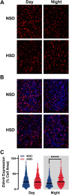
High-salt diet (HSD) increased suprachiasmatic nucleus (SCN) Ednrb expression at night. (A and B) Representative fluorescence in situ hybridization (FISH) images of SCN from normal salt diet (NSD) and HSD-fed mice showing expression of Ednrb (A) or Ednrb (red) plus DAPI (blue, B) expression during the day (Zeitgeber time [ZT] 6) or night (ZT 18). (C) Quantification of Ednrb expression per cell from groups represented in A and B. HSD significantly increased expression of Ednrb at night (time × diet interaction, F(1974.7) = 23.209, < .001; NSD-night vs. HSD-night post hoc, < .001), when Ednrb expression is low in NSD-fed mice, but had no effect during the day (= .372). Solid and dotted lines indicate median and quartiles, respectively. n = 856 (NSD-day), 931 (HSD-day), 470 (NSD-night), and 657 (HSD-night) cells; 4 (NSD-night) or 6 (NSD-day, HSD-day, and HSD-night) mice per group.
Figure 4.
Figure 4.
Endothelin B (ETB) activation increases nighttime suprachiasmatic nucleus (SCN) neuronal activity. (A) Violin plots of spontaneous action potential frequencies of neurons from SCN slices treated with vehicle (water), ETB agonist (IRL-1620, 10 n m) or ET-3 (10 n m). Both treatments significantly increase spontaneous firing rate compared to vehicle (H(2) = 17.842, P < .001). Solid and dotted lines indicate median and quartiles, respectively. n = 133 (vehicle), 146 (IRL-1620), and 130 (ET-3) cells; 3 mice per group. **= .007, ***< .001. (B) Representative loose-patch traces (5 s) from each group. Scale bars: 2 s, 50pA. (C) Percentage of silent (empty bars) versus spiking (filled bars) for cells differed between groups (χ2(2) = 13.529, = .001), with significantly more silent cells in ET-3-treated slices compared to vehicle (< .05).
Figure 5.
Figure 5.
Endothelin B (ETB) antagonist reduces high-salt diet (HSD)-induced suprachiasmatic nucleus (SCN) hyperexcitability at night. (A) Violin plots of spontaneous action potential frequencies of SCN neurons treated with ETB antagonist (A-192621, 1 µm) or vehicle (DMSO, 0.03%) from normal salt diet- (NSD-) or HSD-fed mice. Blocking ETB significantly reduced neuronal activity in neurons from HSD-fed mice (diet × treatment interaction, H(1) = 9.430, P = .002; HSD-veh vs. HSD-antagonist post hoc, P < .001), but did not affect neurons from NSD-fed controls (= 1.00). Solid and dotted lines indicate median and quartiles, respectively. n = 153 (NSD-veh), 177 (NSD-antagonist), 176 (HSD-veh), and 172 (HSD-antagonist) cells; 4 mice per group. (B) Representative loose-patch traces (5 s) from each group. Scale bars: 2 s, 50pA. (C) Percentage of silent (empty bars) versus spiking (filled bars) for cells differed between groups (3-way interaction, χ2(1) = 3.965, = .046), with HSD-veh slices having significantly more spiking cells than all other groups (< .05).
Figure 6.
Figure 6.
Suprachiasmatic nucleus (SCN) neuronal response to endothelin-3 (ET-3) is dependent on monocarboxylate transporter (MCT) lactate transporters. (A) Violin plots of spontaneous action potential frequencies of neurons from SCN slices treated with vehicle (DMSO, 0.2%), MCT inhibitor (2-cyano-3-(4-hydroxyphenyl)-2-propenoic acid [CHC], 200 µm), ET-3 (10 n m), or CHC + ET-3. ET-3 significantly increased SCN firing rate, and this was blocked with CHC (CHC × ET-3 interaction, H(1) = 12.70, < .001). Solid and dotted lines indicate median and quartiles, respectively. n = 135 (veh), 130 (CHC), 134 (ET-3), and 143 (CHC + ET-3) cells; 3 mice per group. ****< .001 compared to all other groups. (B) Representative loose-patch traces (5 s) from each group. Scale bars: 2 s, 50pA. (C) Percentage of silent (empty bars) versus spiking (filled bars) for cells differed between treatment groups (3-way interaction, χ2(1) = 3.971, = .046), with ET-3 slices having significantly more spiking cells than all other groups (P < .05).
Figure 7.
Figure 7.
NMDA receptors mediate suprachiasmatic nucleus (SCN) neuronal response to endothelin-3 (ET-3). (A) Violin plots of spontaneous action potential frequencies of neurons from SCN slices treated with vehicle (water), NMDA blocker (3-(2-carboxyp-iperazin-4-yl)propyl-1-phosphonic acid [CPP], 10 µm), ET-3 (10 n m), or CPP + ET-3. ET-3 significantly increased SCN firing rate, and this was blocked with CPP (CPP × ET-3 interaction, H(1) = 30.018, < .001). Solid and dotted lines indicate median and quartiles, respectively. n = 192 (veh), 195 (CPP), 183 (ET-3), and 187 (CPP + ET-3) cells; 4 mice per group. ****< .001 compared to all other groups. (B) Representative loose-patch traces (5 s) from each group. Scale bars: 2 s, 50pA. (C) Percentage of silent (empty bars) versus spiking (filled bars) for cells differed between treatment groups (3-way interaction, χ2(1) = 18.157, < .001), with ET-3 slices having significantly more spiking cells than all other groups (P < .05).

References

    1. Rosenwasser AM, Turek FW. Neurobiology of circadian rhythm regulation. Sleep Med Clin. 2015;10(4):403–412. - PubMed
    1. Douma LG, Barral D, Gumz ML. Interplay of the circadian clock and endothelin system. Physiology. 2021;36(1):35–43. - PMC - PubMed
    1. Takahashi JS. Molecular components of the circadian clock in mammals. Diabetes Obes Metab. 2015;17(S1):6–11. - PMC - PubMed
    1. Paul JR, Davis JA, Goode LK et al. Circadian regulation of membrane physiology in neural oscillators throughout the brain. Eur J Neurosci. 2020;51(1):109–138. - PMC - PubMed
    1. Gizowski C, Bourque CW. Sodium regulates clock time and output via an excitatory GABAergic pathway. Nature. 2020;583(7816):421–424. - PubMed

MeSH terms

Substances

LinkOut - more resources