. 2025 Mar 7;11(10):eadu0577.
doi: 10.1126/sciadv.adu0577. Epub 2025 Mar 5.

Structural insights into promoter-proximal pausing of RNA polymerase II at +1 nucleosome

Affiliations

Structural insights into promoter-proximal pausing of RNA polymerase II at +1 nucleosome

Masahiro Naganuma et al. Sci Adv. .

Abstract

The metazoan transcription elongation complex (EC) of RNA polymerase II (RNAPII) generally stalls between the transcription start site and the first (+1) nucleosome. This promoter-proximal pausing involves negative elongation factor (NELF), 5,6-dichloro-1-β-d-ribobenzimidazole sensitivity-inducing factor (DSIF), and transcription elongation factor IIS (TFIIS) and is critical for subsequent productive transcription elongation. However, the detailed pausing mechanism and the involvement of the +1 nucleosome remain enigmatic. Here, we report cryo-electron microscopy structures of ECs stalled on nucleosomal DNA. In the absence of TFIIS, the EC is backtracked/arrested due to conflicts between NELF and the nucleosome. We identified two alternative binding modes of NELF, one of which reveals a critical contact with the downstream DNA through the conserved NELF-E basic helix. Upon binding with TFIIS, the EC progressed to the nucleosome to establish a paused EC with a partially unwrapped nucleosome. This paused EC strongly restricts EC progression further downstream. These structures illuminate the mechanism of RNAPII pausing/stalling at the +1 nucleosome.


Figures
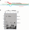
Fig. 1.
Fig. 1.. Effects of NELF and TFIIS on nucleosomal transcription by RNAPII.
(A) Schematic representation of the nucleosomal template. The template DNA, nontemplate DNA, and fluorescently labeled primer RNA are colored green, cyan, and red, respectively. (B) Urea PAGE analysis of RNA transcripts generated by the transcription reactions in the presence or absence of NELF and TFIIS. DSIF is included in all lanes. The experiment was performed in duplicate (fig. S4A).
Fig. 2.
Fig. 2.. Cryo-EM structures of arrested ECs in two alternative NELF-binding modes.
(A) Left panel shows schematic of the cryo-EM sample preparation. The right panel depicts the RNAPII position relative to the nucleosome in obtained complex structures. (B) Overall structure of arrested EC with NELF-binding mode 1 (AEC1-nuc) in three orientations. In the middle and right panels, the nucleosome is omitted for visibility. RNAPII, SPT4/SPT5, and NELF-A/NELF-B/NELF-C/NELF-E are shown as gray, orange/light green, and cyan/green/blue/red surfaces, respectively. Histone proteins, template/nontemplate DNA, and RNA are shown as gray, yellow/orange, and red ribbon models. (C) Overall structure of arrested EC with NELF-binding mode 2 (AEC2-nuc) in three orientations. (D) Comparison of the NELF-AC lobes between modes 1 and 2. (E) Comparison of the NELF-BE lobes between modes 1 and 2. (F) Close-up views of the RNAPII active site in AEC1-nuc and AEC2-nuc. Densities are represented by transparent gray surfaces. The backtracked parts of RNA are indicated. The structures in (B) to (F), represented as cartoons or surfaces, were prepared using PyMOL (Schrödinger; www.pymol.org).
Fig. 3.
Fig. 3.. The contact between NELF-E and the downstream DNA is critical for pausing.
(A) Overall structure of the AEC2-nuc complex (left) and close-up view of the interface between the NELF-E basic helix and the downstream DNA. The Cβ atoms of K23, K27, and K34 are shown as sphere models. The structures, depicted as cartoons, were prepared using PyMOL. (B) Sequence alignment of the N-terminal regions of NELF-E. (C) Transcription assay with the mutant NELF, in which the three Lys residues are replaced by Glu (3E). Urea PAGE analysis of the transcription products is shown. The experiment was performed in triplicate (fig. S4B).
Fig. 4.
Fig. 4.. Cryo-EM structure of PEC with a nucleosome (PEC2-nuc).
(A) The left panel shows schematic of the cryo-EM sample preparation. The right panel depicts the RNAPII position relative to the nucleosome in the obtained complex structure. (B) Overall structure of PEC2-nuc in three orientations. RNAPII, NELF, and DSIF are colored as in Fig. 2C. TFIIS is represented as a violet surface. (C) TFIIS binding site. The density is shown as a transparent gray surface. Although the density of the TFIIS domain III is poor, its model is shown for visibility. (D and E) Close-up views of the nucleosome and its interactions with NELF and RNAPII. (F) Partially unwrapped nucleosome in the complex. (G) Time course of transcription with NELF (the wild type and 3E mutant). Urea PAGE analysis of the transcription products is shown. The experiment was performed in triplicate (fig. S4C). (H) Nucleosome-passage ratio. Band intensities in (G) were quantitated, and the calculated nucleosome-passage ratios (intensity [SHL(–1)]/{intensity [SHL(–1)] + intensity [SHL(–5)]}) were plotted. The mean values ± SD from three independent experiments are shown (n = 3 technical replicates). The structures in (A) to (F), represented as cartoons or surfaces, were prepared using PyMOL. WT, wild type.
Fig. 5.
Fig. 5.. Structural model of promoter-proximal pausing.
NELF and DSIF maintain a stalled EC. The NELF-binding mode is in equilibrium between mode 1 and mode 2. The interaction/conflict between NELF and the +1 nucleosome causes EC backtracking to form arrested ECs (left and middle). When NELF assumes mode 2 (middle), TFIIS can bind RNAPII to reactivate the arrested EC via its RNA-cleavage stimulating function. The EC then advances to the +1 nucleosome and eventually forms a PEC within a partially unwrapped nucleosome (right). NELF and the nucleosome cooperate to strongly restrict the EC progression further downstream, until pause release for productive elongation. In this figure, the DNA/RNA structures are superimposed on the protein structures to indicate their paths. The structures, represented as cartoons or surfaces, were prepared using PyMOL.

References

    1. Jonkers I., Lis J. T., Getting up to speed with transcription elongation by RNA polymerase II. Nat. Rev. Mol. Cell Biol. 16, 167–177 (2015). - PMC - PubMed
    1. Gilmour D. S., Promoter proximal pausing on genes in metazoans. Chromosoma 118, 1–10 (2009). - PubMed
    1. Core L., Adelman K., Promoter-proximal pausing of RNA polymerase II: A nexus of gene regulation. Genes Dev. 33, 960–982 (2019). - PMC - PubMed
    1. Kwak H., Lis J. T., Control of transcriptional elongation. Annu. Rev. Genet. 47, 483–508 (2013). - PMC - PubMed
    1. Kwak H., Fuda N. J., Core L. J., Lis J. T., Precise maps of RNA polymerase reveal how promoters direct initiation and pausing. Science 339, 950–953 (2013). - PMC - PubMed

MeSH terms

LinkOut - more resources