. 2025 Mar 7;11(10):eadt0938.
doi: 10.1126/sciadv.adt0938. Epub 2025 Mar 5.

In-plane hierarchical assembly of two-dimensional molecular crystals toward on-chip multimode optical waveguides

Affiliations

In-plane hierarchical assembly of two-dimensional molecular crystals toward on-chip multimode optical waveguides

Tian-Zhe Feng et al. Sci Adv. .

Abstract

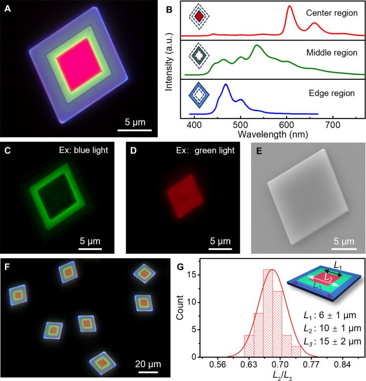
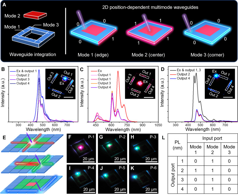
Two-dimensional (2D) organic lateral heterostructures (OLHs) integrating two or more components have important potential applications in optoelectronics. However, the controlled synthesis of 2D OLHs with in-plane tunable emission remains a great challenge owing to the difficulty in the sequential integration of multiple components. Here, a cascaded strategy is demonstrated for the hierarchical assembly of OLHs with in-plane multicolor emission, from red-blue and red-green to lateral red-green-blue (RGB), with a lateral size of ~15 micrometers. The tunable regions of 2D OLHs are realized by synergistic effects of the molecular doping method and the photo-induced oxidation route during the epitaxial growth process. The obtained OLHs can actively achieve full-spectrum light transport from 420 to 720 nanometers depending on different excitation positions and thus function as multimode RGB signal converters. These findings provide insights into the epitaxial growth of OLHs for the development of next-generation organic optoelectronics.


Figures

Fig. 1.
Fig. 1.. Synthesis strategy of 2D OLHs.
(A) Schematic illustration of the self-assembly of a series of 2D heterostructures and the FRET process. (B) FM image of OLH-1 platelets with red and blue emission. (C) FM image of OLH-2 platelets with red and green emission. (D) FM image of OLH-3 platelets with red, green, and blue emission. Scale bars, 20 μm.
Fig. 2.
Fig. 2.. Characterization of OLH-1 platelets.
(A) FM image of a single OLH-1 platelet. Scale bar, 10 μm. (B) PL mapping of the OLH-1 platelets at 450 nm. Scale bar, 10 μm. (C) PL mapping of the OLH-1 platelets at 670 nm. Scale bar, 10 μm. (D) Corresponding micro-area PL spectra collected from the highlighted positions in (B) and (C). (E) FM image of OLH-1 platelets in area 1. Scale bar, 20 μm. (F) L2/L1 ratio distribution of OLH-1 platelets collected from the four areas. (G) Low-magnification TEM image of an OLH-1 platelet. Scale bar, 500 nm. (H and I) Selected area electron diffraction (SAED) patterns of an OLH-1 platelet measured in the blue (H) and red (I) sections. a.u., arbitrary units.
Fig. 3.
Fig. 3.. Characterization of the OLH-2 platelets.
(A) Schematic illustration of the synthesis of OLH-2 platelets. (B) FM image of individual OLH-2 platelets. Scale bar, 5 μm. (C) FM images of corresponding OLH-2 platelets excited with blue light (460 to 490 nm). Scale bar, 5 μm. (D) FM images of corresponding OLH-2 platelets excited with green light (500 to 550 nm). Scale bar, 5 μm. (E) PL spectra collected from the center and edge parts of the OLH-2 platelet. Inset: FM images collected at two different parts under a 375-nm laser beam. (F to I) Low-magnification AFM images of OLH-2 platelets of different heights, which are respectively synthesized at different crystallization temperatures. Scale bars, 10 μm (F) and 5 μm [(G) to (I)]. (J) The AFM height curves of the OLH-2 platelets prepared at different growth temperatures. (K) Temperature-length correlation curve (blue) and temperature-height correlation curve (orange) of OLH-2 platelets.
Fig. 4.
Fig. 4.. Characterization of the OLH-3 platelets.
(A) FM image of single OLH-3 platelet. Scale bar, 5 μm. (B) Corresponding spatially resolved PL spectra of OLH-3 platelets collected from the red, green, and blue regions. (C) FM image of OLH-3 platelet excited with blue light (460 to 490 nm). Scale bar, 5 μm. (D) FM images of OLH-3 platelet excited with green light (500 to 550 nm). Scale bar, 5 μm. (E) SEM image of corresponding OLH-3 platelet. Scale bar, 5 μm. (F) FM image of OLH-3 platelets. Scale bar, 20 μm. (G) L2/L3 ratio distribution of OLH-3 platelets.
Fig. 5.
Fig. 5.. Optical applications and multimode logic gate functions of platelets.
(A) Schematic diagram of three basic optical logic operation modes in OLH-1 platelets. (B to D) Corresponding spatially resolved PL spectra of the input and output signals collected at the excitation (Ex; input) and output positions of the OLH-1 platelet. Insets: FM images of OLH-1 platelets excited by 375-nm UV light at different excitation positions. Scale bars, 10 μm. (E) Schematic diagram of the OLH-3 platelet and different excitation positions. (F to K) FM images of OLH-3 platelets excited by 375-nm UV light at six different positions (including P-1, P-2, P-3, P-4, P-5, and P-6), corresponding to six signal conversion situations. Scale bars, 20 μm. (L) Summarized table for the optical multimode logic gate functions of the obtained OLHs.

References

    1. Geim A. K., Grigorieva I. V., Van der Waals heterostructures. Nature 499, 419–425 (2013). - PubMed
    1. Xu W., Liu W., Schmidt J. F., Zhao W., Lu X., Raab T., Diederichs C., Gao W., Seletskiy D. V., Xiong Q., Correlated fluorescence blinking in two-dimensional semiconductor heterostructures. Nature 541, 62–67 (2017). - PubMed
    1. Choi W., Ahn J., Kim K. T., Jin H. J., Hong S., Hwang D. K., Im S., Ambipolar channel p-TMD/n-Ga2O3 junction field effect transistors and high speed photo-sensing in TMD channel. Adv. Mater. 33, e2103079 (2021). - PubMed
    1. Ma C., Shi Y., Hu W., Chiu M. H., Liu Z., Bera A., Li F., Wang H., Li L. J., Wu T., Heterostructured WS2/CH3NH3PbI3 photoconductors with suppressed dark current and enhanced photodetectivity. Adv. Mater. 28, 3683–3689 (2016). - PubMed
    1. Tan C., Zhang H., Epitaxial growth of hetero-nanostructures based on ultrathin two-dimensional nanosheets. J. Am. Chem. Soc. 137, 12162–12174 (2015). - PubMed

LinkOut - more resources