. 2025 Mar 7;11(10):eadt1763.
doi: 10.1126/sciadv.adt1763. Epub 2025 Mar 5.

Oxytocin induces embryonic diapause

Affiliations

Oxytocin induces embryonic diapause

Jessica L Minder et al. Sci Adv. .

Abstract

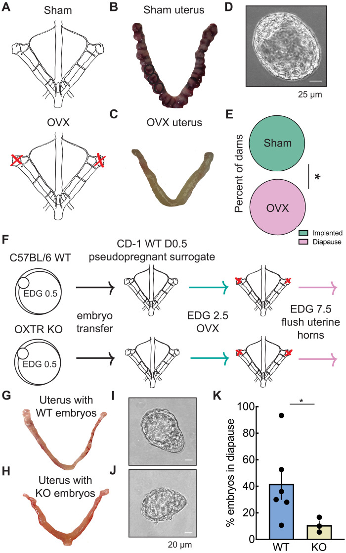
Embryonic development in many species, including case reports in humans, can be temporarily halted before implantation during a process called diapause. Facultative diapause occurs under conditions of maternal metabolic stress such as nursing. While molecular mechanisms of diapause have been studied, a natural inducing factor has yet to be identified. Here, we show that oxytocin induces embryonic diapause in mice. We show that gestational delays were triggered during nursing or optogenetic stimulation of oxytocin neurons simulating nursing patterns. Mouse blastocysts express oxytocin receptors, and oxytocin induced delayed implantation-like dispersion in cultured embryos. Last, oxytocin receptor-knockout embryos transferred into wild-type surrogates had low survival rates during diapause. Our results indicate that oxytocin coordinates timing of embryonic development with uterine progression through pregnancy, providing an evolutionarily conserved mechanism for ensuring successful reproduction.


Figures

Fig. 1.
Fig. 1.. OXTRs help pups survive lactationally-induced diapause.
(A) Experiment outline; dams are introduced to male for breeding immediately after delivery of first litter; males removed 24 hours later to mark initiation of second pregnancy (second litter). Dams are singly housed with pups from first litter until weaning at 3 weeks. (B to D) Gestation in absence (first litter) versus presence (second litter) of lactation. Bars, mean ± SEM. (B) CD-1 gestation: 20.1 ± 0.5 days in absence of nursing (N = 9 litters) versus 27.1 ± 2.5 days (N = 5 litters) while dam was nursing (P = 0.004, Mann-Whitney unpaired two-tailed t test, U = 2). (C) C57BL/6 gestation: 20.3 ± 0.3 days (N = 14 litters) without nursing versus. 28.8 ± 1.8 days (N = 3 litters) with nursing (P = 0.002, Mann-Whitney unpaired two-tailed t test, U = 0). (D) Gestation from breeding C57BL/6 OXTR+/− heterozygous mice: 20.5 ± 0.3 days (N = 6 litters) without nursing versus 30.6 ± 0.8 days (N = 7 litters) when nursing (P = 0.001, Mann-Whitney unpaired two-tailed t test, U = 0). (E) Average % pups with a litter with a given genotype after breeding OXTR+/− mice for litter 1 (WT 19.32%, Het 51.11%, KO 29.57%, N = 10 litters, 67 pups) versus litter 2 when nursing first litter (WT 28.13%, Het 56.94%, KO 14.93%, N = 7 litters, 51 pups; P = 0.0426, one-tailed χ2 test comparing the numbers of wild-type pups to KO + het pups between first and second litters).
Fig. 2.
Fig. 2.. Patterned optogenetic oxytocin neuron activation induces diapause.
(A) Mouse dam nursing frequency. Dam co-housed with litter, recorded for 18 hours with cameras from top, side, and below nest. Nursing bouts occurred every 1 to 3 hours for 20 to 180 min, defined as time spent with 1+ pup visually attached to nipple. (B and C) Photometry from PVN oxytocin neurons when dam out-of-nest or in-nest with pups latched onto nipples. Large dF/F transients evoked during latching. (D) AAV1-EF1a-fl/fl-hChR2(H134R)-mCherry-WPRE channelrhodopsin virus injected bilaterally into PVN in Oxytocin-Cre females, optical fiber implanted midline at PVN. (E) Viral targeting and fiber placement confirmed for optogenetic stimulation between E2.5 to 7.5 in “nursing-like” pattern (20 Hz for 30 min, every 2.5 hours over 5 days). (F) Example uterus from control dam (normal pregnancy, note the implantation sites at E7.5, example sites indicated by black circles) and dam that underwent optogenetic stimulation (note the absence of implantation sites). (G) Top, example diapaused embryo at E7.5 from dam that underwent optogenetic stimulation. Bottom, confirmation that diapaused embryo was viable, resuming development in vitro. Circle, embryo; arrows, expanded trophectoderm cells. (H) Five of six dams underwent diapause in response to nursing-patterned optogenetic stimulation of PVN oxytocin neurons, while 100% of sham-stimulated dams (N = 9 of 9) had normal pregnancy progression (P = 0.002, Fisher’s exact two-tailed test). (I) 100% of oxytocin-Cre dams (N = 6 of 6) had normal pregnancies when oxytocin neurons were chemogenetically activated (DREADDe, hM3D9Gq) via C21 in drinking water; comparison group with 100% normal pregnancies were same genotype expressing empty virus and also receiving C21 in drinking water (N = 5 of 5). (J) 100% of wild-type dams receiving oxytocin injections i.p. terminated pregnancies (N = 6 of 6), compared to four of six wild-types with normal pregnancies receiving saline injections i.p.
Fig. 3.
Fig. 3.. Mouse blastocysts express oxytocin receptors.
(A) Selective monoclonal antibodies to mouse oxytocin receptor (OXTR-1). Left, immmunoblot of HEK cells expressing mouse oxytocin receptors (OXTR) versus control HEK cells (C). Cells lysed, immunoblotted with OXTR-1, showing selective detection in transfected receptor-expressing cells but not untransfected control cells. Red box, region around expected molecular weight of oxytocin receptor (43 kDa). Glyceraldehyde-3-phosphate dehydrogenase, loading controls. Middle and right, validation of monoclonal OXTR-1 antibodies with immunohistochemistry labeling of PVN in wild-type (middle) and OXTR KO (right) dams. (B) Left, OXTR monoclonal antibody labeling on C57BL/6 blastocyst at E3.5 (40×). Middle, DAPI stain of same blastocyst. Right, merge. Dashed circles, absence of OXTRs on actively dividing cells. (C) OXTR-2 polyclonal antibody labeling on C57BL/6 mouse mammary tissue from wild-type (top) and OXTR KO (bottom) dams. (D) Left, OXTR polyclonal antibody labeling on C57BL/6 mouse blastocyst at E3.5 imaged at 40×. Middle, DAPI. Right, merge. Note the absence of OXTRs on actively dividing cell (dashed circle). (E) Quantification of blastocyst OXTR+ expression. Left, number of OXTR+ cells (% of total, defined as ≥5 fluorescent puncta). AB, labeled with OXTR-1 monoclonal antibodies (filled circles) or OXTR-2 polyclonal antibodies (open circle). C, control blastocysts labeled with secondary antibodies only. Right, number of puncta per OXTR+ cell. Mean ± SEM. (F) Left, genetic labeling of OXTRs via tdTomato expression under control of the OXTR promoter, generated by crossing homozygous OXTR cDNA(HA)-Ires-Cre mice to homozygous B6.Cg-Gt(ROSA)26Sortm9(CAG-tdTomato)Hze/J. Blue, DAPI. Blastocyst mounted in fresh agarose, imaged by light sheet microscopy (20×). Right, bright-field image of same blastocyst. Dashed circle, inner cell mass; outer ring of cells is trophectoderm (identified morphologically). Inside of embryo is fluid-filled cavity (blastocoel). OXTRs are expressed by both cell types.
Fig. 4.
Fig. 4.. Mouse blastocyst OXTR signaling induces diapause-like state in vitro.
(A) Mouse embryos harvested at E3.5 and cultured for several days. (B) Mouse blastocysts imaged at 40× harvested on E3.5, treated with either oxytocin (OT) or PBS for 36 hours. Note the expansion of trophectoderm cells in PBS-treated but not OT-treated blastocysts. (C to F) E3.5 blastocysts were imaged every 12 hours for 4 to 5 days after treatment with OT (1 to 10 μM) versus PBS, or mTOR inhibitor INK-128 (100 nM) versus DMSO. All embryos scored here implanted by 5 days. (C) OT-treated CD-1 (top) and C57BL/6 blastocysts (bottom) delayed implantation. By 36 hours, 16 of 33 OT-treated versus 26 of 31 PBS-treated CD-1 blastocysts implanted (P = 0.001, one-tailed χ2 test). (D) OXTR KO blastocysts did not delay implantation after OT treatment (10 μM, n = 12), similar to PBS-treated OXTR KO embryos (n = 11). (E) C57BL/6 blastocysts treated with INK-128 (n = 7) delayed implantation for several days versus DMSO controls (n = 6). (F) OT treatment delayed implantation of CD-1 embryos at 48 and 60 hours but not 72 hours (top). At 60 hours, OT treatment delayed implantation of wild-type but not OXTR KO embryos; at 60 hours, INK-128 treatment also delayed C57BL/6 implantation. Mean ± 95% confidence intervals. (G) Incorporated EU into newly synthesized RNAs of blastocyst treated with PBS versus 1 μM OT for 19.5 hours. (H) 70% less EU incorporation in OT-treated embryos versus PBS treatment (PBS-treated normalized to 1.0 ± 0.1 units, OT-treated had 0.3 ± 0.1 units of EU incorporation, P = 0.004, Student’s unpaired two-tailed t test). Mean ± SEM.
Fig. 5.
Fig. 5.. OXTR signaling helps embryos survive diapause.
(A) Ovariectomized (OVX) females had ovaries removed at E2.5 postmating; ovaries of sham animals remained intact but had similar surgical procedure. (B) Uterus at E7.5 of sham surgery dam that did not undergo diapause. Black circles, highlighted implantation sites. (C) Uterus at E7.5 of OVX dam contains diapaused embryos; note the absence of implantation sites. (D) Diapaused embryo at E7.5 obtained from OVX dam. (E) 100% of sham-surgery dams had normal pregnancy progression (green, N = 4) versus 100% of OVX dams with diapause (pink, N = 3, P = 0.0286, Fisher’s exact two-tailed test). (F) Embryo transfer experiments: either 100% of E0.5 embryos were wild-type C57BL/6 or 100% OXTR KO, and were transferred into oviducts of pseudopregnant day 0.5 CD-1 surrogates. All surrogates underwent diapause induction by ovariectomy on E2.5, treated daily with progesterone (2 mg/kg) from E3.5 to 6.5, uterine horns flushed on E7.5. (G) Uterus from OVX surrogate after wild-type embryo transfer. (H) Uterus from OVX surrogate after OXTR KO embryo transfer. Note absence of implantation sites in (G) and (H). (I and J) Wild-type (I) and OXTR KO (J) diapaused embryos obtained from OVX surrogates after embryo transfer. (K) 30% fewer OXTR KO embryos were recovered after OVX-induced diapause (41.8 ± 11.7% of wild-type embryos entered diapause versus 10.7 ± 3.2% of OXTR KO embryos entering diapause, P = 0.045, Student’s unpaired two-tailed t test). Mean ± SEM.

References

    1. Renfree M. B., Fenelon J. C., The enigma of embryonic diapause. Development 144, 3199–3200 (2017). - PubMed
    1. Lopes L., Desmarais J. A., Murphy B. D., Embryonic diapause and its regulation. Reproduction 128, 669–678 (2004). - PubMed
    1. Renfree M. B., Shaw G., Diapause. Annu. Rev. Physiol. 62, 353–375 (2000). - PubMed
    1. Fenelon J. C., Banerjee A., Murphy B. D., Embryonic diapause: Development on hold. Int.J. Dev. Biol. 58, 163–174 (2014). - PubMed
    1. Norris M. L., Adam C. E., Concurrent lactation and reproductive performance in CFLP mice mated post partum. Lab. Anim. 15, 273–275 (1981). - PubMed

LinkOut - more resources