. 2025 Apr 3;112(4):952-962.
doi: 10.1016/j.ajhg.2025.02.005. Epub 2025 Mar 4.

Bi-allelic variants in MRPL49 cause variable clinical presentations, including sensorineural hearing loss, leukodystrophy, and ovarian insufficiency

Affiliations

Bi-allelic variants in MRPL49 cause variable clinical presentations, including sensorineural hearing loss, leukodystrophy, and ovarian insufficiency

Huw B Thomas et al. Am J Hum Genet. .

Abstract

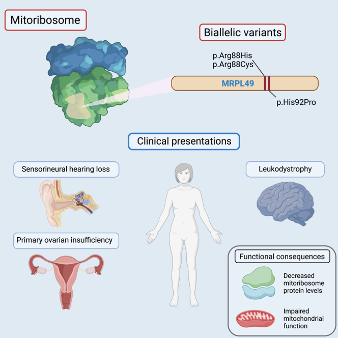
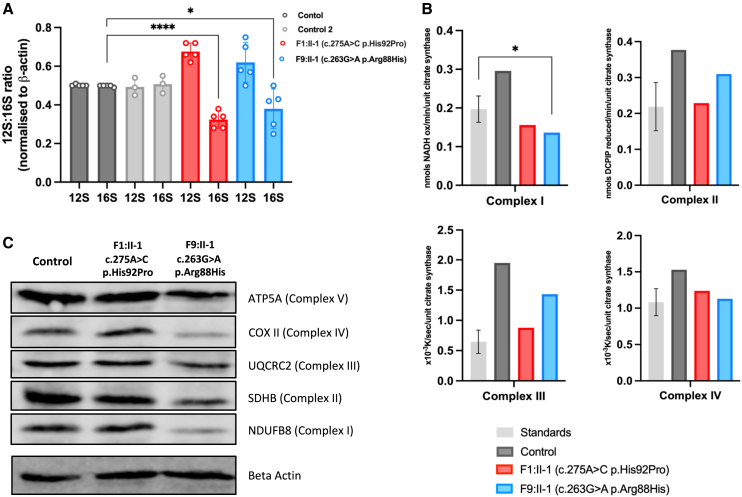
Combined oxidative phosphorylation deficiency (COXPD) is a rare multisystem disorder that is clinically and genetically heterogeneous. Genome sequencing identified bi-allelic MRPL49 variants in individuals from nine unrelated families with presentations ranging from Perrault syndrome (primary ovarian insufficiency and sensorineural hearing loss) to severe childhood onset of leukodystrophy, learning disability, microcephaly, and retinal dystrophy. Complexome profiling of fibroblasts from affected individuals revealed reduced levels of the small mitochondrial ribosomal subunits and a more pronounced reduction of the large mitochondrial ribosomal subunits. There was no evidence of altered mitoribosomal assembly. The reductions in levels of oxidative phosphorylation (OXPHOS) enzyme complexes I and IV are consistent with a form of COXPD associated with bi-allelic MRPL49 variants, expanding the understanding of how disruption of the mitochondrial ribosomal large subunit results in multisystem phenotypes.

Keywords: MRPL49; Perrault syndrome; combined oxidative phosphorylation deficiency; learning disability; leukodystrophy; mitochondria; mitoribosome; primary ovarian insufficiency; rare disease; sensorineural hearing loss.


Conflict of interest statement

Declaration of interests The authors declare no competing interests.

Figures

None
Graphical abstract
Figure 1
Figure 1
Pedigree and clinical data for families with bi-allelic MRPL49 variants (A) Pedigree information for nine unrelated families of individuals with either homozygous or compound heterozygous variants in MRPL49 (GenBank: NM_004927.4) and (B) accompanying clinical details of POI, SNHL, and learning disability shared across probands.
Figure 2
Figure 2
Selected brain MR images demonstrating features of individuals with variants in MRPL49 and an accompanying summary of all imaging features shared across 8 individuals from 6 unrelated families All 8 affected individuals with available brain imaging had symmetrical high T2 signal changes in the globi pallidi (i). This feature progressed to symmetrical cystic changes in two affected individuals with more advanced disease (ii). In 3/8 affected individuals, there was symmetrical high T2 signals and diffusion restriction (not pictured) involving the brain stem, including the substantia nigra (iii) and the dorsal brainstem. In 5/8 affected individuals, there were symmetrical white matter high T2 signal changes, becoming more diffuse and confluent with more advanced disease. There was also evidence of cerebral atrophy in 5/8 affected individuals, particularly involving the cerebellum (iv) but also affecting cortical gray matter and sub-cortical and deep white matter in individuals with more advanced disease (ii and v). The chart summarizes the features noted on imaging of the affected individuals.
Figure 3
Figure 3
In silico modeling of MRPL49 variants (A) Schematic representation of MRPL49 transcript and disease-associated variant locations. All three missense variants are clustered in exon 3, whereas the single frameshift variant is located in exon 2. (B) Evolutionary conservation of MRPL49 affected residues across a broad range of orthologous species. Conserved residues are colored blue, and variant amino acids (arginine 88 and histidine 92) are highlighted in red. (C) Three-dimensional representation of the location of MRPL49 (orange) within the human mitoribosome (PDB: 7QI4). Large (mt-LSU) and small (mt-SSU) units are colored in gray, 16S rRNA in green, and 12S rRNA in yellow.
Figure 4
Figure 4
Functional and molecular assays of fibroblasts from affected individuals reveal significant reductions in levels and activities of mitochondrial respiratory chain complexes I and IV (A) MT-RNR1 (12S) and MT-RNR2 (16S) relative expression levels in fibroblasts from affected individuals and control individuals. Data are expressed as a ratio using relative expression to beta-actin. Error bars represent SD. ∗∗∗∗p < 0.0001 and p = 0.0327; unpaired t test. (B) Mitochondrial respiratory chain enzyme activity assay in control (gray), F1:II-1 (red), and F9:II-1 (blue) fibroblasts. (C) Western blot analysis of proteins associated with each of the mitochondrial oxidative phosphorylation complexes in fibroblasts from F1:II-1, F9:II-1, and control individuals. Image is representative of three separate experiments.
Figure 5
Figure 5
Complexome analysis of mitochondrial ribosomes and OXPHOS complexes from control and F1:II-1- and F9:II-1-derived fibroblasts Enriched mitochondria were solubilized with digitonin, separated by BNE, and analyzed by mass spectrometry (MS)-based complexome profiling. Protein abundance profiles of each mitoribosomal subunit and OXPHOS complex were generated by averaging the intensity-based absolute quantification (iBAQ) values of all their individual subunits identified by MS. Resultant profiles are illustrated as heatmaps and two-dimensional (2D) profile plots against the apparent molecular mass. (A) Abundance profiles of mitoribosomal small (mt-SSU) and large (mt-LSU) subunits showing a visible decrease in the fibroblasts from affected individuals. (B) Abundance profiles of the five mitochondrial OXPHOS complexes consistently showing a drop in their content, particularly for complexes I and IV. CI, CII, CIII, CIV, and CV stand for complexes I, II, III, IV, and V, respectively. S1, respiratory supercomplex formed by complexes I, III, and IV; SCs, higher-order respiratory complexes (respirasomes).

Update of

References

    1. Liu M., Spremulli L. Interaction of mammalian mitochondrial ribosomes with the inner membrane. J. Biol. Chem. 2000;275:29400–29406. doi: 10.1074/jbc.M002173200. - DOI - PubMed
    1. Koc E.C., Burkhart W., Blackburn K., Moyer M.B., Schlatzer D.M., Moseley A., Spremulli L.L. The large subunit of the mammalian mitochondrial ribosome. Analysis of the complement of ribosomal proteins present. J. Biol. Chem. 2001;276:43958–43969. doi: 10.1074/jbc.M106510200. - DOI - PubMed
    1. Brown A., Amunts A., Bai X.C., Sugimoto Y., Edwards P.C., Murshudov G., Scheres S.H.W., Ramakrishnan V. Structure of the large ribosomal subunit from human mitochondria. Science. 2014;346:718–722. doi: 10.1126/science.1258026. - DOI - PMC - PubMed
    1. Greber B.J., Boehringer D., Leibundgut M., Bieri P., Leitner A., Schmitz N., Aebersold R., Ban N. The complete structure of the large subunit of the mammalian mitochondrial ribosome. Nature. 2014;515:283–286. doi: 10.1038/nature13895. - DOI - PubMed
    1. Ferrari A., Del’Olio S., Barrientos A. The Diseased Mitoribosome. FEBS Lett. 2021;595:1025–1061. doi: 10.1002/1873-3468.14024. - DOI - PMC - PubMed

Supplementary concepts