. 2025 Mar 5;16(1):2204.
doi: 10.1038/s41467-025-57412-6.

Structural basis for transcription activation through cooperative recruitment of MntR

Affiliations

Structural basis for transcription activation through cooperative recruitment of MntR

Haoyuan Shi et al. Nat Commun. .

Abstract

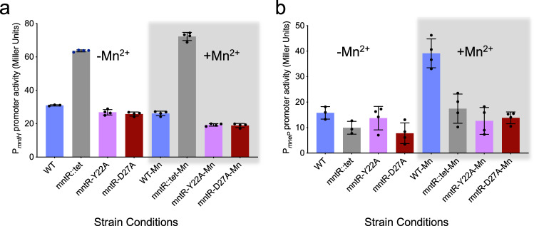
Bacillus subtilis MntR is a dual regulatory protein that responds to heightened Mn2+ availability in the cell by both repressing the expression of uptake transporters and activating the expression of efflux proteins. Recent work indicates that, in its role as an activator, MntR binds several sites upstream of the genes encoding Mn2+ exporters, leading to a cooperative response to manganese. Here, we use cryo-EM to explore the molecular basis of gene activation by MntR and report a structure of four MntR dimers bound to four 18-base pair sites across an 84-base pair regulatory region of the mneP promoter. Our structures, along with solution studies including mass photometry and in vivo transcription assays, reveal that MntR dimers employ polar and non-polar contacts to bind cooperatively to an array of low-affinity DNA-binding sites. These results reveal the molecular basis for cooperativity in the activation of manganese efflux.


Conflict of interest statement

Competing interests: The authors declare no relevant financial or non-financial competing interests.

Figures

Fig. 1
Fig. 1. Structure of 4xMntR2-P84 complex.
a Cryo-EM structure of MntR2-P84 complex showcasing the organization of the four MntR dimers (tan, slate blue, aquamarine blue, and purple) with respect to P84 (orange). The P84 sequence is displayed below highlighting the four 18-bp MntR dimer binding sites (site 1-4) in color. The black box highlights the RNA polymerase (RNAP) binding site (−35). b An alternate view of the 4xMntR2-P84 structure after a 90° rotation compared to (a) looking down the DNA helical axis. c A close-up view of the boxed region from (a) showcasing the cryo-EM density for some of the amino acids in the MntR dimer-dimer and MntR-DNA interface from the 3.09 Å 2xMntR2-P84 map.
Fig. 2
Fig. 2. MntR-DNA interactions.
a A portion of the cryoEM structure of the 2xMntR2-P84 complex highlighting a single dimer of MntR in surface representation (tan) bound to an 18-bp operator region on P84 (orange). Mn2+ ions are presented as yellow spheres. The buried surface area of an MntR monomer is highlighted in blue interacting with a major and minor groove of P84 highlighted in gray. b A schematic presenting the interactions between amino acids of MntR (blue) with the nucleotides of P84 (gray) in the boxed region in (a). The base pair at −7/ + 7 position is always a TA base pair (highlighted in dark blue in (a) and (b)) in all 8 half sites in the 4xMntR2-P84. The label, @N, indicates an interaction with the backbone amide nitrogen. His35, Pro36, Ser37 and Lys41 interact with the base edges in the major groove and Tyr57 interacts within the minor groove.
Fig. 3
Fig. 3. Interdimer interactions observed in the 2xMntR2-P84 structure.
a A cartoon representation of the cryo-EM structure of the 2xMntR2-P84 complex with two MntR dimers highlighted in green and blue, with each dimer bound to 4 Mn2+ ions (yellow spheres). The MntR dimers are interacting with a portion of P84 (orange). Tyr22 and Asp27 are highlighted in the cartoon. b A schematic representing a summary of interactions observed between the two adjacent dimers of MntR in the 2xMntR2-P84 structure. Polar interactions such as dipole-dipole interactions, H-bonds, and salt-bridges are highlighted by black lines and hydrophobic or vdW interactions are highlighted by orange lines. The dotted lines represent weaker interactions between residues that are separated by a distance ~4–5 Å. Note that the side chain of Tyr22 and Val 61 are interacting with the backbone of Leu60 and Lys20, respectively. c Expanded views of the network of interdimer contacts around the conserved Asp27 (left) and Tyr22 (right). The DNA surface in the left box is colored by charge distribution, while amino acids in the right box are shown as sticks and semi-transparent van der Waals surfaces. The black dotted lines represent polar interactions including salt bridges and H-bonds.
Fig. 4
Fig. 4. Complex formation of WT and mutant MntR with P84 in solution.
Mass photometry data on (a) WT MntR, (b) Y22A MntR and (c) D27A MntR alone (gray) and in complex with the P84 (colored) is presented. The gray broken arrow indicates the expected mass of the 4xMntR2-P84 complex. MP data on P84 alone (light brown) is presented in (a). df endogenous tryptophan FSEC data was collected on (d) WT MntR, (e) Y22A MntR and (f) D27A MntR alone (gray) and in complex with P84 (colored). The gray broken arrow indicates the expected retention volume of the 4xMntR2-P84 complex. Source data are provided as a Source Data file.
Fig. 5
Fig. 5. Binding studies on WT and mutant MntR with C84.
Mass photometry data on (a) WT MntR, (b) Y22A MntR and (c) D27A MntR alone (gray) and in complex with C84 (colored). The gray broken arrow indicates the expected mass of the 4xMntR2-C84 complex. MP data on C84 alone (light brown) is presented in (a). df endogenous tryptophan FSEC data on (d) WT MntR, (e) Y22A MntR and (f) D27A MntR alone (gray) and in complex with the C84 (colored). The gray broken arrow indicates the expected retention volume of the 4xMntR2-C84 complex. Source data are provided as a Source Data file.
Fig. 6
Fig. 6. Mutations affecting interdimer contacts affect transcription activation but not repression.
β-Galactoside reporter gene assays in B. subtilis to study the effect of Y22A (pink) and D27A (red) mutations in MntR on the activity of (a) the Mn2+ repressed mntH promoter and (b) Mn2+ activated mneP promoter. Promoter activities were measured in the presence (gray box) and absence of Mn2+ ions. The gray bars in the histograms represent the mntR::tet null mutant of B. subtilis and the blue bars represent wild type (WT) B. subtilis. Promoter activity was averaged over four (n = 4) independent replicates and the standard deviation is presented as error bars. Source data are provided as a Source Data file.

Update of

References

    1. Chandrangsu, P., Rensing, C. & Helmann, J. D. Metal homeostasis and resistance in bacteria. Nat. Rev. Microbiol15, 338–350 (2017). - PMC - PubMed
    1. Osman, D. et al. Bacterial sensors define intracellular free energies for correct enzyme metalation. Nat. Chem. Biol.15, 241–249 (2019). - PMC - PubMed
    1. Capdevila, D. A., Edmonds, K. A. & Giedroc, D. P. Metallochaperones and metalloregulation in bacteria. Essays Biochem.61, 177–200 (2017). - PMC - PubMed
    1. Baksh, K. A. & Zamble, D. B. Allosteric control of metal-responsive transcriptional regulators in bacteria. J. Biol. Chem.295, 1673–1684 (2020). - PMC - PubMed
    1. Gilston, B. A. et al. Structural and mechanistic basis of zinc regulation across the e. coli Zur regulon. PLoS Biol.12, e1001987 (2014). - PMC - PubMed

MeSH terms

LinkOut - more resources