. 2025 Mar;639(8055):691-699.
doi: 10.1038/s41586-025-08689-6. Epub 2025 Mar 5.

Genome duplication in a long-term multicellularity evolution experiment

Affiliations

Genome duplication in a long-term multicellularity evolution experiment

Kai Tong et al. Nature. 2025 Mar.

Abstract

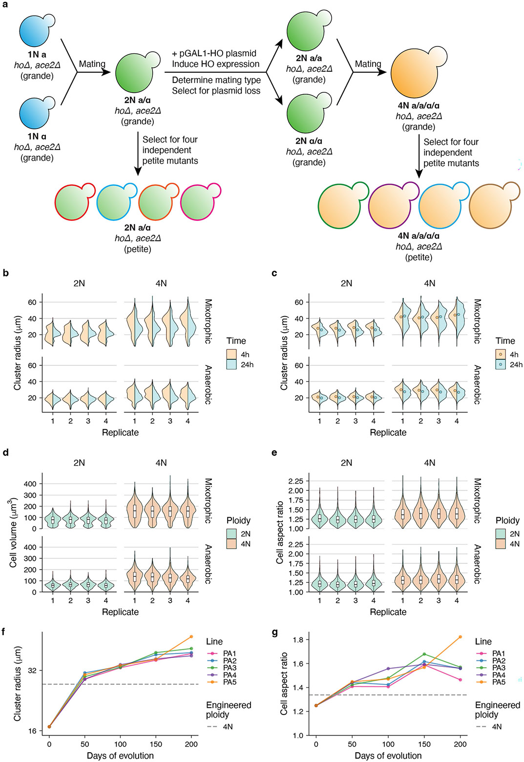
Whole-genome duplication (WGD) is widespread across eukaryotes and can promote adaptive evolution1-4. However, given the instability of newly formed polyploid genomes5-7, understanding how WGDs arise in a population, persist, and underpin adaptations remains a challenge. Here, using our ongoing Multicellularity Long Term Evolution Experiment (MuLTEE)8, we show that diploid snowflake yeast (Saccharomyces cerevisiae) under selection for larger multicellular size rapidly evolve to be tetraploid. From their origin within the first 50 days of the experiment, tetraploids persisted for the next 950 days (nearly 5,000 generations, the current leading edge of our experiment) in 10 replicate populations, despite being genomically unstable. Using synthetic reconstruction, biophysical modelling and counter-selection, we found that tetraploidy evolved because it confers immediate fitness benefits under this selection, by producing larger, longer cells that yield larger clusters. The same selective benefit also maintained tetraploidy over long evolutionary timescales, inhibiting the reversion to diploidy that is typically seen in laboratory evolution experiments. Once established, tetraploidy facilitated novel genetic routes for adaptation, having a key role in the evolution of macroscopic multicellular size via the origin of evolutionarily conserved aneuploidy. These results provide unique empirical insights into the evolutionary dynamics and impacts of WGD, showing how it can initially arise due to its immediate adaptive benefits, be maintained by selection and fuel long-term innovations by creating additional dimensions of heritable genetic variation.


Conflict of interest statement

Competing interests: The authors declare no competing interests.

Figures

Extended Data Fig. 1 ∣
Extended Data Fig. 1 ∣. Images of the ancestors and evolved isolates in the MuLTEE.
a,b, Representative cluster-level (a) and cell-level (b) images of PM/PA t0 and PM/PA1–5 t200, t400, t600, and t1000 isolates. The images of PM/PA t0 are reused for five replicate populations. Scale bars, 200 μm (a) and 10 μm (b).
Extended Data Fig. 2 ∣
Extended Data Fig. 2 ∣. Phenotypic characterization of the ancestors and evolved isolates in the MuLTEE.
a-d, Violin plots showing the distributions of cluster radius (a,b, where b is weighted by cluster volume), cell volume (c), and cell aspect ratio (d) in PM/PA t0 and PM/PA1–5 t200, t400, t600, and t1000 isolates (on average, n = 789 clusters (a,b) and 1,288 cells (c,d) measured per sample). The distributions of PM/PA t0 are reused for five replicate populations. For a,b, we measured cluster radius at 4 h (exponential phase) and 24 h(stationary phase) after transferring the culture to fresh media, and the 24-hour measurements (corresponding to the states of the cultures right before settling selection) are used throughout the paper unless otherwise noted. For b, filled circles show biomass-weighted mean cluster radius (the 24-hour values are the same as the values in Fig. 1c). For c,d, boxes, IQR; center lines, median; whiskers, values within 1.5 × IQR of the first and third quartiles.
Extended Data Fig. 3 ∣
Extended Data Fig. 3 ∣. Imaging-based method for measuring ploidy level of snowflake yeast.
a, Overview of imaging and image analysis workflow. Snowflake yeast clusters are crushed into a single layer of cells and imaged at the brightfield channel and fluorescent channel, with the latter showing the nuclear DNA stained by propidium iodide (PI). The nuclei in the fluorescent image are segmented and filtered to get single round nuclei, outlined in cyan. The fluorescent image is also nuclei-cleared and brightness/contrast-enhanced to show the background fluorescence in the cytoplasm, and the cytoplasm is segmented, shown in white, for background subtraction. The total fluorescence intensity of PI in each nucleus is quantified and background-subtracted. Scale bar, 20 μm. b, Distribution of the nuclear PI intensity (arbitrary unit) of the engineered diploid and tetraploid mixotrophic clusters (n = 14,276 and 10,031 nuclei, respectively), as a validation for this ploidy measurement method. Since asynchronous, exponential-phase cultures are used for ploidy measurements, each strain shows two peaks that correspond to G1- and G2-phase nuclei of the actively-dividing cells, and the G2 peak has double the fluorescent intensity of the G1 peak. Also, the G2 peak of diploid clusters aligns nicely with the G1 peak of tetraploid clusters.
Extended Data Fig. 4 ∣
Extended Data Fig. 4 ∣. Copy number variation of the ancestors and evolved isolates in the MuLTEE.
a,b, Estimated copy number of each 1-kb non-overlapping bin in each chromosome in PM/PA t0 and PM/PA1-5 t200, t400, t600, and t1000 isolates (a, PM; b, PA). Estimated bin copy numbers above 12 are shown as 12, indicated by little triangles. Red horizontal line, baseline ploidy of each strain (i.e., 2 for PM/PA t0 and 4 for all evolved isolates). Red arrowhead, incidence of segmental aneuploidy.
Extended Data Fig. 5 ∣
Extended Data Fig. 5 ∣. Genetic construction and phenotypic characterization of diploid and tetraploid clusters.
a, Procedure for engineering isogenic grande diploid and tetraploid clusters, from each of which four independent petite mutants were isolated. Isolating multiple petite mutants is important because petite mutations are not isogenic and may confound ploidy-phenotype map. Grande and petite clusters correspond to mixotrophic and anaerobic conditions, respectively. b-e, Violin plots showing the distributions of cluster radius (b,c, where c is weighted by cluster volume), cell volume (d), and cell aspect ratio (e) in engineered diploid and tetraploid clusters under mixotrophic and anaerobic conditions (on average, n = 922 clusters (b,c) and 2,458 cells (d,e) measured per sample). Four biological replicates were measured for the mixotrophic condition, and the four independent petite mutants (each with one biological replicate) were measured for the anaerobic condition. For b,c, we measured cluster radius at 4 h (exponential phase) and 24 h (stationary phase) after transferring the culture to fresh media, and the 24-hour measurements are used throughout the paper unless otherwise noted. For c, filled circles show biomass-weighted mean cluster radius (the 24-hour values are the same as the values in Fig. 3e). For d,e, boxes, IQR; center lines, median; whiskers, values within 1.5 × IQR of the first and third quartiles. f,g, Comparison of the biomass-weighted mean cluster radius (f) and mean cell aspect ratio (g) of the engineered petite tetraploid clusters (mean of the four independent petite mutants, the same values as those in Fig. 1c,e) to the PA t0 and PA1-5 t50, t100, t150, and t200 populations (data from Bozdag et al.).
Extended Data Fig. 6 ∣
Extended Data Fig. 6 ∣. Label-free method for distinguishing engineered diploid and tetraploid clusters in competition assays.
a, Brightfield image of a snowflake yeast cluster (an engineered tetraploid mixotrophic cluster is shown) (top), whose bright cells in the cluster edges are detected (bottom). Scale bar, 30 μm. On average, 381 clusters were segmented per sample using this approach. b, Mean area of the five largest cells detected in a cluster can be used to distinguish between the engineered tetraploid and diploid clusters under both mixotrophic and anaerobic conditions, with the dashed line indicating the manually-chosen decision boundary.
Extended Data Fig. 7 ∣
Extended Data Fig. 7 ∣. Experimental evolution of the MuLTEE ancestors and t1000 isolates with selection against larger size.
a, Experimental setup. We evolved PM/PA t0 and PM/PA1-5 t1000 isolates, each with four replicate populations (A, B, C, D), under selection against larger size for 70 days (~500 generations) by growing them on agar in 24-well plates with daily dilution. b, Distributions of cluster radius (weighted by cluster volume) in the ancestral (“anc”) and evolved populations (on average, n = 406 clusters measured per population). Vertical thick solid line, biomass-weighted mean cluster radius of each population. Vertical dashed line, biomass-weighted mean cluster radius of PM/PA t0. Color codes for the evolved ploidy in each t0 population and in each t1000 population are the same as those in Fig. 3h,i, respectively, and the ancestral populations are colored in gray.
Extended Data Fig. 8 ∣
Extended Data Fig. 8 ∣. Experimental evolution of the MuLTEE ancestors, t1000 isolates, and engineered tetraploids with minimal selection.
We evolved PM/PA t0, PM/PA1-5 t1000 isolates, and engineered mixotrophic and anaerobic tetraploids, each with three replicate populations (A, B, C), with minimal selection for 56 days or 28 bottlenecks, by growing them on agar with picking and streaking single colonies every two days. a, Distributions of cellular DNA contents in the ancestral (“anc”) and evolved populations (on average, n = 15,848 cells measured per population). Color code for ploidy change categories is the same as that in Fig. 3j, and the ancestral populations are colored in light gray.
Extended Data Fig. 9 ∣
Extended Data Fig. 9 ∣. Point mutation changes in donut-to-spread transitions.
Two donut-to-spread transitions with near-triploidization were excluded, and mutation allele frequency refers to the corrected allele frequency, calculated by dividing estimated allele copy number with copy number of the chromosome that carries the mutation. LOH, loss of heterozygosity. a,b, Number of mutations in each category of change in allele frequency in all donut-to-spread transitions combined, colored by change in allele copy number (a), and how change in allele frequency is associated with change in chromosome copy number (b). a,b share the color code. c, Number of mutations in each category of change in allele frequency in each donut-to-spread transition, colored by mutation impact. d, Percentage of mutations in each mutation impact category in each donut background, whose total number of mutations is indicated in the brackets. c,d share the color code. e, Comparison of the distribution of mutation impacts, between the mutations that were gained in all donut-to-spread transitions combined and the mutations randomly introduced into yeast genome, as well as between the mutations that underwent loss, LOH, increase, decrease, or maintenance in terms of allele frequency in all donut-to-spread transitions combined and the mutations in all donut backgrounds combined. Number of mutations is indicated in the brackets. P values were calculated by two-sided chi-squared test. f, For each donut-to-spread transition, the percentage of high/moderate-impact mutations in the mutations that increased or decreased in allele frequency is on average not significantly larger than the percentage of high/moderate-impact mutations in the donut background (for increase and decrease, respectively, P = 0.891 and 0.442, t22 = −1.27 and 0.147, one-tailed one-sample Student’s t-test), and is largely explained by random sampling of mutations in the donut background (simulation with random seed = 1). Values are mean ± s.d. (n = 23 donut-to-spread transitions).
Fig. 1 ∣
Fig. 1 ∣. Increased cell volume and aspect ratio drive the evolution of larger multicellular groups in the 1,000-day MuLTEE.
a, Experimental setup of the MuLTEE. b, Representative cluster-level (top two rows) and cell-level (bottom two rows) images of the ancestor (t0) and day 1,000 (t1,000) isolates from the five replicate populations evolved under PM and PA conditions. Scale bars, 200 μm (cluster-level images) and 10 μm (cell-level images). ce, Evolutionary dynamics of biomass-weighted mean cluster radius (Methods; c), mean cell volume (d) and mean cell aspect ratio (e), showing the values of PM/PA t0 and PM/PA1–5 t200, t400, t600 and t1,000 isolates (on average, n = 886 clusters (c) and 1,288 cells (d,e) measured for each of the 42 strains), with the grey dotted and dashed lines representing artificially constructed diploid and tetraploid strains (mean value of the four biological replicates in Fig. 3e,b,c), respectively. fh, Heat maps showing the biophysical simulations of how cell volume and cell aspect ratio affect the mean cluster radius (f), cell number per cluster (g) and cell packing fraction (fraction of the cluster volume occupied by cells; h) of clusters at fragmentation (n = 50 simulated clusters per pair of parameter values).
Fig. 2 ∣
Fig. 2 ∣. Tetraploidy rapidly emerged and was maintained in all PMs and PAs; PAs subsequently evolved extensive aneuploidy.
a, Violin plot of the distribution of allele frequencies (each determined as the proportion of reads containing the mutant allele) of novel point mutations identified in PM/PA1–5 t200, t400, t600 and t1,000 isolates, with each dot representing one point mutation. b, Evolutionary dynamics of DNA content, showing that of PM/PA t0 and the strains used in panel a (on average, n = 11,456 cells measured for each of the 42 strains). c, Ridge plot of the distribution of cellular DNA contents in the PM/PA t0 and PM/PA1–5 t50 and t100 populations (on average, n = 12,922 cells measured for each of the 22 populations). As we used asynchronous cultures for ploidy measurements, a single genotype in a population would form two distinct DNA-content peaks, corresponding to its G1-phase and G2-phase cells. d, Heat maps of the karyotypes of PM/PA t0 and the strains used in panel a, determined based on chromosome coverages from the whole-genome sequencing data.
Fig. 3 ∣
Fig. 3 ∣. Tetraploidy confers immediate phenotypic effects that are beneficial under selection for larger size, driving the origin and maintenance of tetraploidy despite its genomic instability.
a,d, Cell-level (a) and cluster-level (d) images of the engineered diploid and tetraploid clusters under mixotrophic and anaerobic conditions. Scale bars, 5 μm (a) and 50 μm (d). b,c,e, Mean cell volume (b), mean cell aspect ratio (c) and biomass-weighted mean cluster radius (e) of the strains in panels a,d. f, Relationship of the changes in cell volume and cell aspect ratio due to tetraploidization, under mixotrophic and anaerobic conditions. Values are means of the four replicates or petite mutants in panels b,c. g, Per day selection rate of competing engineered tetraploid clusters against diploid counterparts with and without settling selection for 3 days, under mixotrophic and anaerobic conditions. For b,c,e,g, values are mean ± s.e.m. (n = 4 biological replicates for mixotrophic condition or 4 independent petite mutants for anaerobic condition; on average, 2,458 cells (b,c) and 1,077 clusters (e) were measured per replicate or petite mutant, and 381 clusters (g) were measured per sample). P values were calculated by two-tailed Welch’s t-test. h,i, Distribution of cellular DNA contents in the ancestral (Anc) and evolved populations, initiated with PM/PA t0 (h) and PM/PA1–5 t1,000 isolates (i) with four replicate populations (A–D), after selecting against larger size by evolving on agar for 70 days (on average, n = 14,231 cells measured per population). j, Ploidy (G1 peak) changes in the evolved populations, initiated with PM/PA t0, PM/PA1–5 t1,000 isolates, and engineered mixotrophic and anaerobic tetraploids, each with three replicate populations, after evolving with minimal selection for 56 days or 28 bottlenecks (on average, n = 15,848 cells measured per population). Cohen’s d = 0.8 is used as the cut-off for large and small ploidy changes. Grey dashed line indicates no ploidy change.
Fig. 4 ∣
Fig. 4 ∣. Changes in colony morphology allow screening for spontaneous losses of macroscopic multicellularity in PAs.
a, Evolved macroscopic isolates (PA3 t1,000 is shown) form ‘donut’ colonies and occasionally ‘spread’ colonies that cannot form macroscopic clusters. Scale bars, 5 mm. bd, Biomass-weighted mean cluster radius (b), mean cell volume (c) and mean cell aspect ratio (d) of the 25 D–S pairs (each connected by a line) from nine macroscopic isolate backgrounds. The boxes denote the interquartile range (IQR), the centre lines indicate the median, and the whiskers denote the values within 1.5× the IQR of the first and third quartiles. P values were calculated by two-tailed paired Student’s t-test (on average, n = 605 clusters (b) and 1,294 cells (c,d) measured per strain). e,f, Relationship of how cell volume and aspect ratio changed in D–S transitions, with donut and spread strains separated by a linear boundary generated by support vector machine (e), and with each D–S transition connected by an arrow pointing from donut to spread (f).
Fig. 5 ∣
Fig. 5 ∣. Aneuploidy promotes the origin of macroscopic multicellularity in PAs.
a,b, Karyotypes of D–S pairs (a) and karyotype changes in D–S transitions (b). Inside the heatmap (b), dashed lines indicate two D–S transitions with near-triploidization that were excluded from subsequent analyses. Symbols beside the heatmap (b) indicate cases of convergent karyotype changes where two D–S pairs came from the same background (magenta), different backgrounds but the same PA line (green) or different lines (blue), with each case in a different symbol shape. c, For each background, chromosomes with copy number changes in the evolution (evo; diamonds) and loss (bars, showing number of occurrences) of macroscopic size, with the diamond and the bar connected by a dashed line when the copy number of a chromosome changes in both the evolution and loss of macroscopic size. d, Histogram of the number of chromosomes with copy number changes in each D–S pair, coloured by background. e, Changes in cell volume and aspect ratio compared between D–S pairs with convergent karyotype changes, with symbol colour–shape code consistent with panel b and line colour–shape code similarly assigned. fi, Aggregating all backgrounds in panel c, the number of times each chromosome changes its copy number in the loss (f,g) and evolution (h,i) of macroscopic size, coloured by background (f,h; share a colour legend with panel d) or direction of change (g,i).

Update of

References

    1. Otto SP The evolutionary consequences of polyploidy. Cell 131, 452–462 (2007). - PubMed
    1. Selmecki AM et al. Polyploidy can drive rapid adaptation in yeast. Nature 519, 349–352 (2015). - PMC - PubMed
    1. Van de Peer Y, Mizrachi E & Marchal K The evolutionary significance of polyploidy. Nat. Rev. Genet 18, 411–424 (2017). - PubMed
    1. Vittoria MA, Quinton RJ & Ganem NJ Whole-genome doubling in tissues and tumors. Trends Genet. 39, 954–967 (2023). - PMC - PubMed
    1. Comai L The advantages and disadvantages of being polyploid. Nat. Rev. Genet 6, 836–846 (2005). - PubMed

LinkOut - more resources