

The effect of IGF-1 on cartilage injury in bone marrow mesenchymal stem cells through the BMP2-Smad1/5 signaling pathway
- PMID: 40045149
- PMCID: PMC11978553
- DOI: 10.1007/s11626-025-01015-4
The effect of IGF-1 on cartilage injury in bone marrow mesenchymal stem cells through the BMP2-Smad1/5 signaling pathway
Abstract
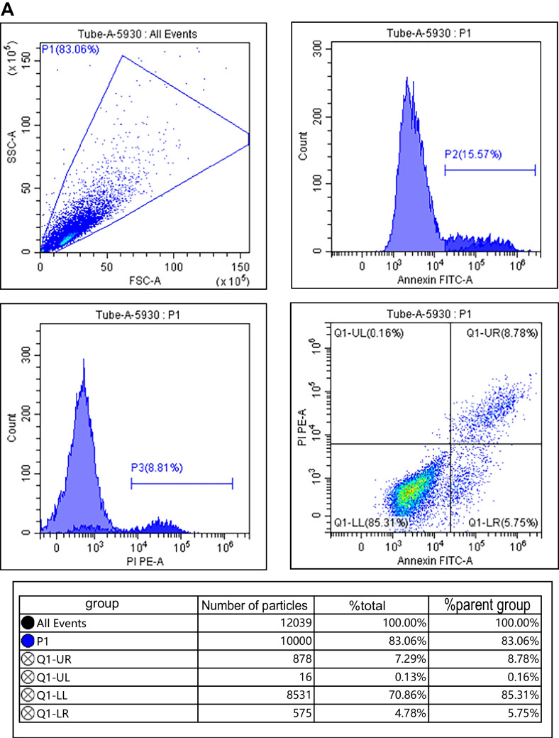
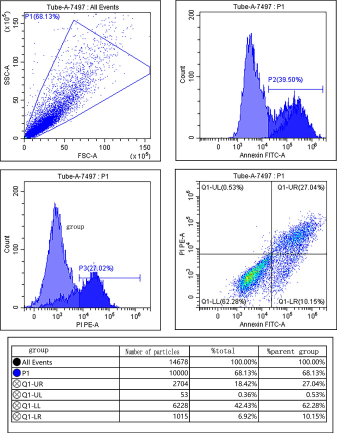
The objective of this study is to analyze the effect of insulin-like growth factor-1 (IGF-1) in bone marrow mesenchymal stem cells (BMSCs) on cartilage injury and explore the regulatory mechanism of IGF-1 on the bone morphogenetic protein 2 (BMP2)-Smad1/5 signaling pathway. We cultivated rat BMSCs in vitro and observed their cell morphology using an inverted microscope. Flow cytometry was used to identify the surface antigen expression of BMSCs. IL-1β is used to induce rat chondrocyte ATDC5 to construct a cartilage injury model. We integrated IGF-1 overexpressed BMSCs, empty vector transfected BMSCs, and BMSCs with IL-1, respectively. IL-1β-induced ATDC5 cells were co-cultured for 24 h. We recorded them as BMSCs + IGF-1 group, BMSCs + empty vector group, BMSCs group, and normal cultured ATDC5 cells as the control group. qRT-PCR and Western blot were used to detect IGF-1 mRNA and protein levels in each group. CCK-8 experiment and flow cytometry were used to detect cell proliferation and apoptosis in each group. ELISA is used to detect the levels of TNF-α, IL-8, and IL-6. Western blot was used to detect protein levels of Bax, Bcl-2, Cleaved Caspase-3, Aggrescan, Col II, MMP-1, MMP-13, BMP2, and p-Smad1/5 in each group. Fifty rats were randomly divided into a control group, a model group, a BMSCs group, a BMSCs + empty body group, and a BMSCs + IGF-1 group using a random number table method, with 10 rats in each group. We evaluated cartilage repair using the O'Driscoll scoring system and Mankin's scoring system. HE staining was used to observe pathological changes in cartilage tissue. qRT-PCR and Western blot were used to detect the expression levels of cartilage repair-related genes OC, GSK-3β, and Runx2 in various cartilage tissues. Overexpression of IGF-1 in BMSCs could enhance IL-1β-induced ATDC5 cell survival rate and the protein level of Bcl-2; reduce apoptosis rate and the protein levels of Bax and Cleaved Caspase-3; decrease the levels of IL-6, TNF-α, and IL-8; increase the protein levels of BMP2, p-Smad1/5, Aggrescan, and Col II; and reduce the protein levels of MMP-1 and MMP-13 (P < 0.05). Compared with the model group, the O'Driscoll score in the BMSCs group, the BMSCs + empty body group, and the BMSCs + IGF-1 group was increased; Mankin's score was decreased; and the expression levels of OC, GSK-3β, and Runx2 were decreased (P < 0.05). Compared with the BMSCs group and BMSCs + empty body group, the O'Driscoll score in the BMSCs + IGF-1 group was increased, Mankin's score was decreased, and the expression levels of OC, GSK-3β, and Runx2 were decreased (P < 0.05). Overexpression of IGF-1 in BMSCs could inhibit IL-1β-induced chondrocyte apoptosis, promote cell proliferation, reduce the secretion of inflammatory factors, alleviate chondrocyte damage, and promote cartilage tissue repair. Its mechanism may be related to the activation of the BMP2-Smad1/5 signaling pathway.
Keywords: Bone marrow mesenchymal stem cells; Bone morphogenetic protein 2; Chondrocytes; Insulin-like growth factor-1; Osteoarthritis; Smad1/5.
© 2025. The Author(s).
Conflict of interest statement
Declarations. Conflict of interest: None.
Figures


References
-
- Ding N, Li E, Ouyang XB et al (2021) The therapeutic potential of bone marrow mesenchymal stem cells for articular cartilage regeneration in osteoarthritis[J]. Curr Stem Cell Res Ther 16(7):840–847. 10.2174/1574888X16666210127130044 - PubMed
-
- He Q, Yin H et al (2019) Establishment and verification of rat models of early knee osteoarthritis[J]. Chinese J Tissue Eng Res 23(27):4338–4343. 10.3969/j.issn.2095-4344.1382
MeSH terms
Substances
LinkOut - more resources
Full Text Sources
Research Materials
Miscellaneous
