. 2025 Mar 5;22(1):67.
doi: 10.1186/s12974-025-03378-7.

Development of cerebral microhemorrhages in a mouse model of hypertension

Affiliations

Development of cerebral microhemorrhages in a mouse model of hypertension

Danny F Xie et al. J Neuroinflammation. .

Abstract

Cerebral microhemorrhages (CMH) are the pathological substrate for MRI-demonstrable cerebral microbleeds, which are associated with cognitive impairment and stroke. Aging and hypertension are the main risk factors for CMH. In this study, we investigated the development of CMH in a mouse model of aging and hypertension. Hypertension was induced in aged (17-month-old) female and male C57BL/6J mice via angiotensin II (Ang II), a potent vasoconstrictor. We investigated the vascular origin of CMH using three-dimensional images of 1-mm thick brain sections. We examined Ang II-induced CMH formation with and without telmisartan, an Ang II type 1 receptor (AT1R) blocker. To evaluate the effect of microglia and perivascular macrophages on CMH formation, mice were treated with PLX3397, a selective colony-stimulating factor 1 receptor (CSF1R) inhibitor, to achieve microglial and macrophage depletion. Iba-1 and CD206 labeling were used to study the relative contributions of microglia and macrophages, respectively, on CMH formation. CMH quantification was performed with analysis of histological sections labeled with Prussian blue. Vessels surrounding CMH were primarily of capillary size range (< 10 μm in diameter). Ang II-infused mice exhibited elevated blood pressure (p < 0.0001) and CMH burden (p < 0.001). CMH burden was significantly correlated with mean arterial pressure in mice with and without Ang II (r = 0.52, p < 0.05). Ang II infusion significantly increased Iba-1 immunoreactivity (p < 0.0001), and CMH burden was significantly correlated with Iba-1 in mice with and without Ang II (r = 0.32, p < 0.05). Telmisartan prevented elevation of blood pressure due to Ang II infusion and blocked Ang II-induced CMH formation without affecting Iba-1 immunoreactivity. PLX3397 treatment reduced Iba-1 immunoreactivity in Ang II-infused mice (p < 0.001) and blocked Ang II-induced CMH (p < 0.0001). No significant association between CMH burden and CD206 reactivity was observed. Our findings demonstrate Ang II infusion increases CMH burden. CMH in this model appear to be capillary-derived and Ang II-induced CMH are largely mediated by blood pressure. In addition, microglial activation may represent an alternate pathway for CMH formation. These observations emphasize the continuing importance of blood pressure control and the role of microglia in hemorrhagic cerebral microvascular disease.

Keywords: Aging; Angiotensin II; Cerebral microbleeds; Cerebral microhemorrhages; Hypertension; Microglial activation; Telmisartan.


Conflict of interest statement

Declarations. Ethics approval and consent to participate: All experimental procedures were conducted in accordance with the NIH Guide for the Care and Use of Laboratory Animals and were approved by the Institutional Animal Care and Use Committee at the University of California, Irvine. Consent for publication: Not applicable. Competing interests: The authors declare no competing interests.

Figures

Fig. 1
Fig. 1
Three-dimensional microscopy of cleared, thick brain sections enables simultaneous visualization of CMH and cerebral vasculature. Top row: Confocal transmission images of Prussian-blue positive deposits. Bottom row: Confocal fluorescence images of lectin-Dylight-649 labeled brain vasculature. Regions with no visible vessels correspond to locations where a Prussian-blue positive deposit is present. Images displayed represent various depths within a CMH. This example shows a CMH extending across a tissue region ~ 200 µm thick. The rightmost image is a maximum intensity projection of the entire tissue region. Scale bars are 100 μm
Fig. 2
Fig. 2
The majority of vessels nearest to a CMH have inner diameters less than 10 µm. For each Prussian-blue positive deposit, the diameters of the five nearest vessels to the centroid of each deposit were quantified. This finding suggests that CMH formation primarily occurs near capillaries
Fig. 3
Fig. 3
Angiotensin II infusion significantly increased mean arterial pressure, and this was eliminated by telmisartan administration. (A) The experimental timeline involved a 4-week infusion of Ang II (1000 ng/kg/min) or a control vehicle (PBS) via an Alzet pump. Telmisartan, a blocker of the Ang II type 1 receptor (AT1R), was administered at 0.5 mg/kg/day in drinking water for the same duration. (B) Blood pressure measurements (mmHg) were taken before and after the pump implantation using the tail-cuff technique. These data indicate a clear effect of telmisartan in blocking Ang II-induced hypertension in this mouse model. Data shown are mean ± SEM. n = 12–15 per sex per group. ****p < 0.0001
Fig. 4
Fig. 4
Ang II infusion promotes CMH formation. (A) Images showing Prussian blue-positive deposits, indicative of CMH. Color balance was adjusted for visualization. Analysis was performed on raw images. Scale bar = 100 µm. (B) The number of Prussian blue-positive deposits, indicating CMH, was significantly higher in mice with Ang II infusion compared with control mice with PBS infusion, while telmisartan blocked AngII-induced increase in CMH (AngII-Tel vs PBS-Tel). (C) In animals with and without Ang II-induced hypertension (PBS-CTL and AngII-CTL), the number of CMH was positively correlated with MAP (r = 0.52). Data shown are mean ± SEM. n = 12–15 per sex per group. ***p < 0.001
Fig. 5
Fig. 5
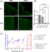
Ang II infusion increases Iba-1 immunoreactivity. (A) Images of Iba-1 immunohistochemistry. Color balance was adjusted for visualization. Analysis was performed on raw images. Scale bar = 100 µm. (B) Immunoreactivity of Iba-1, a marker for microglia and macrophages, was significantly higher in Ang II-infused mice. Telmisartan treatment did not affect Iba-1 immunoreactivity. (C) In animals with and without Ang II-induced hypertension (PBS-CTL and AngII-CTL), the number of CMH was positively correlated with Iba-1 immunoreactivity (r = 0.32). Data shown are mean ± SEM. n = 12–15 per sex per group. ***p < 0.001 and ****p < 0.0001
Fig. 6
Fig. 6
Ang II infusion led to elevated blood pressure with and without PLX3397 diet. (A) In this study, the effect of microglial depletion on Ang II-induced CMH formation was examined. Mice were given a PLX3397 diet for 7 weeks, including 3 weeks before and 4 weeks during hypertension induction with Ang II infusion (1000 ng/kg/min). (B) Both regular chow and PLX3397 diet groups showed an increase in mean arterial pressure due to Ang II infusion. Final MAP was lower in mice with Ang II and PLX3397 than mice with Ang II alone. Data shown are mean ± SEM. n = 3–5 per sex per group. *p < 0.05 and ****p < 0.0001
Fig. 7
Fig. 7
PLX3397 diet significantly reduced Iba-1 immunoreactivity. (A) Images of Iba-1 immunohistochemistry. Color balance was adjusted for visualization. Analysis was performed on raw images. Scale bar = 100 µm. (B) Immunoreactivity of Iba-1 was significantly lower in mice on the PLX3397 diet than those on regular chow. The increase in immunoreactivity induced by Ang II was eliminated in mice on the PLX3397 diet. Data shown are mean ± SEM. n = 3–5 per sex per group. **p < 0.01 and ****p < 0.0001
Fig. 8
Fig. 8
PLX3397 diet significantly reduced CMH number in mice with Ang II-induced hypertension. (A) Images showing Prussian blue-positive deposits, indicative of CMH. Color balance was adjusted for visualization. Analysis was performed on raw images. Scale bar = 100 µm. (B) As shown previously, CMH number per cm2 is elevated in response to Ang II infusion. However, PLX3397 diet inhibited the formation of CMH induced by Ang II. Data shown are mean ± SEM. n = 3–5 per sex per group. **p < 0.01 and ***p < 0.001
Fig. 9
Fig. 9
CD206 immunoreactivity is unaffected by Ang II and is significantly reduced by PLX3397 diet. (A) Images showing CD206 immunofluorescence, indicative of perivascular macrophages. Scale bar = 50 µm. (B) Ang II infusion does not affect CD206 immunoreactivity, while PLX3397 diet significantly reduces CD206 immunoreactivity. (C) In animals with and without Ang II-induced hypertension (PBS-CTL and AngII-CTL), the number of CMH was positively correlated with Iba-1 immunoreactivity (r = 0.51). A nonsignificant (p = 0.35) association was observed between CD206 immunoreactivity and CMH number per cm2. Data shown are mean ± SEM. n = 3–5 per sex per group. ***p < 0.001 and ****p < 0.0001
Fig. 10
Fig. 10
Schematic of the relationship between Ang II and CMH. Ang II infusion leads to hypertension and microglial activation. Increased blood pressure results in increase CMH formation, while microglial activation may also lead to CMH

References

    1. Haller S, Vernooij MW, Kuijer JPA, Larsson EM, Jäger HR, Barkhof F. Cerebral microbleeds: Imaging and clinical significance. Radiology. 2018;287(1):11–28. 10.1148/radiol.2018170803. - PubMed
    1. Vernooij MW, Van Der Lugt A, Ikram MA, Wielopolski PA, Niessen WJ, Hofman A, et al. Prevalence and risk factors of cerebral microbleeds: the Rotterdam scan study. Neurology. 2008;70(14):1208–14. 10.1212/01.wnl.0000307750.41970.d9. - PubMed
    1. Akoudad S, Wolters FJ, Viswanathan A, De Bruijn RF, Van Der Lugt A, Hofman A, et al. Association of cerebral microbleeds with cognitive decline and dementia. JAMA Neurol. 2016;73(8):934–43. 10.1001/jamaneurol.2016.1017. - PMC - PubMed
    1. Wilson D, Charidimou A, Ambler G, Fox ZV, Gregoire S, Rayson P, et al. Recurrent stroke risk and cerebral microbleed burden in ischemic stroke and TIA. Neurology. 2016;87(14):1501–10. 10.1212/WNL.0000000000003183. - PMC - PubMed
    1. Bokura H, Saika R, Yamaguchi T, Nagai A, Oguro H, Kobayashi S, et al. Microbleeds are associated with subsequent hemorrhagic and ischemic stroke in healthy elderly individuals. Stroke. 2011;42(7):1867–71. 10.1161/STROKEAHA.110.601922. - PubMed

MeSH terms