. 2025 Mar 5;14(1):31.
doi: 10.1186/s40164-025-00614-x.

Bone marrow sympathetic neuropathy is a hallmark of hematopoietic malignancies and it involves severe ultrastructural damage

Affiliations

Bone marrow sympathetic neuropathy is a hallmark of hematopoietic malignancies and it involves severe ultrastructural damage

Aurora Bernal et al. Exp Hematol Oncol. .

Abstract

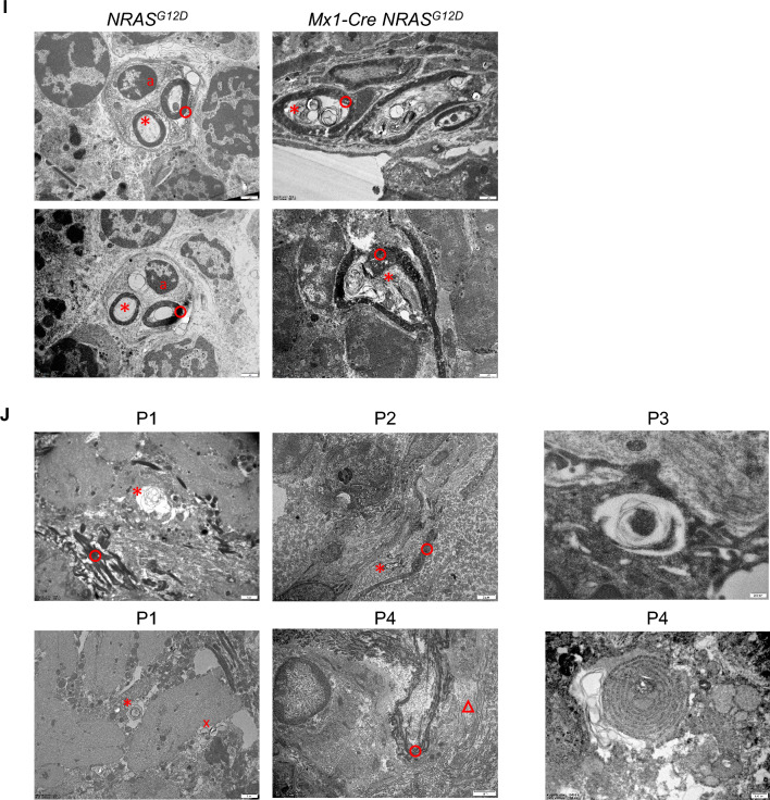
The hematopoietic stem cell (HSC) niche in the bone marrow (BM) supports HSC function, fate and numbers [1]. Sympathetic fibres innervate the BM and are components of the hematopoietic stem and progenitor cell (HSPC) niche [2]. Neuropathy of the HSPC niche is present and essential for disease development in experimental models of JAK2V617F+ myeloproliferative neoplasms (MPN) and MLL-AF9+ acute myeloid leukemia (AML), and it is present in the BM of human MPN and AML patients [3-6]. Neuropathy contributes to mutant HSC expansion and represents an effective therapeutic target to block disease progression in JAK2V617F+ MPN mice [3]. The sympathomimetic agonist mirabegron restored nestin+ cells and reduced reticulin fibrosis in MPN patients [7]. Here, we show that neuropathy of the HSPC niche emerges in two additional experimental models of hematological disease including pre-leukemic myelopoiesis driven by NRASG12D and lymphoma/lymphoblastic leukemia driven by p53 deletion. Neuropathy involves severe ultrastructural damage in NRASG12D+ mice and AML patients as shown by electron microscopy. When further reinforced chemically, neuropathy has a profound impact on the experimental NRASG12D mouse model, promoting myeloid bias, reducing HSPC numbers and inducing changes in the stem cell microenvironment that include reduced numbers of mesenchymal stromal cells (MSC) and increased presence of morphologically abnormal blood vessels in BM. Together, BM neuropathy is a prevalent factor in hematopoietic malignancies that involves important degradation of sympathetic fibres and contributes to disease in a different manner depending on the driver mutation. This should be taken in consideration in the clinic, given that chemotherapy induces neuropathy of the HSC niche [8] and it is the most frequent first line treatment for AML, acute lymphoblastic leukemia and MPN patients.

Keywords: Hematological cancers; Peripheral nervous system; Stem cell niche; Sympathetic fibres; Transmission electron microscopy.


Conflict of interest statement

Declarations. Ethics approval and consent to participate: Animal experiments related to this work were approved by the Norwegian Food and Safety Authority under protocol numbers 7141, 7922, 7960, 8043, 8231, 8408, 9005, 21740 and 24735. Human studies were approved by the Regional Committee for Medical Research Ethics North Norway (REC North 2015/1082). Written informed consent was obtained in accordance with the Norwegian legislation and the Declaration of Helsinki. Participants received no compensation. Competing interests: The authors declare no competing interests.

Figures

Fig. 1
Fig. 1
Bone marrow neuropathy is a hallmark of disease in mouse models of both myeloid and lymphoid malignancies, and it involves severe ultrastructural damage to myelin and axons in pre-leukemic NRAS-G12D+ primary mutant mice and acute myeloid leukemia (AML) patients. C57BL/6J female mice transplanted with bone marrow (BM) nucleated cells obtained from polyinosine-polycytosine (pI-pC)-induced control (NRASG12D) or NRAS-G12D+ (Mx1-Cre NRASG12D) mice, and analyzed at 40 weeks of age, 32 weeks after the transplant (n = 3–4 per group). (A) Representative immunostaining of tyrosine hydroxylase (TH, red) to visualize sympathetic fibres in BM; nuclei were counterstained with DAPI (blue); scale bar, 100 µm. (B) Quantification of TH+ sympathetic fibres in BM diaphysis (%). (C-F) Time-course analysis of TH+ sympathetic fibres and of glial fibrillary acidic protein (GFAP)+ ensheathing Schwann cells in BM diaphysis from control (NRASG12D) or NRAS-G12D+ (Mx1-Cre NRASG12D) primary mutant female mice, induced with pI-pC at 7–27 weeks of age. (C) Quantification of TH+ sympathetic fibres and of GFAP+ ensheathing Schwann cells in BM diaphysis (%), 6 weeks after pI-pC induction (n = 5–6 per group). (D) Quantification of TH+ sympathetic fibres and of GFAP+ ensheathing Schwann cells in BM diaphysis (%), 24 weeks after pI-pC induction (n = 3–5 per group). (E) Representative immunostaining of TH (red, upper panel) to visualize sympathetic fibres and of GFAP (red, lower panel) to visualize ensheathing Schwann cells in BM; nuclei were counterstained with DAPI (blue); scale bar, 500 µm. Magnification is shown for better appreciation of TH+ sympathetic fibres, and (F) Quantification in BM diaphysis (%), 38 weeks after pI-pC induction (n = 5–7 per group). (G-H) Wild-type (p53+/+) or p53−/− female and male mice, analyzed at 12 weeks of age (n = 6–9 per group). (G) Representative immunostaining of TH (red, upper panel) to visualize sympathetic fibres in BM; nuclei were counterstained with DAPI (blue); scale bar, 250 µm. (H) Quantification in BM diaphysis (%). *p < 0.05, **p < 0.01, ***p < 0.001 unpaired two-tailed t test. (I) Representative transmission electron microscopy (TEM) images of GFAP+ BM sections from control (NRASG12D) or NRAS-G12D+ (Mx1-Cre NRASG12D) male mice, analyzed at 29 weeks of age, 20 weeks after pI-pC induction (n = 2–3 per group). (J) Representative TEM images of GFAP+ BM sections from AML patients (n = 4, P1-P4). a, Schwann cell nucleus; o, myelin sheath; *, axon; △, collagen fibres; x, tubule-vesicular elements. Scale bar indicated in the images.
Fig. 1
Fig. 1
Bone marrow neuropathy is a hallmark of disease in mouse models of both myeloid and lymphoid malignancies, and it involves severe ultrastructural damage to myelin and axons in pre-leukemic NRAS-G12D+ primary mutant mice and acute myeloid leukemia (AML) patients. C57BL/6J female mice transplanted with bone marrow (BM) nucleated cells obtained from polyinosine-polycytosine (pI-pC)-induced control (NRASG12D) or NRAS-G12D+ (Mx1-Cre NRASG12D) mice, and analyzed at 40 weeks of age, 32 weeks after the transplant (n = 3–4 per group). (A) Representative immunostaining of tyrosine hydroxylase (TH, red) to visualize sympathetic fibres in BM; nuclei were counterstained with DAPI (blue); scale bar, 100 µm. (B) Quantification of TH+ sympathetic fibres in BM diaphysis (%). (C-F) Time-course analysis of TH+ sympathetic fibres and of glial fibrillary acidic protein (GFAP)+ ensheathing Schwann cells in BM diaphysis from control (NRASG12D) or NRAS-G12D+ (Mx1-Cre NRASG12D) primary mutant female mice, induced with pI-pC at 7–27 weeks of age. (C) Quantification of TH+ sympathetic fibres and of GFAP+ ensheathing Schwann cells in BM diaphysis (%), 6 weeks after pI-pC induction (n = 5–6 per group). (D) Quantification of TH+ sympathetic fibres and of GFAP+ ensheathing Schwann cells in BM diaphysis (%), 24 weeks after pI-pC induction (n = 3–5 per group). (E) Representative immunostaining of TH (red, upper panel) to visualize sympathetic fibres and of GFAP (red, lower panel) to visualize ensheathing Schwann cells in BM; nuclei were counterstained with DAPI (blue); scale bar, 500 µm. Magnification is shown for better appreciation of TH+ sympathetic fibres, and (F) Quantification in BM diaphysis (%), 38 weeks after pI-pC induction (n = 5–7 per group). (G-H) Wild-type (p53+/+) or p53−/− female and male mice, analyzed at 12 weeks of age (n = 6–9 per group). (G) Representative immunostaining of TH (red, upper panel) to visualize sympathetic fibres in BM; nuclei were counterstained with DAPI (blue); scale bar, 250 µm. (H) Quantification in BM diaphysis (%). *p < 0.05, **p < 0.01, ***p < 0.001 unpaired two-tailed t test. (I) Representative transmission electron microscopy (TEM) images of GFAP+ BM sections from control (NRASG12D) or NRAS-G12D+ (Mx1-Cre NRASG12D) male mice, analyzed at 29 weeks of age, 20 weeks after pI-pC induction (n = 2–3 per group). (J) Representative TEM images of GFAP+ BM sections from AML patients (n = 4, P1-P4). a, Schwann cell nucleus; o, myelin sheath; *, axon; △, collagen fibres; x, tubule-vesicular elements. Scale bar indicated in the images.
Fig. 2
Fig. 2
Enforced neuropathy worsens pre-leukemic phenotype in NRAS-G12D+ primary mutant mice. (A) In vivo intraperitoneal (i.p.) administration with 6-hydroxydopamine (6-OHDA) to induce chemical sympathectomy or vehicle in NRAS-G12D+ (Mx1-Cre NRASG12D) female mice aged 25–49 weeks for a total of 16 weeks, starting 14 weeks after polyinosine-polycytosine (pI-pC) induction. (B) Fraction of CD11b+Gr-1+f4/80+ monocytes and B220+ B lymphocytes in total white blood cells (WBC) analyzed by fluorescence-activated cell sorting (FACS), after 12 weeks of treatment (n = 5–6 per group). (C) Fraction of CD11b+ myeloid cells, monocytes and B lymphocytes in total spleen cells (TSP), analyzed by FACS, after 16 weeks of treatment (n = 3–6). (D) Fraction of CD11b+ myeloid cells, monocytes and B lymphocytes in bone marrow nucleated cells (BMNC) analyzed by FACS, after 16 weeks of treatment (n = 3–6). (E) Total BM (TBM) number of Linc-Kit+Sca-1+ (LSK) cell subsets: LSK CD34Flt3CD48CD150+, hematopoietic stem cells (HSC); LSK CD34+Flt3CD48CD150+, multipotent progenitors 1 (MPP1); LSK CD34+Flt3CD48+CD150+ (MPP2); LSK CD34+Flt3CD48+CD150 (MPP3); LSK CD34+Flt3+CD48+CD150 (MPP4); LSK CD34+Flt3CD48CD150 (MPP5); LSK CD34Flt3CD48CD150 (MPP6) analyzed by FACS, after 16 weeks of treatment (n = 3–6). (F) Gene expression analysis of Cebpa, Mki67, Bcl2 and p53 relative to Gapdh by digital droplet PCR after reverse transcription of total mRNA extracted from FACS-sorted LSK, after 16 weeks of treatment (n = 3–6). (G) TBM number of CD45CD31Ter-119CD63+ mesenchymal stromal cells analyzed by FACS, after 16 weeks of treatment (n = 3–5). (H) Representative immunostaining of endomucin (red) to visualize sinusoids and arterioles in BM, 16 weeks after treatment; scale bar, 500 µm. Magnification is shown for better appreciation of blood vessels; scale bar, 50 µm (n = 2–4). *, sinusoids; arrows, arterioles. *p < 0.05, **p < 0.01 unpaired two-tailed t test

References

    1. Pinho S, Frenette PS. Haematopoietic stem cell activity and interactions with the niche. Nat Rev Mol Cell Biol. 2019;20:303–20. - PMC - PubMed
    1. Mendez-Ferrer S, Lucas D, Battista M, Frenette PS. Haematopoietic stem cell release is regulated by circadian oscillations. Nature. 2008;452:442–7. - PubMed
    1. Arranz L, Sanchez-Aguilera A, Martin-Perez D, Isern J, Langa X, Tzankov A, Lundberg P, Muntion S, Tzeng YS, Lai DM, et al. Neuropathy of haematopoietic stem cell niche is essential for myeloproliferative neoplasms. Nature. 2014;512:78–81. - PubMed
    1. Hanoun M, Zhang D, Mizoguchi T, Pinho S, Pierce H, Kunisaki Y, Lacombe J, Armstrong SA, Dührsen U, Frenette PS. Acute myelogenous leukemia-induced sympathetic neuropathy promotes malignancy in an altered hematopoietic stem cell niche. Cell Stem Cell. 2014;15:365–75. - PMC - PubMed
    1. Ting S, Mankai J, Xinyue D, Huan D, Wenjing G, Yuchen G, Rongfeng F, Xiaofan L, Yueting H, Wei L, et al. Multilevel defects in the hematopoietic niche in essential thrombocythemia. Haematologica. 2020;105:661–73. - PMC - PubMed

LinkOut - more resources