. 2025 Mar 5;22(1):48.
doi: 10.1186/s12984-025-01587-y.

Impact of an upper limb motion-driven virtual rehabilitation system on residual motor function in patients with complete spinal cord injury: a pilot study

Affiliations

Impact of an upper limb motion-driven virtual rehabilitation system on residual motor function in patients with complete spinal cord injury: a pilot study

Yanqing Xiao et al. J Neuroeng Rehabil. .

Abstract

Background: Assessing residual motor function in motor complete spinal cord injury (SCI) patients using surface electromyography (sEMG) is clinically important. Due to the prolonged loss of motor control and peripheral sensory input, patients may struggle to effectively activate residual motor function during sEMG assessments. The study proposes using virtual reality (VR) technology to enhance embodiment, motor imagery (MI), and memory, aiming to improve the activation of residual motor function and increase the sensitivity of sEMG assessments.

Methods: By Recruiting a sample of 12 patients with AIS A/B and capturing surface electromyographic signals before, druing and after VR training, RESULTS: Most patients showed significant electromyographic improvements in activation frequency and or 5-rank frequency during or after VR training. However, one patient with severe lower limb neuropathic pain did not exhibit volitional electromyographic activation, though their pain diminished during the VR training.

Conclusions: VR can enhance the activation of patients' residual motor function by improving body awareness and MI, thereby increasing the sensitivity of sEMG assessments.

Keywords: Residual motor control ability; Spinal cord injury; VR; sEMG.


Conflict of interest statement

Declarations. Ethics approval and consent to participate: The study procedures were approved by the Medical Ethics Committee of China Rehabilitation Research Center (approval No. 2020-014-1, April 1, 2020) and were conducted with the informed consent of all participants. Consent for publication: In the experiment, participants agreed to have their experimental data used for publication, and this part of the agreement is written in the informed consent form(approval No. 2020-014-1, April 1, 2020) for the experiment. Competing interests: The authors declare no competing interests.

Figures

Fig. 1
Fig. 1
VR System Equipment and Principles: (A) System Equipment; (B) Schematic Diagram of the Whole Experimental Device. (C) Patients with SCI performed VR training in supine position
Fig. 2
Fig. 2
Comparison of 5-rank activation frequencies was performed before VR training,during VR training and after VR training. In comparison to the before VR training, there was a notable and statistically significant increase in the number of 5-rank acitvation frequencies during and after VR training. However, no statistically significant difference was observed in the number of 5-rank acitvation frequencies between the during and after VR training conditions
Fig. 3
Fig. 3
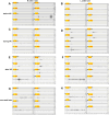
sEMG recordings of P3 were obtained before,during (for 5 min),and after VR training. Before VR training, activation was observed in only the L BF during the R ankle task (A), while no muscle channels exhibited activation during the L ankle task (B). During VR training, no muscle activation was observed during the R ankle task (C); however, five muscle channels (RRF, RBF, RTIB, RGAS, LTIB) exhibited activation during the L ankle task (D). After VR training, five muscle channels (RBF, RTIB, RGAS, LBF, LTIB)exhibited activation during the R ankle task (E) and one muscle channel (LTIB) were activated during L ankle tasks (F). During the follow-up visit one week later, a total of seven task frequencies during both the R and L ankle tasks, four channel (RBF, RTIB, RGAS, LTIB) were activated during R ankle task (G) and three channles (RTIB, RGAS, LTIB) were activated during L ankle task (H)
Fig. 4
Fig. 4
sEMG recordings before VR training,during VR training (for 10 min),and after VR training for P6. Before VR training, there was no observed muscle activation during either the R or L ankle tasks (A, B). During VR training, the R TIB exhibited activation during the R ankle task (C), whereas no muscle activation was observed during the L ankle task (D). After VR training, muscle channels exhibited activation during both the R and L ankle tasks, resulting in a total of three task frequencies, both the R TIB and R GAS were activated during the R ankle task (E), whereas only the R TIB showed activation during the L ankle task (F). Figure G represents an amplified sEMG of the R TIB activated during the R ankle task in VR training. Notably, around 365 s, an escalating burst frequency in the R GAS channel can be observed, demonstrating sustained control until approximately 480 s
Fig. 5
Fig. 5
sEMG recordings of P8 before VR training,during VR training (for 10 min),and after VR training. Before VR training, no muscle activation was observed during both the R and left ankle tasks (A, B). However, during VR training, the R RF and R GAS exhibited activation specifically during the R ankle task (C), while the sEMG signal of the R TIB indicated an sEMG artifact, implying non-volitional muscle activity. Notably, during the L ankle task, activation of the R GAS was observed (D). After VR training, the R RF exhibited activation during the R ankle task (E), whereas no muscle activation was observed during the L ankle task (F). Figure G represents an enlarged sEMG signal of the R RF activated during the R ankle task in VR training, Figure H depicts an enlarged sEMG signal of the R GAS activated during the R ankle task, and Figure I illustrates an enlarged sEMG signal of the R GAS activated during the R ankle task, demonstrating intermittent continuous control in all three muscle channels

Similar articles

References

    1. Kakulas BA. Pathology of spinal injuries. Cent Nerv Syst Trauma. 1984;1(2):117–26. - PubMed
    1. Kakulas A. The applied neurobiology of human spinal cord injury: a review. Spinal Cord. 1988;26(6):371–9. - PubMed
    1. Kakulas B. Neuropathology: the foundation for new treatments in spinal cord injury. Spinal Cord. 2004;42(10):549–63. - PubMed
    1. Bunge RP, Puckett W, Becerra J, et al. Observations on the pathology of human spinal cord injury. A review and classification of 22 new cases with details from a case of chronic cord compression with extensive focal demyelination. Adv Neurol. 1993;59:75–89. - PubMed
    1. Dimitrijevic M, Faganel J, Lehmkuhl D, Sherwood A. Motor control in man after partial or complete spinal cord injury. Adv Neurol. 1983;39:915–26. - PubMed

LinkOut - more resources