Clinical Trial
. 2025 Aug;80(8):2225-2239.
doi: 10.1111/all.16514. Epub 2025 Mar 6.

Non-Allergic Urticarial Skin Reactions Associated With MOv18 IgE, a First-In-Class IgE Antibody Recognising Folate Receptor Alpha

Affiliations
Clinical Trial

Non-Allergic Urticarial Skin Reactions Associated With MOv18 IgE, a First-In-Class IgE Antibody Recognising Folate Receptor Alpha

Chara Stavraka et al. Allergy. 2025 Aug.

Abstract

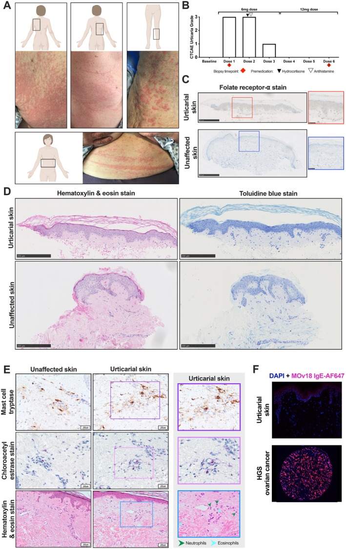
Background: IgE antibodies directed against cancer antigens have demonstrated potent anti-tumour effects in pre-clinical studies. MOv18 IgE, the first-in-class IgE recognising the cancer antigen folate receptor alpha (FRα), showed preliminary signs of efficacy in a Phase I trial. Treatment was well tolerated, with the most common adverse event being transient urticarial skin reactions. We investigated immunological and allergic response parameters associated with urticarial skin reactions in MOv18 IgE-treated patients.

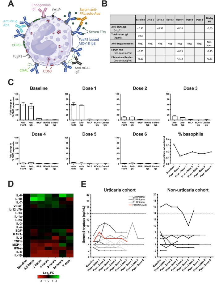
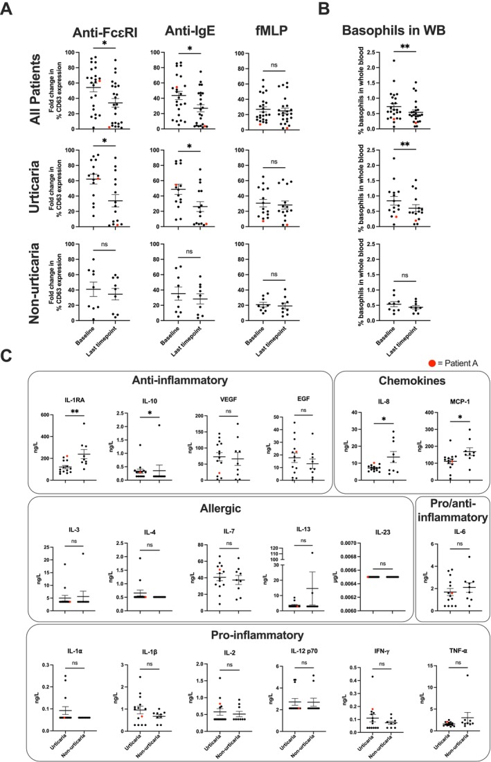
Methods: Expression of target antigen, FRα, and MOv18 IgE reactivity with FRα or any component in human skin was studied by immunohistochemistry, immunofluorescence and immuno-mass spectrometry. We conducted transcriptomic analyses in paired lesional and non-lesional skin biopsies from a patient who developed an urticarial skin reaction. Systemic immunological markers including cytokines, β-tryptase and basophil activation states were interrogated throughout the trial and contemporaneously with the skin reaction.

Results: Of the 24 IgE-treated patients, 62.5% developed transient urticarial skin reactions, with onset during the first infusion, diminishing with consecutive infusions and no β-tryptase elevation nor clinical features indicating allergic aetiology. No FRα expression or MOv18 IgE binding to human skin was identified. Lesional skin biopsies from a patient given the highest antibody dose revealed scattered eosinophils, neutrophils and mast cell degranulation, but no increased immune cell infiltration. Transcriptomic analysis indicated pro-inflammatory, but not allergic, pathway activation. No systemic allergic or hypersensitivity mediators or basophil activation were detected.

Conclusions: Urticarial skin reactions following MOv18 IgE treatment were unlikely to result from allergic mechanisms or skin antigen recognition. The clinical presentation is consistent with infusion-related reactions commonly observed with monoclonal antibody treatments.

Trial registration: EudraCT number: 2014-000070-19; ClinicalTrials.gov identifier: NCT02546921, registered 11/Sept/2015.

Keywords: AllergoOncology; IgE antibodies; folate receptor alpha; transcriptomic analysis; urticarial skin reactions.


Conflict of interest statement

J.S. and S.N.K. are founders and shareholders of Epsilogen Ltd. H.J.B. is employed through a fund provided by Epsilogen Ltd. J.C. has been employed through a fund provided by Epsilogen Ltd. S.N.K., J.S., D.H.J. and H.J.B. declare patents on antibodies for cancer. R.K. is the Lead Investigator for the Phase IB/II MOv18 trial and has received travel support from Epsilogen Ltd. All other authors declare no conflicts of interest.

Figures

FIGURE 1
FIGURE 1
Readily manageable urticarial adverse events were associated with higher doses of MOv18 IgE treatment. (A) Panel of representative images of urticarial skin reactions, seen in a patient who received a 500 μg dose (purple) and a patient who received a 1.5 mg dose (orange) from the larger cohort of MOv18 IgE‐treated patients. (B) Proportion and CTCAE urticarial grade per dosing cohort of patients treated with MOv18 IgE (urticarial: n = 15; total n = 24). Intra‐patient dose escalation was performed with this patient (n = 1) receiving 3 doses at 6 mg, followed by 3 doses at 12 mg. Number of patients treated, and number of patients who experienced urticaria, outlined per dosing cohort shown in Table S1. (C) Per dose CTCAE urticarial grade of patients that experienced urticarial symptoms (n = 15). The dotted line signifies when the patient stopped receiving MOv18 IgE treatment. Black and white arrows signify when the patient was given premedication with hydrocortisone and/or antihistamines, respectively.
FIGURE 2
FIGURE 2
FRα is not expressed in the skin and immuno‐mass spectrometry (IMS) of MOv18 antibody clone pulldown demonstrated no skin‐associated binding. (A) Normal skin tissues stained with commercially available anti‐FRα antibody were negative for FRα staining (n = 48 across two TMAs). Further details of TMA sample map and demographic information included in Figure S1, Tables S2 and S3. (B) HGS ovarian tumour and normal ovarian control tissues were positively and negatively stained, respectively. (C) High FOLR1 mRNA expression in primary ovarian tumours (n = 419), but no or low expression in normal ovaries (n = 88), normal fallopian tube (n = 5) and normal skin (n = 556) (data extracted from Human Protein Atlas, v20.proteinatlas.org) [20, 21]. (D) Schematic of immuno‐mass spectrometry (IMS) experimental procedure and analysis pipeline for discovery of skin (healthy skin tissues lysate; n = 3) and IGROV1 cell lysate antigens, using MOv18 IgG and isotype control (NIP IgG) (upper row). Number of peptides found following pipeline filtering criteria in skin and IGROV1 lysates (dotted line signifying the cut‐off for which results are believed to be true, lower left). Peak area of peptides found in skin and IGROV1 lysates (lower right). Figure generated on BioRender. (E) Tables of peptide sequences observed for peptides identified in skin lysates (upper) and IGROV1 cell lysates (lower).
FIGURE 3
FIGURE 3
No evidence of FRα, immune cell infiltration or mast cell activation in urticarial and unaffected skin from a patient treated with the highest MOv18 IgE dose levels (Patient A). (A) Panel of images of the CTCAE grade 3 urticarial reaction observed in a patient during their first infusion of MOv18 IgE at 6 mg: Upper left back (upper left image), upper right back (upper centre image), left calf (upper right image) and abdomen (lower image). (B) Decreasing CTCAE urticarial grade was observed in a patient who experienced symptoms during treatment with MOv18 IgE. No urticarial symptoms were observed following intra‐dose escalation to a higher dose (12 mg). Red diamonds represent the biopsy collection timepoints for urticarial (timepoint 1) and unaffected (timepoint 2) skin samples analysed subsequently. (C) Staining for FRα in urticarial and unaffected skin indicated the absence of FRα in both tissues. Red and blue boxes correspond to respective zoomed‐in section of tissues. Biopsy timepoints as outlined in B. (D) Haematoxylin and eosin (H&E; left) and toluidine blue (right) staining show the absence of immune cell and mast cell infiltration, respectively, in comparison to unaffected tissue. Biopsy timepoints outlined in B. (E) Mast cell tryptase (MCT, upper row) and chloroacetyl esterase staining (CAE, middle row) shows evidence of enhanced mast cell degranulation in urticarial skin, which is not present in unaffected skin; higher magnification view of urticarial skin (right; purple and pink box, respectively). H&E staining (lower row) provides evidence of scattered neutrophils and eosinophils in urticarial skin (lower right) which is not present in unaffected skin; zoomed‐in section of urticarial tissue (right, blue box). (F) MOv18 IgE labelled with AlexaFluor647 (MOv18 IgE‐AF647, pink) did not bind to urticaria skin (upper) but did bind high‐grade serous ovarian tissue (lower).
FIGURE 4
FIGURE 4
Gene expression and enriched pathways in urticarial tissue of a patient treated with the highest MOv18 IgE dose levels (Patient A). (A) Significantly differentially expressed genes were identified between urticarial and unaffected skin, using the limma package, ranked according to fold change. (B) Calculated enrichment of gene sets was evaluated within Hallmark, with selected example pathways shown, ranked according to fold change. (C) Top 50 most differentially expressed (FDR‐corrected) genes are shown for each selected pathway. p ≤ 0.05 for significant genes within the selected Hallmark pathways.
FIGURE 5
FIGURE 5
Diminishing CTCAE urticarial grade was associated with diminishing ex vivo basophil activation in a patient who experienced urticarial symptoms during MOv18 IgE treatment at the highest doses. (A) Overview of the potential mechanisms of basophil activation within the circulation: Anti‐drug antibodies (ADAs) could cross‐link FcɛRI‐bound MOv18 IgE; shed monovalent serum folate receptor alpha (FR) could form complexes with serum anti‐FR⍺ auto‐antibodies (auto‐Abs) and MOv18 IgE; and ⍺GAL (galactose‐alpha‐1,3‐galactose) found on the surface of SP2/0 produced MOv18 IgE may be cross‐linked by an anti‐⍺GAL IgE Abs. Figure generated on BioRender. (B) Table summarising levels of circulating mediators measured in Patient A. (C) In Patient A, no basophil activation (% CD63 expression) was triggered by MOv18 IgE or control IgE, however, diminishing IgE‐mediated (anti‐FcɛRI and anti‐IgE) ex vivo basophil activation was observed through treatment doses. The proportion of basophils in whole blood decreased from baseline levels following the first dose of MOv18 IgE, recovering slightly through subsequent doses (lower right). (D) Log2 foldchange (Log2FC) in median circulating cytokine levels post‐dose 1 (0.5, 2, 4, 6, 24 h and 7 days post‐dose) relative to normalised baseline values for Patient A. Overall increases in IL‐6, IL‐10, IL‐7 and VEGF were observed, with no change in IL‐12 p70, IL‐13, IL‐1⍺, IL‐23, IL‐3, IL‐4 and an overall decrease in EGF, IL1RA, IL‐2, TNF‐⍺, MCP‐1, IFN‐, IL‐8 and IL‐1β. (E) Serum β‐tryptase at baseline and throughout treatment in urticaria cohort (left; n = 14; colour denotes highest grade of CTCAE urticaria experienced by patient; red = Patient A who experienced grade 3 urticaria) and non‐urticaria (right; n = 9) patients. β‐tryptase levels in patients who experienced urticaria (urticaria cohort) (in the absence of anaphylaxis), consistently stayed below the upper limit of normal (14 ng/mL; dotted line) and no association with the grade of urticaria was observed. This is also consistent for patients who did not experience urticaria (non‐urticaria cohort); however, one patient was consistently above the upper limit of normal at baseline and through treatment—this was without any significant hypersensitivity or anaphylaxis experienced by this patient.
FIGURE 6
FIGURE 6
Reduced IgE‐mediated ex vivo basophil activation propensity and reduced circulating basophil levels basophils in patients who experienced urticarial symptoms through MOv18 IgE treatment. (A) Significantly decreased ex vivo basophil activation via IgE‐mediated mechanisms (anti‐FcɛRI and anti‐IgE) was observed in all patients (upper row) and in those patients who experienced any CTCAE grade urticaria following treatment with MOv18 IgE (middle row). Stimulation (% CD63 expression) was as measured via the basophil activation test at baseline and at the last available timepoint during MOv18 IgE treatment. No significant changes were observed in ex vivo basophil activation by a non‐IgE‐mediated (fMLP) stimulus in the whole patient cohort and in patients who experienced urticaria (upper and middle row: right). No significant changes in basophil activation were observed by IgE‐mediated and non‐IgE‐mediated stimuli in patients who did not experience urticaria through treatment with MOv18 IgE (lower row). All patients: n = 24, urticaria patients: n = 15, and non‐urticaria patients: n = 9. Patient A's datapoints highlighted in red. (B) Proportion of basophils in whole blood significantly significant decreased in all patients (upper, n = 24), and in those patients that experienced urticaria through treatment with MOv18 IgE (middle, n = 15). No significant changes in % of basophils in blood proportion were observed in those patients who did not experience any urticaria during treatment (lower, n = 9). Patient A's datapoints highlighted in red. (C) Circulating cytokine and chemokine levels at baseline, prior to treatment with MOv18 IgE, in urticaria (n = 14) and non‐urticaria (n = 9) patients. Urticaria patients show significant differences in anti‐inflammatory cytokines (IL‐1RA, IL‐10) and chemokines (IL‐8, MCP‐1). No differences in allergic or pro‐inflammatory cytokines. Patient A's datapoint highlighted in red. Data are shown as mean ± SEM. Wilcoxon matched‐pairs rank test (A: Upper and middle row; B: Upper and middle), paired t‐test (A: lower row, B: lower), unpaired t‐test (C: IL‐1RA, VEGF; EGF, IL‐8, MCP‐1, IL‐7, IL‐6, IL‐1α, IL‐1β, IFN‐α), Mann–Whitney test (C: IL‐10, IL‐3, IL‐4, IL‐13, IL‐23, IL‐2, IL12 p70, TNF‐α): *p ≤ 0.05, **p ≤ 0.01.

References

    1. Weiner L. M., Surana R., and Wang S., “Monoclonal Antibodies: Versatile Platforms for Cancer Immunotherapy,” Nature Reviews. Immunology 10, no. 5 (2010): 317–327. - PMC - PubMed
    1. Rudman S. M., Josephs D. H., Cambrook H., et al., “Harnessing Engineered Antibodies of the IgE Class to Combat Malignancy: Initial Assessment of FcvarepsilonRI‐Mediated Basophil Activation by a Tumour‐Specific IgE Antibody to Evaluate the Risk of Type I Hypersensitivity,” Clinical and Experimental Allergy 41, no. 10 (2011): 1400–1413. - PubMed
    1. McCraw A. J., Chauhan J., Bax H. J., et al., “Insights From IgE Immune Surveillance in Allergy and Cancer for Anti‐Tumour IgE Treatments,” Cancers 13, no. 17 (2021): 4460, 10.3390/cancers13174460. - DOI - PMC - PubMed
    1. Karagiannis P., Singer J., Hunt J., et al., “Characterisation of an Engineered Trastuzumab IgE Antibody and Effector Cell Mechanisms Targeting HER2/Neu‐Positive Tumour Cells,” Cancer Immunology, Immunotherapy 58, no. 6 (2009): 915–930. - PMC - PubMed
    1. Gould H. J., Mackay G. A., Karagiannis S. N., et al., “Comparison of IgE and IgG Antibody‐Dependent Cytotoxicity In Vitro and in a SCID Mouse Xenograft Model of Ovarian Carcinoma,” European Journal of Immunology 29, no. 11 (1999): 3527–3537. - PubMed

Publication types

Associated data