

MRI identifies disrupted cerebral development in medulloblastoma patients
- PMID: 40046337
- PMCID: PMC11879392
- DOI: 10.1093/braincomms/fcaf090
MRI identifies disrupted cerebral development in medulloblastoma patients
Abstract
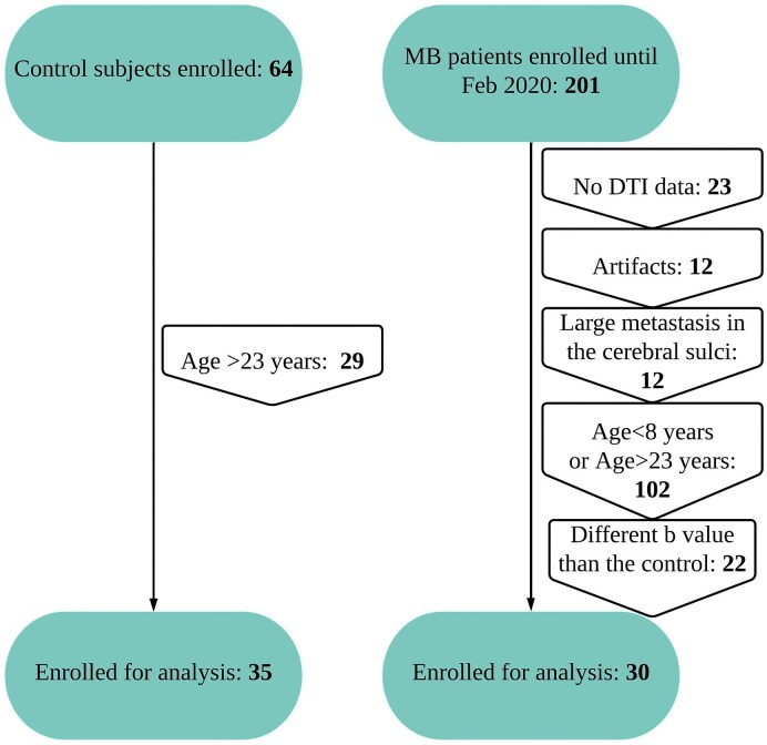
Cognitive decline in survivors of medulloblastoma is commonly attributed to radiation- and chemotherapy-induced brain microstructural alterations. Factors preceding this adjuvant therapy, such as disrupted brain development or resection surgery, may affect brain microstructure but have not been thoroughly explored in medulloblastoma. The aim of this study was to assess cortical thickness and microstructural integrity of the cerebrum prior to adjuvant therapy in medulloblastoma patients. Cross-sectional image data were acquired of medulloblastoma patients (n = 30) after surgery but before adjuvant therapy and compared with data from healthy controls (n = 35) matched for age range (12-22 years). Biomarkers of microstructural integrity include fractional anisotropy, mean diffusivity, axial diffusivity and radial diffusivity. Thickness, surface area and volume were estimated for parcels of neocortex to evaluate potential morphology differences. Participants with medulloblastoma showed increased diffusivity parameters (mean, axial and radial diffusivity) and decreased fractional anisotropy, within nearly all white and grey matter parcels of the cerebrum, compared with healthy controls. Medulloblastoma participants additionally showed decreased cortical thickness in sub-regions of frontal, parietal, temporal and paracentral cortex. Broad cerebral microstructural alterations in medulloblastoma patients following surgery but before initiation of radiation or chemotherapy suggest that cerebellar insult, by tumour development or tumour resection, likely contributes to compromised integrity of cerebral grey and white matter. Locations of cortical thinning suggest that cerebellar insult may impair normal growth in cerebral regions responsible for executive function, language and attention-cognitive domains typically affected in medulloblastoma survivors.
Keywords: DTI; MRI; brain development; medulloblastoma.
© The Author(s) 2025. Published by Oxford University Press on behalf of the Guarantors of Brain.
Conflict of interest statement
The authors report no competing interests.
Figures


References
-
- Northcott PA, Robinson GW, Kratz CP, et al. Medulloblastoma. Nat Rev Dis Primers. 2019;5(1):11. - PubMed
-
- Riggs L, Bouffet E, Laughlin S, et al. Changes to memory structures in children treated for posterior fossa tumors. J Int Neuropsychol Soc. 2014;20(2):168–180. - PubMed
-
- Scantlebury N, Bouffet E, Laughlin S, et al. White matter and information processing speed following treatment with cranial-spinal radiation for pediatric brain tumor. Neuropsychology. 2016;30(4):425–438. - PubMed
LinkOut - more resources
Full Text Sources