. 2025 Mar;21(3):e70030.
doi: 10.1002/alz.70030.

Genetic and pharmacologic enhancement of SUMO2 conjugation prevents and reverses cognitive impairment and synaptotoxicity in a preclinical model of Alzheimer's disease

Affiliations

Genetic and pharmacologic enhancement of SUMO2 conjugation prevents and reverses cognitive impairment and synaptotoxicity in a preclinical model of Alzheimer's disease

Luana Fioriti et al. Alzheimers Dement. 2025 Mar.

Erratum in

Abstract

Introduction: Amyloid beta oligomers (Aβos) are toxic to synapses and key to the progression of Alzheimer's disease (AD) and amyloid pathology, representing a target for therapeutic strategies.

Methods: Amyloid and small ubiquitin modifier 2 (SUMO2) transgenics were analyzed by electrophysiology and behavioral testing. A recombinant analogue of SUMO2, SBT02, was generated and assessed for brain penetration and the ability to mitigate amyloid pathology.

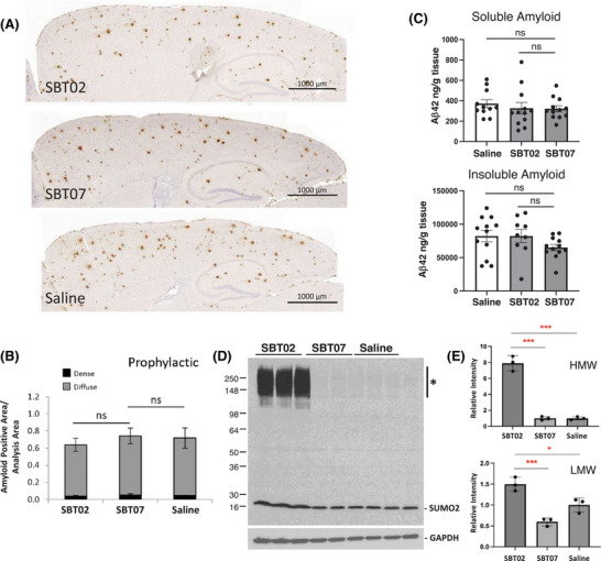
Results: Elevated SUMO2 expression prevents cognitive and synaptic impairment in a mouse model of AD amyloid pathology. Systemic administration of SBT02 resulted in high brain bioavailability and prophylactically halted the progression of AD-associated deficits. SBT02 also restored cognition and synaptic function in late-stage amyloid load. Mechanistically, SUMO2 and SBT02 do not alter amyloid processing or clearance and mitigate synaptotoxicity in the presence of high amyloid loads.

Discussion: SBT02 is a promising therapeutic strategy to counteract and reverse the toxic effects of Aβos in AD.

Highlights: Genetic overexpression of human SUMO2 prevents the long-term potentiation (LTP) impairments and cognitive deficits in amyloid precursor protein (APP) transgenics without affecting amyloid pathology. A recombinant analogue of human SUMO2, termed SBT02, when administered systemically, displays high brain bioavailability and has no adverse effects at high doses. Prophylactic treatment of APP transgenics with SBT02 prior to the development of amyloid pathology results in the prevention of synaptic and behavioral dysfunction. SBT02 also reverses pre-existing LTP and cognitive impairments when administered to APP transgenics with advanced and severe pathology. SBT02 has no impact on amyloid pathology, indicating a mechanism of action on synaptic resistance to Aβ toxicity.

Keywords: SUMOylation; amyloid transgenic mouse model; behavioral analyses; biologic therapy; cognition; drug development; electrophysiology; preclinical testing; small ubiquitin‐like modifiers.


Conflict of interest statement

L.F., O.A., and P.E.F. are co‐inventors on a patent application for the use of SUMO2 mimetic peptides in Alzheimer's disease. N.W., E.K.A., S.M., H.T., K.S., K.H., H.Y., A.S., E.A., F.O., A.M., and T.K. have nothing to disclose. Author disclosures are available in the Supporting Information.

Figures

FIGURE 1
FIGURE 1
Elevated SUMO2 protects against amyloid‐induced cognitive decline and defects in synaptic plasticity in vivo. (A) Immunoblot analysis of selected brain regions from transgenic mice expressing human SUMO2 (Ob, Cx, Cb, Hip) showing increased levels of free SUMO2 and no significant increases in high‐molecular‐weight conjugates. Ct and HA‐tagged SUMO2 Tf HEK293 cells were used as positive controls. (B) SUMO2 overexpression protected against LTP impairment of APP mice (two‐way ANOVA: Non‐Tg vs APP: F (1,17) = 9.960, p = 0.0058; APP vs SUMO2‐APP: F (1,17) = 5.713, p = 0.0287; APP vs SUMO2: F (1,15) = 13.91, p = 0.002). (C) fEPSP of the last 20 min of the LTP curve (one‐way ANOVA followed by Bonferroni's comparisons: F (3,33) = 5.107, p = 0.0052; Non‐Tg vs APP: p = 0.0162; APP vs SUMO2‐APP: p = 0.0272; APP vs SUMO2: p = 0.003). (D) Impairment of associative memory in APP mice measured through contextual fear conditioning was prevented by SUMO2 overexpression with SUMO2‐APP transgenics comparable to Non‐Tg mice (one‐way ANOVA followed by Bonferroni's multiple comparisons: F (3,59) = 5.153, p = 0.0031; Non‐Tg vs APP: p = 0.018; APP vs SUMO2 APP: p = 0.0125; APP vs SUMO2: p = 0.0015). No differences were found in baseline freezing between the different groups (one‐way ANOVA for all groups: F (3,59) = 0.9068, p = 0.4433. **p < 0.01; *p < 0.05. Data are mean ± SEM. ANOVA, analysis of variance; APP, amyloid precursor protein; Cb, cerebellum; Ct, control untransfected; Cx, cortex; Hip, hippocampus; fEPSP, field excitatory post‐synaptic potential; LTP, long‐term potentiation; Non‐Tg, non‐transgenic; Ob, olfactory bulb; SEM, standard error of the mean; SUMO, small ubiquitin modifier; Tf, transfected.
FIGURE 2
FIGURE 2
Amyloid loads and SUMO2 distribution in brain. (A) Representative images showing amyloid immunohistochemistry in the APP and SUMO2‐APP transgenics displaying comparable plaque densities. (B) Quantitative image analysis of amyloid plaques revealed comparable levels in APP and SUMO2‐APP transgenic mice indicating that SUMO2 overexpression had no effect on the amounts of dense plaques (black) and diffuse (gray) amyloid deposits, which were modestly increased in the SUMO2‐APP mice (*p < 0.05; Tukey's multiple comparisons test after one‐way ANOVA). (C) Quantification of soluble and insoluble Aβ42 as determined by sandwich ELISA indicated no significant difference in the Cb and C/H extracts; ns, Dunnett's multiple comparison test after one‐way ANOVA. (D) Immunoblotting for SUMO2 demonstrated an increase in SUMO2 higher‐molecular‐weight conjugated proteins (*) in the SUMO2‐APP transgenics and elevated free SUMO2 in synaptic compartments in SUMO2‐APP transgenics as compared to APP mice. Elevated SUMO2 was also observed in isolated synaptosomes suggesting a targeting to synapses and potential impact on activity. Aβ, amyloid beta; ANOVA, analysis of variance; APP, amyloid precursor protein; Cb, cerebellum; C/H, cortex‐hippocampus; ELISA, enzyme‐linked immunosorbent assay; ns, not significant; SUMO, small ubiquitin modifier.
FIGURE 3
FIGURE 3
Pharmacokinetics of the active SUMO2 biologic. SBT02 levels were examined following IV or SubQ administration. Wild‐type C57BL/6 mice were administered a single injection of N‐terminally His‐tagged SBT02 (20 mg/kg), and samples were analyzed at various time points. (A) Quantification of the plasma using His‐ELISA assays showed a rapid elevation in SBT02 following IV injection that peaked at ≈30 min and was completely cleared from the periphery after 4 h. (B) SubQ injected SBT02 peaked at 1 h and gradually decreased with detectable levels of ≈15 ng/mL at 4 h post‐injection. Peak plasma levels were higher in the IV‐injected group at ≈115 ng/mL as compared to SubQ injected mice at ≈40 ng/mL plasma, indicating that IV administered SBT02 results in higher plasma concentrations but these are reduced more rapidly than in mice receiving SubQ‐injected biologic. (C) SBT02 concentrations in the cortex following IV administration gradually increased and reached a maximum of ≈60 ng/g tissue 24 h post‐injection. (D) SubQ‐injected SBT02 rapidly increased in the cortex and reached a maximal level of ≈125 ng/g tissue at the 4 h time point, and significant levels were observed 120 h post‐injection. n = 3 mice per time point, and data are mean ± SEM. ELISA, enzyme‐linked immunosorbent assay; IV, intravenous; SEM, standard error of the mean; SubQ, subcutaneous; SUMO, small ubiquitin like modifier.
FIGURE 4
FIGURE 4
Recombinant biologic, SBT02, mimicking SUMO2 activity prevents impairments in synaptic and cognitive function. (A) Schematic of the prophylactic therapy time course for treatments with SUMO2 biologics. (B) APP mice exhibit LTP deficits in comparison to littermates, Non‐Tg mice (F (1,25) = 11.66; p = 0.0022). SBT02 prevented the LTP impairment in APP mice as compared to inactive SBT07 (F (1,21) = 7.662; p = 0.0115) or saline (F (1,22) = 10.19; p = 0.042) and exhibited potentiation similar to that of Non‐Tg mice receiving comparable treatments (F (1,21) = 0.006; p = 0.9401). (C) fEPSP of the last 20 min (one‐way ANOVA followed by Bonferroni's multiple comparisons: F (5,74) = 5.06, p = 0.0005; APP + saline vs Non‐Tg + saline: p = 0.0021; APP + SBT02 vs APP + SBT07: p = 0.028; APP + SBT02 vs APP + saline: p = 0.0328). (D) Deficit in associative memory as determined by contextual fear conditioning was prevented by SBT02 administration as compared to SBT07‐ or saline‐treated APP animals (one‐way ANOVA followed by Bonferroni's multiple comparisons: F (5,120) = 5.940, p < 0.0001; APP + saline vs Non‐Tg + saline: p = 0.0353; APP + SBT02 vs APP + SBT07: p = 0.012; APP + SBT02 vs APP + saline: p = 0.002). No differences were found in baseline freezing between the different groups (one‐way ANOVA for all groups: F (5,120) = 2.186, p = 0.06). **p < 0.01; *p < 0.05. Data are mean ± SEM. ANOVA, analysis of variance; APP, amyloid precursor protein; fEPSP, field excitatory post‐synaptic potential; LTP, long‐term potentiation; Non‐Tg, non‐transgenic; SUMO, small ubiquitin modifier.
FIGURE 5
FIGURE 5
Amyloid loads and SUMO conjugation in treated APP transgenics. Representative immunocytochemistry images for the prophylactic treatments showing (A) similar plaque densities in APP mice treated with SBT02, SBT07, or saline. Sections were stained for Aβ, indicating comparable amyloid plaque loads in the cortex and hippocampus. Amyloid plaque loads were assessed by immunohistochemistry and image analysis of the cortex and hippocampus. (B) Dense plaque cores (black) and diffuse halos (gray) for APP‐Tg mice treated prophylactically and examined at the 6‐month end point (n = 3; 5 sections/animal) or (C) ELISA quantification of soluble and insoluble Aβ42 in cortex‐hippocampus indicated; ns, Tukey's multiple comparisons after one‐way ANOVA. (D) SUMO2 immunoblotting revealed an increase in HMW conjugates (*) and free SUMO2‐positive bands for whole brain homogenates from SBT02‐treated APP transgenics relative to SBT07‐ and saline‐treated mice. (E) Quantification of the SUMO2 immunoreactivity indicated an ≈8‐fold increase in HMW SUMO2‐positive conjugates in animals that received SBT02 from 3 months of age until being sacrificed at 6 months (prevention therapy); the LMW free SUMO2 was also elevated in the SBT02 animals consistent with the treatment. Data are mean ± SEM, *; p < 0.05, ***; p < 0.001 by Tukey's multiple comparison test following ordinary one‐way ANOVA. Aβ, amyloid beta; ANOVA, analysis of variance; APP, amyloid precursor protein; ELISA, enzyme‐linked immunosorbent assay; HMW, high molecular weight; LMW, low molecular weight; ns, not significant; SEM, standard error of the mean; SUMO, small ubiquitin like modifier.
FIGURE 6
FIGURE 6
SBT02 reverses cognitive and synaptic impairments in late‐stage disease. (A) Timelines for the reversal study with SBT02/SBT07 and treatment of APP transgenics with pre‐existing amyloid pathology and LTP/cognitive impairments at 6 months (moderate amyloid load) until 9 months of age (severe amyloid load). (B) LTP impairment in APP mice (APP + saline vs Non‐Tg + saline: F (1,27) = 8.138, p = 0.0082) was reverted by SBT02 treatment as compared to animals receiving inactive SBT07 (F (1,29) = 11.16; p = 0.0023) or saline (F (1,25) = 7.433; p = 0.0082), and exhibited potentiation similar to that of Non‐Tg mice receiving comparable treatments (F (1,27) = 0.3956, p = 0.5347). (C) fEPSP of the last 20 min (one‐way ANOVA followed by Bonferroni's multiple comparisons: F (5,82) = 4.638, p = 0.0009; APP + saline vs Non‐Tg + saline: p = 0.0328; APP + SBT02 vs APP + SBT07: p = 0.0054; APP + SBT02 vs APP + saline: p = 0.0381). (D) Associative memory deficits in APP mice (APP + saline vs Non‐Tg + saline: p = 0.002), assessed by contextual fear conditioning were reversed by SBT02 relative to transgenics treated with SBT07 (p = 0.037) or saline (p = 0.02) (one‐way ANOVA for all groups: F (5,98) = 7.183, p < 0.0001. No difference was found in baseline activity (one‐way ANOVA for all groups: F (5,98) = 1.719, p = 0.137). **p < 0.01; *p < 0.05. Data are mean ± SEM. ANOVA, analysis of variance; APP, amyloid precursor protein; fEPSP, field excitatory post‐synaptic potential; LTP, long‐term potentiation; Non‐Tg, non‐transgenic; SEM, standard error of the mean.
FIGURE 7
FIGURE 7
Amyloid loads and SUMO2 conjugation in the reversal treatment. (A) Representative immunocytochemistry images for the reversal mice showing comparable plaque densities in APP transgenes treated with SBT02, SBT07, or saline. (B) Quantitative image analysis of the cortex and hippocampus indicated no changes in dense plaque cores (black) or diffuse halos (gray) for APP‐Tg mice examined at the late stage of pathology (9 months of age; n = 3; five sections/animal) receiving the active SBT02, inactive SBT07, or saline (Tukey's multiple comparisons test after one‐way ANOVA, ns). (C) Immunoblot indicating the increase in SUMO2‐positive conjugates and monomers in the SBT02‐treated animals. (D) Quantification of the free and conjugated SUMO2‐positive proteins. ANOVA, analysis of variance; APP, amyloid precursor protein; ns, not significant; SUMO, small ubiquitin like modifier; Tg, transgenic.
FIGURE 8
FIGURE 8
Synaptic markers in STB02‐treated APP transgenic mice. (A) Immunoblot for the post‐synaptic PSD‐95 from brain homogenates (cortex‐hippocampus combined) indicates an increase in SBT02‐treated mice as compared to mice treatment with SBT07 or saline. (B) Quantification of PSD‐95 immunoreactivity and the increased levels in SBT02‐treated mice. (C) Immunoblot for the presynaptic Homer1 from brain homogenates (cortex‐hippocampus combined) indicating an increase in SBT02‐treated mice as compared to mice treatment with SBT07 or saline. (D) Quantification of Homer1 immunoreactivity and the significantly increased levels in SBT02‐treated mice. (E) Immunoblot for synaptophysin in the treated mice indicates an increase similar to that of the mice administered SBT02. (F) Quantification of the synaptophysin levels showing a statistical significance increase in the SBT02‐treated animals. ***p < 0.001; **p < 0.01; *p < 0.05, by Tukey's multiple comparison test following ordinary one‐way ANOVA. ANOVA, analysis of variance; APP, amyloid precursor protein.

References

    1. Karran E, De Strooper B. The amyloid hypothesis in Alzheimer disease: new insights from new therapeutics. Nat Rev Drug Discovery. 2022;21(4):306‐318. doi: 10.1038/s41573-022-00391-w - DOI - PubMed
    1. Tzioras M, McGeachan RI, Durrant CS, Spires‐Jones TL. Synaptic degeneration in Alzheimer disease. Nat Rev Neurol. 2023;19(1):19‐38. doi: 10.1038/s41582-022-00749-z - DOI - PubMed
    1. Henley JM, Seager R, Nakamura Y, Talandyte K, Nair J, Wilkinson KA. SUMOylation of synaptic and synapse‐associated proteins: an update. J Neurochem. 2021;156(2):145‐161. doi: 10.1111/jnc.15103 - DOI - PMC - PubMed
    1. Chang HM, Yeh ETH. Sumo: from bench to bedside. Physiol Rev. 2020;100(4):1599‐1619. doi: 10.1152/physrev.00025.2019 - DOI - PMC - PubMed
    1. Gong L, Yeh ETH. Characterization of a family of nucleolar SUMO‐specific proteases with preference for SUMO‐2 or SUMO‐3. J Biol Chem. 2006;281(23):15869‐15877. doi: 10.1074/jbc.M511658200 - DOI - PubMed

MeSH terms

Substances

LinkOut - more resources