. 2025 Mar 6;135(10):e186705.
doi: 10.1172/JCI186705. eCollection 2025 May 15.

Acute kidney injury triggers hypoxemia by lung intravascular neutrophil retention that reduces capillary blood flow

Affiliations

Acute kidney injury triggers hypoxemia by lung intravascular neutrophil retention that reduces capillary blood flow

Yohei Komaru et al. J Clin Invest. .

Abstract

Sterile acute kidney injury (AKI) is common in the clinic and frequently associated with unexplained hypoxemia that does not improve with dialysis. AKI induces remote lung inflammation with neutrophil recruitment in mice and humans, but which cellular cues establish neutrophilic inflammation and how it contributes to hypoxemia is not known. Here we report that AKI induced rapid intravascular neutrophil retention in lung alveolar capillaries without extravasation into tissue or alveoli, causing hypoxemia by reducing lung capillary blood flow in the absence of substantial lung interstitial or alveolar edema. In contrast to direct ischemic lung injury, lung neutrophil recruitment during remote lung inflammation did not require cues from intravascular nonclassical monocytes or tissue-resident alveolar macrophages. Instead, lung neutrophil retention depended on the neutrophil chemoattractant CXCL2 released by activated classical monocytes. Comparative single-cell RNA-Seq analysis of direct and remote lung inflammation revealed that alveolar macrophages were highly activated and produced CXCL2 only in direct lung inflammation. Establishing a CXCL2 gradient into the alveolus by intratracheal CXCL2 administration during AKI-induced remote lung inflammation enabled neutrophils to extravasate. We thus discovered important differences in lung neutrophil recruitment in direct versus remote lung inflammation and identified lung capillary neutrophil retention that negatively affected oxygenation by causing a ventilation-perfusion mismatch as a driver of AKI-induced hypoxemia.

Keywords: Inflammation; Monocytes; Nephrology; Neutrophils; Pulmonology.


Conflict of interest statement

Conflict of interest: The authors have declared that no conflict of interest exists.

Figures

Figure 1
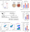
Figure 1. AKI induces intravascular neutrophil accumulation in the lungs.
(A) Serum BUN levels 24 hours after sham or AKI surgery in WT mice. n = 3 per group. ****P < 0.0001, by unpaired, 2-tailed Student’s t test. (B) Representative image of mouse lung before and after perfusion with PBS. Total neutrophil counts in sham versus AKI lungs were quantified by flow cytometry (FACS) with or without prior lung perfusion. Neutrophils/CD45+ cells: nonperfused, 36.0% versus 60.5%; perfused, 39.4% versus 57.6%, in both sham versus AKI. n = 3 per group. **P < 0.01 and ***P < 0.001, by 2-way ANOVA with Šidák’s multiple-comparison test. (C) Schematic of the in vivo neutrophil extravasation assay using 2 different anti-Ly6G Abs coupled to different fluorescence labels (APC or BV421). Sac, sacrifice. (D) FACS quantification of lung total neutrophils after sham operation, AKI (remote lung inflammation), or i.t. LPS (direct lung injury). n = 4–6 per group. ***P < 0.001, by 1-way ANOVA with Tukey’s multiple-comparison test. (E) Identification of extravasated neutrophils by FACS. Ly6G fluorescence (APC vs. BV421) was analyzed in CD45+CD11b+Gr1hi cells in sham-operated animals (left), AKI animals (middle), and animals subjected to direct lung injury by LPS i.t. (left). Single-positive (BV421+) cells are extravascular; double-positive cells (APC+BV421+) cells are intravascular. (F) FACS quantification of intravascular lung neutrophils (after PBS perfusion) for sham, AKI, or LPS i.t. n = 4–6 per group. **P < 0.01 and ****P < 0.0001, by 1-way ANOVA with Tukey’s multiple-comparison test. All data represent the mean ± SD.
Figure 2
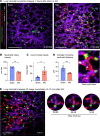
Figure 2. Intravascular lung capillary “neutrophil train” formation after AKI.
(A) Intravital 2-photon imaging of sham-treated and AKI lungs 2 hours after AKI using Ccr2gfp/+ mice and in vivo staining of neutrophils; CCR2+ monocytes (green), Ly6G+ neutrophils (red). Blood flow was assessed with 1 μm beads (white), and lung capillary circulation was labeled by i.v. injection of quantum dots (purple). The magnified inset image shows a neutrophil train in a lung capillary, illustrating vessel-occlusive accumulation of neutrophil trains attached to CCR2+ monocyte “locomotives.” Full videos are available in the supplemental materials. Scale bars: 100 μm; original magnification, x3.25 (inset). (BD) Quantification of intravital imaging videos: speed of neutrophil rolling (B), the number of static beads (C), and the average distance between CCR2+ monocytes and neutrophils (D). n = 4 per group. *P < 0.05 and **P < 0.01, by unpaired, 2-tailed Student’s t test. (E) Lung intravital time-lapse 3D image immediately after AKI (5–10 minutes). Magnified images on the right illustrate the process of neutrophil (red) train formation in the presence of a CCR2+ monocyte (green). Scale bar: 100μm A full time-lapse video is available as Supplemental Video 3. Scale bar: 100 μm; original magnification, x3.25 (inset). All data represent the mean ± SD (BE).
Figure 3
Figure 3. Hypoxemia is observed early after AKI in the absence of impaired ventilation or overt lung edema.
(A and B) Arterial blood gas analysis and lung H&E staining with alveolar wall thickness measurements after sham operation or 2–6 hours after AKI. Arterial blood samples were collected directly from the ascending aorta under mechanical ventilation using 100% O2. Scale bar: 200 μm. n = 4–9 per group. ***P < 0.001, by 1-way ANOVA with Tukey’s multiple-comparison test. (CE) Neutrophil depletion experiment. n = 5–6 per group. (C) Quantification of circulating neutrophils (CD11b+Ly6Cint cells) following 2 injections of anti-Ly6G Ab. Ly6G was not used as a neutrophil marker in FACS analysis due to epitope protection by the Ly6G Ab injected for neutrophil depletion. (D) Arterial blood gas analysis 6 hours after AKI. (E) Quantification of lung fluorescence beads injected 10 minutes prior to sacrifice as a surrogate marker for stagnation of lung microcirculation. Scale bar: 100 μm. *P < 0.05, ***P < 0.001, and ****P < 0.0001 compared with sham or control, by unpaired, 2-tailed Student’s t test. All data represent the mean ± SD.
Figure 4
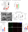
Figure 4. Rapid lung capillary neutrophil capture is enhanced by decreased neutrophil deformability but not classical neutrophil–endothelial cell interactions.
(A) scRNA-Seq analysis (see Supplemental Figure 2) showing expression of the adhesion molecule ICAM1 in lung cell types. (B) Anti-CD18 Ab blockade versus IgG control in C57BL/6 mice: quantification of lung neutrophils by Ly6G immunofluorescence staining after AKI. n = 5–6/group. (C) Representative EM images of sham versus AKI lung obtained with quantification. Arrows indicate empty vessel lumina, while black arrowheads point to deformable neutrophils in sham or nondeformable neutrophils in AKI lung; in the AKI lung, an outlined arrowhead points to a monocyte locomotive leading a nondeformable neutrophil train. Right lower image shows crawling or arrested neutrophils (black arrowheads). Scale bar: 10 μm. Plot on the left shows the neutrophil count presented as the number of neutrophils per total nuclei in each image. Plot on the right shows the ratio of short versus long diameter in each neutrophil as an indicator of its morphology. The short/long ratio closer to 1.0 is indicative of a more rounded, nondeformable shape. Each colored dot represents mean data from an individual animal (n = 3/group), while each gray dot represents each observation (image or neutrophil). **P < 0.05 and ****P < 0.0001, by unpaired, 2-tailed Student’s t test (B and C). (D) Quantification of F-actin in circulating neutrophils by flow cytometry. Using fluorescence-labeled phalloidin, the geometric MFI was analyzed and compared between sham and AKI groups. n = 4/group. *P < 0.05, by unpaired, 2-tailed Student’s t test. (E) F-actin staining of leukocytes isolated from circulating blood 2 and 4 hours after sham or AKI. Scale bar: 40 μm. Percentage of F-actin polymerized Ly6G+ cells (neutrophils) are quantified in graph. n = 4/group. **P < 0.01 and ****P < 0.0001, by 2-way ANOVA with Šidák’s multiple-comparison test. All data represent the mean ± SEM.
Figure 5
Figure 5. Neither nonclassical monocytes nor alveolar macrophages drive capillary neutrophil retention.
(A) Quantification of blood monocytes in WT versus NR4A1-KO mice (Nr4a1–/–). n = 3 per group. **P < 0.01, by unpaired, 2-tailed Student’s t test. (B) Serum BUN levels 24 hours after AKI in WT and NR4A1-KO mice. n = 6 per group. (C) Quantification of lung neutrophils after AKI measured by immunofluorescence staining. n = 6 per group. (D) Schematic of in vivo alveolar macrophage depletion model using diphtheria toxin and CD169-DTR heterozygous mice. (E) Serum BUN levels 24 hours after AKI in CD169-DTR heterozygous mice and their littermate controls. n = 6 per group. (F) Quantification of lung neutrophils after AKI measured by immunofluorescence staining in CD169-DTR heterozygous mice and their littermate controls. n = 6 per group. Statistical significance in B, C, E, and F was determined by unpaired, 2-tailed Student’s t test. All data represent the mean ± SD.
Figure 6
Figure 6. CCR2+ classical monocytes and CXCL2/CXCR2 signaling drive lung capillary neutrophil retention after AKI.
(A) Schematic of classical monocyte depletion using anti-CCR2 Ab in vivo. (B) Lung monocytes after pretreatment with anti-CCR2 Ab or control IgG. n = 6 per group. (C) Serum BUN indicating kidney injury level on day 1 after AKI. n = 6 per group. (D) Lung immunofluorescence staining after AKI in control versus anti-CCR2 Ab–treated animals. Shown are alveolar and interstitial macrophages (CD68+, red) and neutrophils (Ly6G+, green). Hoechst 33342 dye (blue) was used to visualize nuclei. Scale bar: 100 μm. n = 6 per group. (E) Total number of cell-cell communications inferred by CellChat based on the scRNA-Seq analysis for sham-operated versus AKI mice (also see Supplemental Figure 2). (F) Cell-cell communications from monocytes or macrophages to neutrophils were predicted at single ligand-receptor resolution (unbiased); the neutrophil chemoattractant CXCL2 in monocytes and macrophages and their receptor CXCR2 in neutrophils was predicted to be significantly increased in AKI versus sham groups and are underlined. (G) Lung immunofluorescence staining for Ly6G+ neutrophils (green) from WT C57BL/6 mice injected with anti-CXCL2 or control Ab and subjected to AKI. Scale bar: 100 μm. n = 4–5 per group. The graph shows a quantification of lung neutrophils detected by immunofluorescence. Data represent the mean ± SD. *P < 0.05 and **P < 0.01, by unpaired, 2-tailed Student’s t test.
Figure 7
Figure 7. Comparative analysis of lung scRNA-Seq data reveals that remote lung injury as compared with direct lung injury lacks alveolar macrophage activation and alveolar macrophage release of neutrophil chemoattractants.
(A) Comparison of inflammatory genes using scRNA-Seq analysis. A lung syngeneic transplant dataset was analyzed as a model of direct lung injury and compared with our AKI dataset (both reflect warm IRI). Expression levels of inflammatory molecules in neutrophils and alveolar macrophages are shown. The dot size denotes the percentage of cells expressing each gene, and the color scale represents the average gene expression levels. CTRL, control. (B) DEG analysis in alveolar macrophages: downregulated genes in post-AKI lung compared with direct lung injury were analyzed using GO and MSigDB Hallmark pathway analysis. GO terms and pathways related to inflammation and associated with activation of macrophages are underlined. Nega. reg., negative regulation; Pos. reg., positive regulation. (C) FACS and quantification of extravasated neutrophils 24 hours after AKI. Either mouse CXCL2 protein (0.01 μg/g) or vehicle in 50 μL sterile saline was administered i.t. immediately after AKI surgery. n = 4 mice per group. ***P < 0.001, by unpaired, 2-tailed Student’s t test. Data represent the mean ± SD.
Figure 8
Figure 8. Schematic summary.
AKI induces rapid intravascular neutrophil train formation in lung alveolar capillaries, a form of neutrophil retention. Rapid retention is enhanced by decreased deformability secondary to F-actin polymerization (submembrane F-actin bands) in circulating neutrophils that impedes their lung capillary passage. CCR2+ classical monocytes are required for neutrophil train formation and release CXCL2 to attract neutrophils into trains. Neutrophil train formation reduces alveolar capillary blood flow and is associated with thrombosis (thrombi contain both platelets and fibrin). This capillary perfusion defect leads to reduced oxygenation due to a ventilation perfusion mismatch, a scenario that differs from infectious inflammatory lung diseases, such as bacterial pneumonia or pulmonary alveolar edema in which ventilation is affected.

Update of

References

    1. Demirjian S, et al. Model to predict mortality in critically ill adults with acute kidney injury. Clin J Am Soc Nephrol. 2011;6(9):2114–2120. doi: 10.2215/CJN.02900311. - DOI - PMC - PubMed
    1. Chertow GM, et al. Prognostic stratification in critically ill patients with acute renal failure requiring dialysis. Arch Intern Med. 1995;155(14):1505–1511. doi: 10.1001/archinte.1995.00430140075007. - DOI - PubMed
    1. Wang HE, et al. Acute kidney injury and mortality in hospitalized patients. Am J Nephrol. 2012;35(4):349–355. doi: 10.1159/000337487. - DOI - PMC - PubMed
    1. Teixeira JP, et al. Pulmonary consequences of acute kidney injury. Semin Nephrol. 2019;39(1):3–16. doi: 10.1016/j.semnephrol.2018.10.001. - DOI - PubMed
    1. Faubel S, Edelstein CL. Mechanisms and mediators of lung injury after acute kidney injury. Nat Rev Nephrol. 2016;12(1):48–60. doi: 10.1038/nrneph.2015.158. - DOI - PubMed

MeSH terms