. 2025 Apr 25;388(6745):eads8412.
doi: 10.1126/science.ads8412. Epub 2025 Apr 25.

Structural mechanism of LINE-1 target-primed reverse transcription

Affiliations

Structural mechanism of LINE-1 target-primed reverse transcription

George E Ghanim et al. Science. .

Abstract

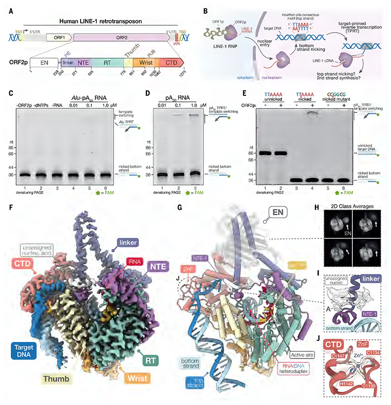
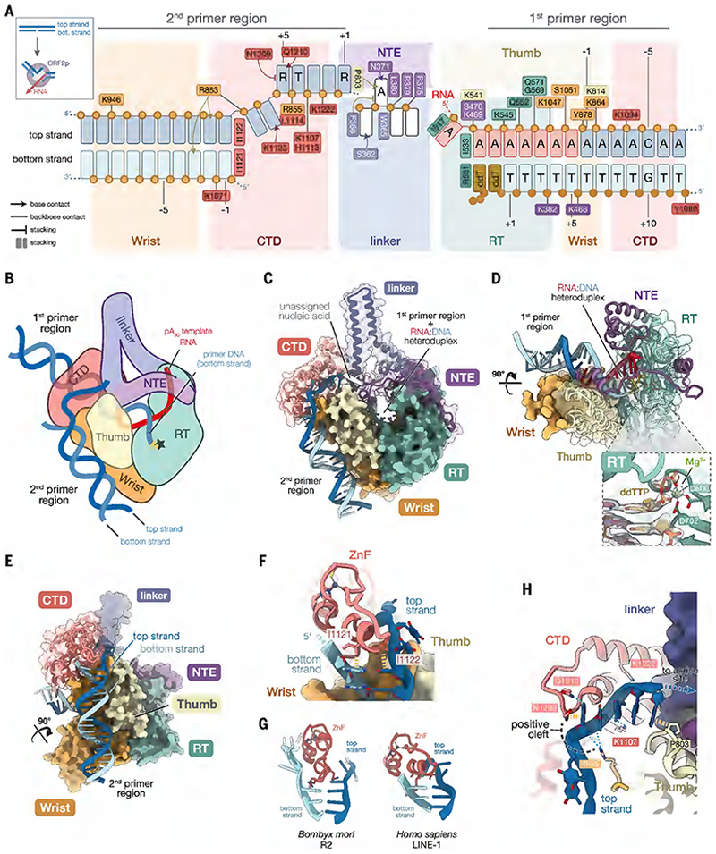
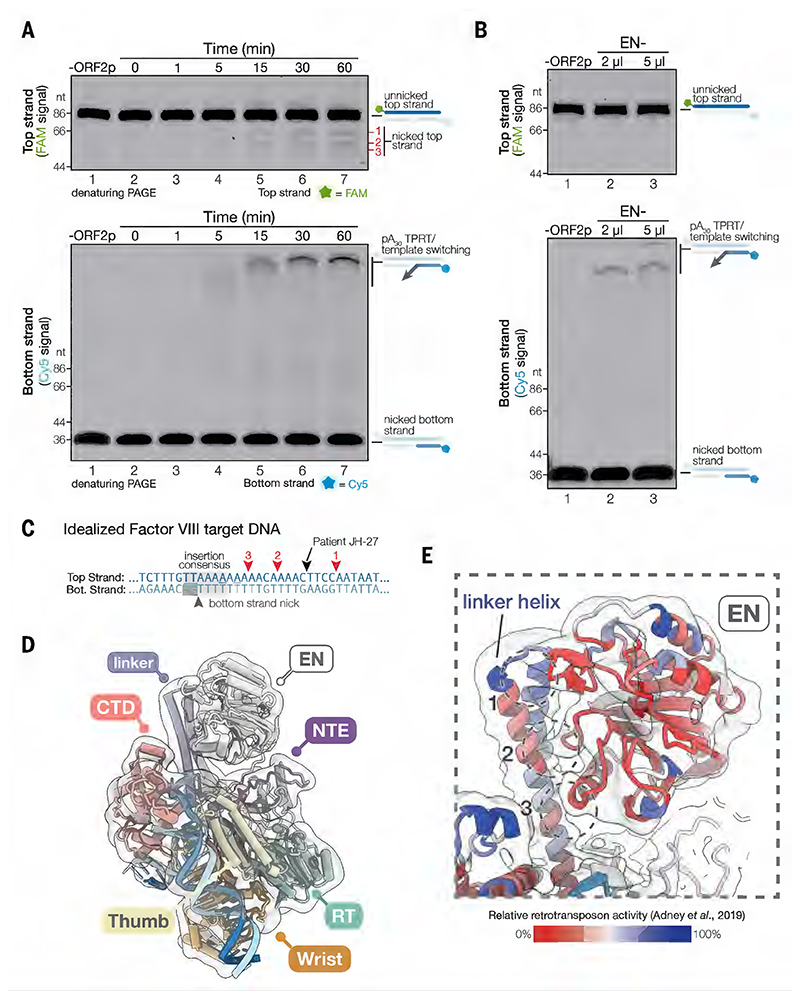
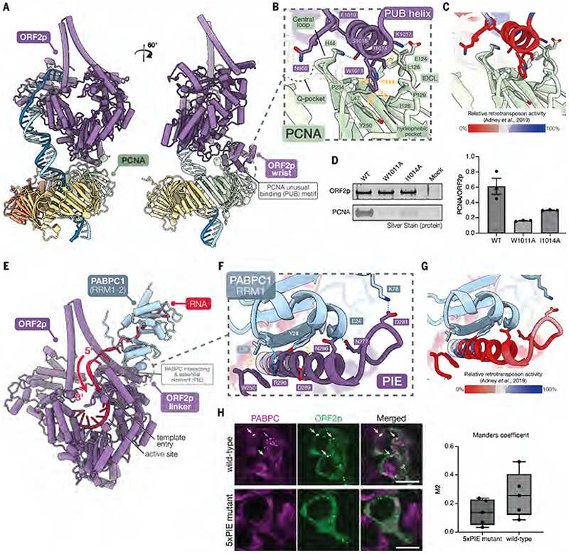
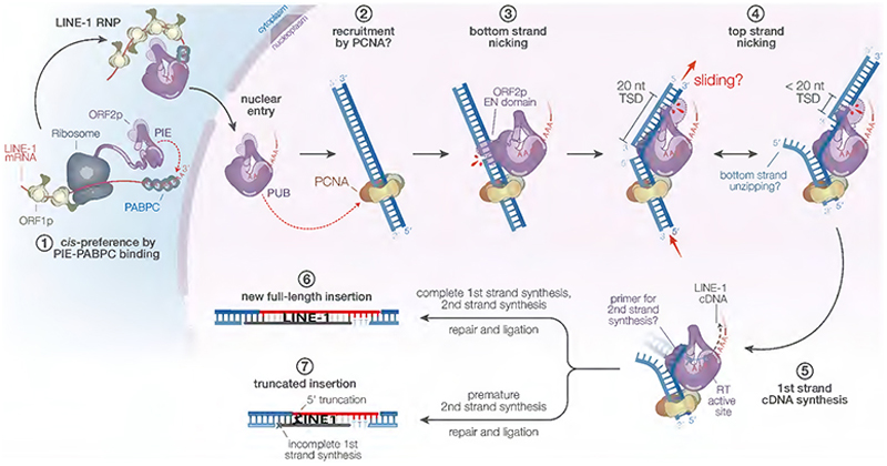
Long interspersed element-1 (LINE-1) retrotransposons are the only active autonomous transposable elements in humans. They propagate by reverse transcribing their messenger RNA into new genomic locations by a process called target-primed reverse transcription (TPRT). In this work, we present four cryo-electron microscopy structures of the human LINE-1 TPRT complex, revealing the conformational dynamics of open reading frame 2 protein (ORF2p) and its extensive remodeling of the target DNA for TPRT initiation. We observe nicking of the DNA second strand during reverse transcription of the first strand. Structure prediction identifies high-confidence binding sites for LINE-1-associated factors-namely proliferating cell nuclear antigen (PCNA) and cytoplasmic poly(A)-binding protein 1 (PABPC1)-on ORF2p. Together with our structural data, this suggests a mechanism by which these factors regulate retrotransposition and supports a model for TPRT that accounts for retrotransposition outcomes observed in cells.


Conflict of interest statement

Competing interests: Authors declare that they have no competing interests.

Figures

Fig. 1
Fig. 1. Cryo-EM structure of the LINE-1 TPRT complex.
(A) Organization of the human LINE-1 retrotransposon and domains of ORF2p. TSD, target site duplication; EN, endonuclease; PIE, PABC interacting and essential element; linker, EN linker; NTE, N-terminal extension; RT, reverse transcriptase; PUB, PCNA unusual binding site; CTD, C-terminal segment domain. (B) Schematic of LINE-1 retrotransposition. (C) Denaturing gel showing target primed reverse transcription (TPRT) activity with an Alu RNA template. (D) Denaturing gel showing TPRT activity with a 30 nt poly(A) RNA (pA30). (E) Denaturing gel showing the effects of unnicked, pre-nicked, or mutated pre-nicked target DNA substrate on TPRT activity. (F) Composite 2.3 Å cryo-EM reconstruction of the LINE-1 TPRT complex. Linker and target DNA densities were blurred to highlight flexible features. (G) Atomic model of the LINE-1 TPRT complex. ZnF, zinc finger. (H) 2D class averages showing flexibility of the EN domain. (I) Unassigned nucleic acid contacts the NTE. Cryo-EM density is shown as a transparent surface and blurred to highlight flexible features. (J) CTD zinc finger. Cryo-EM density is shown as a transparent surface.
Fig. 2
Fig. 2. The target DNA is unzipped and broken across the domains of ORF2p.
(A) Schematic of ORF2p interactions with the target DNA. Top and bottom strands are numbered relative to their respective cleavage sites. Inset shows a simplified schematic of target DNA remodeling accompanying TPRT complex formation. (B) Cartoon of the TPRT complex. Green star indicates RT active site. (C) Structure highlighting interactions with the two target DNA regions. (D) Structure surrounding the 1st primer region of the target DNA. Inset, view of ddTTP in the RT active site; cryo-EM density is shown as a transparent surface. (E) Structure surrounding the 2nd primer region of the target DNA. (F) CTD ZnF unzips the target DNA. Yellow dashed lines indicate stacking interactions. (G) Comparison of target DNA unzipping by BmR2 ZnF (35). (H) Interactions with the melted top strand and ORF2p. Blue dashed lines indicate hydrogen bonding.
Fig. 3
Fig. 3. The top strand is nicked with reverse transcription.
(A) Top strand nicking correlates with bottom strand TPRT. Denaturing gel of TPRT assay time course with doubly fluorescently labeled target DNA, visualized by FAM fluorescence to show top strand nicking (top) or by Cy5 fluorescence to show bottom strand TPRT products (bottom). (B) EN- (D145A) mutant blocks top strand nicking and reduces bottom strand TPRT. (C) Schematic of target DNA nicking. Red numbered triangles indicate the mapped cleavage sites of the top strand nicked products in (A). (D) Cryo-EM reconstruction with the EN domain resolved. Atomic model fit into the density is shown. Cryo-EM map was lowpass filtered to 8 Å. (E) Detailed view of EN-linker domain contacts. Retrotransposition efficiencies from trialanine scanning substitutions (50) are mapped onto the structure. Cryo-EM map was lowpass filtered to 5 Å.
Fig. 4
Fig. 4. Cellular factors facilitate nucleic acid binding.
(A) AlphaFold 3 prediction of the ORF2p-PCNA complex. (B) Detailed view of the predicted interaction between the PUB motif and PCNA. IDCL, interdomain connector loop. (C) Effect of PUB trialanine substitutions on retrotransposition efficiency. The same view as in (B) is shown, with retrotransposition efficiencies from trialanine scanning substitutions (50) mapped onto the structure. (D) Silver-stained SDS-PAGE from ORF2p pulldown experiments with PUB site mutants (left panel) and quantification of the pulldown experiments (right panel). The experiments were performed in triplicate (n = 3). Values represent PCNA band intensity normalized to ORF2p band intensity. Error bars represent standard error of the mean (SEM). (E) AlphaFold 3 prediction of the ORF2p-PABPC1 complex. RRM1-2, RNA recognition motif 1 and 2. (F) Detailed view of the interactions between the PIE region of ORF2p and PABPC1. RRM, RNA recognition motif. (G) Effect of PIE trialanine substitutions on retrotransposition efficiency. The same view as in (F) is shown, with retrotransposition efficiencies from trialanine scanning substitutions (50) mapped onto the structure. (H) Immunofluorescence staining of ORF2p (wild-type or 5xPIE mutant; green) and PABPC (magenta). Arrows indicate examples of ORF2p cytoplasmic puncta co-localized with PABPC, which were not observed in the ORF2p 5xPIE mutant. 5xPIE mutant, M272A, N277A, D281A, N286A and R296A. Scale bar: 10 µm. Quantification of Manders coefficients (n = 10, right).
Fig. 5
Fig. 5. Model for TPRT and LINE-1 retrotransposition.
1, Co-translational ORF2p PIE-PABPC binding establishes cis-preference. 2, After RNP formation and nuclear entry, PCNA recruits the LINE-1 RNP to a target DNA with the appropriate architecture for retrotransposition. 3, EN domain nicks the bottom strand at a site resembling the EN cleavage consensus motif. 4, Sliding and unzipping of the target DNA allows the EN domain to nick the top strand at a suitable site and explains the observed distribution of target site duplication (TSD) lengths. The timing of top strand nicking is unclear. 5, First-strand cDNA synthesis initiates after the bottom strand is passed to the RT active site and anneals with the poly(A) tail. 6, Complete first-strand synthesis followed by template jumping to the exposed top strand initiates second-strand synthesis and results in a new full-length insertion. 7, Premature second-strand synthesis before first-strand has completed would lead to a new 5' truncated insertion.

Similar articles

References

    1. Boeke JD, Garfinkel DJ, Styles CA, Fink GR. Ty elements transpose through an RNA intermediate. Cell. 1985;40:491–500. - PubMed
    1. Lander ES, I. H. G. S. Consortium. Linton LM, Birren B, Nusbaum C, Zody MC, Baldwin J, Devon K, Dewar K, Doyle M, FitzHugh W, et al. Initial sequencing and analysis of the human genome. Nature. 2001;409:860–921. - PubMed
    1. Brouha B, Schustak J, Badge RM, Lutz-Prigge S, Farley AH, Moran JV, Kazazian HH. Hot L1s account for the bulk of retrotransposition in the human population. Proc Natl Acad Sci. 2003;100:5280–5285. doi: 10.1073/pnas.0831042100. - DOI - PMC - PubMed
    1. Beck CR, Collier P, Macfarlane C, Malig M, Kidd JM, Eichler EE, Badge RM, Moran JV. LINE-1 Retrotransposition Activity in Human Genomes. Cell. 2010;141:1159–1170. doi: 10.1016/j.cell.2010.05.021. - DOI - PMC - PubMed
    1. Sassaman DM, Dombroski BA, Moran JV, Kimberland ML, Naas TP, DeBerardinis RJ, Gabriel A, Swergold GD, Kazazian HH. Many human L1 elements are capable of retrotransposition. Nat Genet. 1997;16:37–43. - PubMed