. 2025 May 13;9(9):2285-2299.
doi: 10.1182/bloodadvances.2024015061.

DNMT3A regulates murine megakaryocyte-biased hematopoietic stem cell fate decisions

Affiliations

DNMT3A regulates murine megakaryocyte-biased hematopoietic stem cell fate decisions

Sarah M Waldvogel et al. Blood Adv. .

Abstract

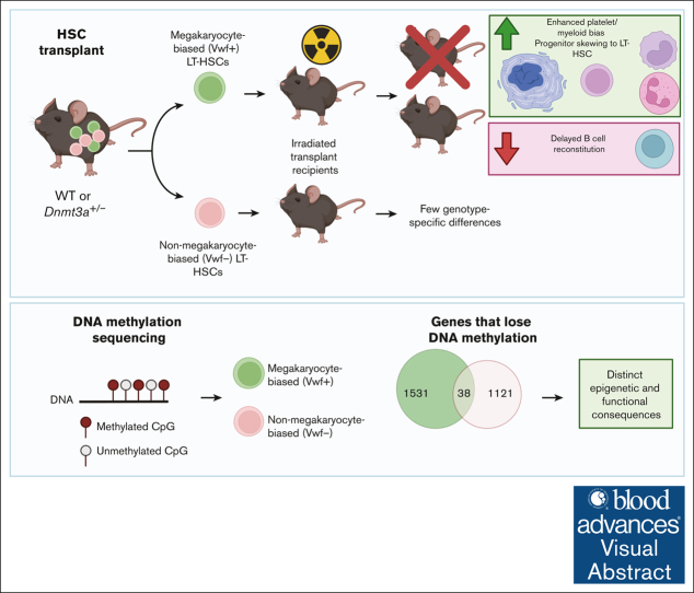
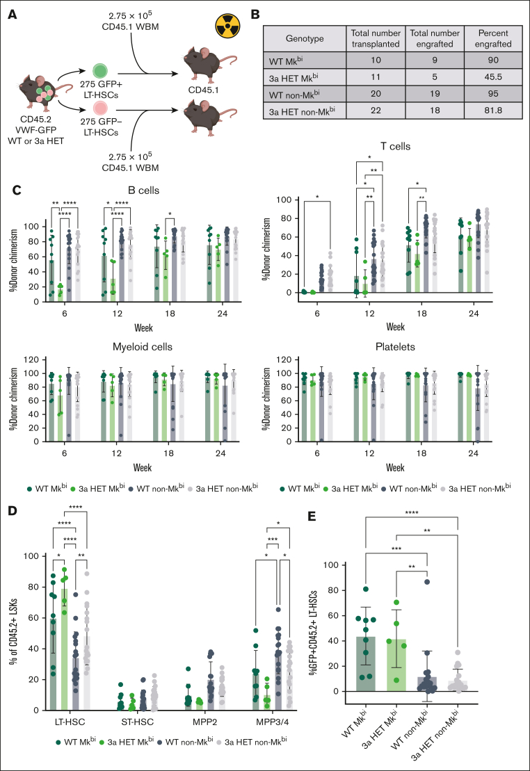
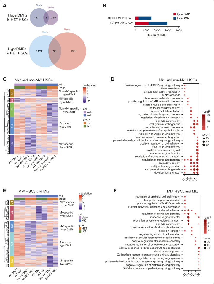
Hematopoietic stem cells (HSCs) are defined by their capacity to regenerate all main components of peripheral blood, but individual HSCs exhibit a range of preferences for generating downstream cell types. Their propensities are thought to be epigenetically encoded, but few differential regulatory mechanisms have been identified. In this work, we explored the role of DNA methyltransferase 3A (DNMT3A) in the megakaryocyte-biased HSC population, which is thought to reside at the top of the hematopoietic hierarchy. We demonstrate that heterozygous loss of DNMT3A (Dnmt3a+/-) in these megakaryocyte-biased HSCs has distinct consequences compared with the rest of the HSC pool. These megakaryocyte-biased HSCs become delayed in their lymphoid-repopulating ability but can ultimately regenerate all lineages. We further demonstrate that Dnmt3a+/- mice have increased numbers of megakaryocytes in the bone marrow. Analysis of DNA methylation differences between wild-type (WT) and Dnmt3a+/- HSC subsets, megakaryocyte-erythroid progenitors, and megakaryocytes revealed that DNA methylation is eroded in the mutants in a cell type-specific fashion. Although transcriptional differences between WT and Dnmt3a+/- megakaryocyte-biased HSCs are subtle, the pattern of DNA methylation loss in this HSC subset is almost completely different from that in non-megakaryocyte-biased HSCs. Together, our findings establish the role of epigenetic regulation in the fate of megakaryocyte-biased HSCs and their downstream progeny and suggest that the outcomes of DNMT3A loss might vary depending on the identity of the HSC that acquires the mutation.


Conflict of interest statement

Conflict-of-interest disclosure: The authors declare no competing financial interests.

Figures

None
Graphical abstract
Figure 1.
Figure 1.
Reduced DNMT3A results in LT-HSC skewing, but fewer are Mkbi. (A) Experimental design. WBM (3 × 106 cells) was transplanted into lethally irradiated CD45.1 recipients. The transplanted WBM was composed of 80% CD45.1 cells and 20% test cells, either VWF-GFP 3a WT or VWF-GFP Dnmt3a+/– (“3aHET”). Test cells were CD45.2. After 5 months, bone marrow from all primary recipients of the same sex and genotype was pooled and transplanted into lethally irradiated CD45.1 secondary recipients. Blood was sampled every 4 weeks for a total of 12 weeks. Primary transplant included 15 mice per genotype, including both male and female mice; secondary transplant included 7 WT and 8 3aHET female mice. (B) PB donor chimerism in WT and 3aHET secondary transplant recipients over 12 weeks (n = 7 WT and 8 3aHET female mice). Two-way analysis of variance (ANOVA) with Šídák multiple comparisons test. All comparisons not significant (ns) with a P value of >.05. Data are represented as the mean ± standard deviation (SD). (C) PB donor chimerism within the B cell, myeloid, T cell, and platelet lineages in WT and 3aHET secondary transplant recipients at 12 weeks (n = 7 WT and 8 3aHET female mice). Two-way ANOVA with Šídák multiple comparisons test. All comparisons ns with a P value of >.05. Data are represented as the mean ± SD. (D) Quantification of the percentage of test CD45.2+ LSK cells composed of LT-HSCs and MPP3/4 in WT and 3aHET secondary transplant recipients. Flow cytometry counts within each of the 4 groups were summed, and the percentage was calculated from the total (n = 7 WT and 8 3aHET female mice). Two-way ANOVA with Šídák multiple comparisons test. ∗P ≤ .05; ∗∗P ≤ .01. Data are represented as the mean ± SD. (E) Flow cytometry gating strategy for the data represented in panel D. Lineage-depleted bone marrow cells were gated as ckit+ Lineage–negative Sca1+ CD45.2+ and then divided into LT-HSCs (CD150hiCD48lo), ST-HSCs (CD150loCD48lo), MPP2 (CD150hiCD48hi), and MPP3/4 (CD150loCD48hi). (F) WBM donor chimerism within the B cell, myeloid, and T cell lineages of WT and 3aHET secondary transplant recipients at 12 weeks (n = 7 WT and 8 3aHET female mice). Two-way ANOVA with Šídák multiple comparisons test. All comparisons ns with a P value of >.05. Data are represented as the mean ± SD. (G) The percentage of test CD45.2+ LT-HSCs that are Mkbi in the WT and 3aHET secondary transplant recipients. Mann-Whitney test; ∗P ≤ .05 (n = 7 WT and 8 3aHET female mice). Data are represented as the mean ± SD. ST-HSC, short-term HSC; MPP3/4, multipotent progenitor-3/4.
Figure 2.
Figure 2.
3aHET Mkbi HSCs are poorly transplantable and exhibit delayed B cell reconstitution. (A) Experimental design. Mkbi (GFP+) and non-Mkbi (GFP) LT-HSCs were sorted from VWF-GFP 3a WT or VWF-GFP 3aHET CD45.2 donors. A total of 275 LT-HSCs (GFP+ or GFP) were mixed with 275 000 supporting CD45.1 WBM cells and transplanted into lethally irradiated CD45.1 recipients. Blood was collected from the recipients every 6 weeks for 24 weeks. Cells from male and female donors were pooled separately and transplanted into male recipients. The data shown are from a combination of 2 transplants performed at separate times with separate donor pools. (B) The number of mice that were transplanted with HSCs and the outcome for each group combined from 2 separate transplants. The total number engrafted, and the percent engrafted indicate mice that survived to 6 weeks after transplant and exhibited >0.5% donor chimerism in the PB or bone marrow. A single mouse in the Mkbi 3aHET group survived long term with <0.5% donor chimerism. All other mice that failed to engraft died within ∼3 weeks after transplant. (C) The percent donor chimerism in the PB B cells, T cells, myeloid cells, and platelets from 6 to 24 weeks after transplant in the HSC transplant recipients. Two-way ANOVA with Šídák multiple comparisons test. ∗P ≤ .05; ∗∗P ≤ .01; ∗∗∗P ≤ .001; ∗∗∗∗P ≤ .0001. Data are represented the mean ± SD. (D) The composition of the CD45.2+ LSK compartment with regard to the indicated stem and progenitor cells as gated in Figure 1E. Two-way ANOVA with Šídák multiple comparisons test. ∗P ≤ .05; ∗∗P ≤ .01; ∗∗∗P ≤ .001; ∗∗∗∗P ≤ .0001. Data are represented as the mean ± SD. (E) The percent of Mkbi CD45.2+ LT-HSCs in Mkbi vs non-Mkbi transplant recipients. One-way ANOVA with Tukey multiple comparisons test. ∗P ≤ .05; ∗∗P ≤ .01; ∗∗∗P ≤ .001; ∗∗∗∗P ≤ .0001. Data are represented as the mean ± SD. ST-HSC, short-term HSC; MPP3/4, multipotent progenitor-3/4.
Figure 3.
Figure 3.
3aHET Mkbi HSCs display enhanced transcriptional signatures of low output and Mk-bias. (A) Uniform Manifold Approximation and Projection plot of cell type distribution for WT and 3aHET single-cell RNAseq data sets from 2 pooled mice per genotype 10 months after WBM transplant (data set from Reyes et al40). CD45.2+ LSKs were sorted for sequencing. Immunological Genome Project reference cell type LT-HSCs, SC.LT34F (CD34FLK2 LT-HSCs; CD34Flk2Linc-kit+Sca1+) are highlighted in blue, and cells expressing >0 Vwf transcripts are highlighted in red. (B) Gene set enrichment analysis interaction terms from differentially expressed genes between 3aHET and WT Vwf+ LT-HSCs. An adjusted P value of < .05 was set as the threshold for significantly related genes, displaying the top 7 significant pathways. The gene list was generated by ranking the log2FC in decreasing order. (C) Fast gene set enrichment analysis plot of differentially expressed genes in 3aHET and WT Vwf+ HSCs compared with the Mk-biased output HSC signature. A total of 1000 gene permutations were used to calculate statistical significance, and an adjusted P value of <.05 was set for statistical significance of the gene sets. (D) Fast gene set enrichment analysis plot of differentially expressed genes in 3aHET and WT Vwf+ HSCs compared with the low-output HSC signature. One-thousand gene permutations were used to calculate statistical significance, and an adjusted P value of <.05 was set for statistical significance of the gene sets. adj, adjusted; NES, normalized enrichment score; Pos, positive; TM, transmembrane.
Figure 4.
Figure 4.
Transcriptional cell identity is determined more by intrinsic HSC bias than genotype. (A) Heat map showing the expression levels of key marker genes from bulk RNAseq of Mkbi and non-Mkbi HSCs from pooled WT and 3aHET mice. Mkbi and non-Mkbi HSCs were sorted from a pool of male and female mice for each genotype (n = 3-4 mice per pool; aged 10-12 months). Expression type = transcript per million, samples ordered by Euclidean hierarchical clustering, transformed by row-wise z score. (B) Venn diagram showing the genes upregulated in Mkbi HSCs compared with non-Mkbi HSCs. A total of 203 genes are unique to the WT Mkbi vs WT non-Mkbi comparison, 31 genes are unique to the 3aHET Mkbi vs 3aHET non-Mkbi comparison, and 197 genes are upregulated in both. The top Gene Ontology-Biological Process (GO-BP) terms for genes uniquely upregulated in WT (blue, 1) and uniquely upregulated in 3aHET (orange, 2) are shown ranked by P value. (C) Venn diagram showing the genes downregulated in Mkbi HSCs compared with non-Mkbi HSCs. A total of 152 genes are unique to the WT Mkbi vs WT non-Mkbi comparison, 10 genes are unique to the 3aHET Mkbi vs 3aHET non-Mkbi comparison, and 42 genes are downregulated in both. The top GO-BP terms for genes uniquely downregulated in WT (blue, 3) and uniquely downregulated in 3aHET (orange, 4) are shown ranked by P value.
Figure 5.
Figure 5.
Germ line 3aHET animals display phenotypic abnormalities at the Mk level. (A) Quantification of Mks per μm in WT vs 3aHET mice via immunofluorescence imaging of bone marrow sections from mouse femurs (n = 4 female mice per group; aged 4 months); and the entire length of 1 femur was imaged. ∗∗P ≤ .01. Data are represented as the mean ± SD. (B) Representative images of WT and 3aHET Mks from frozen femur sections (red = α-CD41, AF647; green = α-Laminin, AF488; scale bar = 100 mm). Imaged on the Zeiss LSM900 confocal microscope with the Plan-Apo 20× objective and displayed as an orthogonal projection of the stack using the Zen Blue software. The channels were overlaid, the images were cropped, and scale bars were applied in ImageJ. (C) Quantification of the composition of the LSK compartment in WT vs 3aHET germ line animals (n = 7 WT and 9 3aHET, both male and female mice; aged 2-4 months). Two-way ANOVA with Šídák multiple comparisons test. ∗∗P ≤ .01. Data are represented as the mean ± SD. (D) The percent of GFP+ HSCs in germ line 3aHET animals. Young animals included 7 WT and 9 3aHET, both male and female mice; aged 2 to 4 months. Unpaired t test. P > .05 (ns). Data are represented as the mean ± SD. Old animals included 7 WT and 6 3aHET, both male and female mice; aged 9 to 12 months. Unpaired t test. P > .05 (ns). Data are represented as the mean ± SD. (E) The percent of GFP+ HSCs in germ line 3aHET animals 7 days after treatment with PBS or 5FU (n = 3 WT mice and 4 3aHET mice per group, both male and female mice; aged 2-10 months; ages matched between groups). Two-way ANOVA with Šídák multiple comparisons test. P > .05 (ns). Data are represented as the mean ± SD. (F) Volcano plot showing the differentially expressed proteins, as determined by mass spectrometry, between WT (–log2FC) and 3aHET (+log2FC) platelets (n = 4 per group, including 2 males and 2 females; aged 2 months). (G) Gene ontology analysis of platelet RNAseq showing pathways downregulated in the 3aHET samples (n = 4 males per group; aged 3 months). ns, not significant; PBS, phosphate-buffered saline; ST-HSC, short term HSC.
Figure 6.
Figure 6.
3aHET Mkbi and non-Mkbi HSCs, MEPs, and Mks exhibit unique hypomethylation patterns. (A) Shared and unique regions of decreased methylation (hypo-) and increased methylation (hyper-) in 3aHET compared with WT from Mkbi and non-Mkbi HSCs. Mkbi and non-Mkbi HSCs were sorted from a pool of male and female mice for each genotype (n = 3-4 mice per pool; aged 10-12 months). DNA was isolated from the same samples used for bulk RNAseq in Figure 4. (B) Total number of DMRs in 3aHET MEPs and Mks compared with WT (n = 2 female mice per group; aged 6 months). (C) Integrative clustering of DMRs from Mkbi and non-Mkbi HSCs in 3aHET compared with WT. (D) Functional enrichment of the clusters from panel C. (E) Integrative clustering of DMRs from Mkbi HSCs and Mks in 3aHET compared with WT. (F) Functional enrichment of the clusters from panel E. C1, cluster 1; HET, 3aHET; TGF, transforming growth factor; VEGFR, vascular endothelial growth factor receptor.

References

    1. Dykstra B, Kent D, Bowie M, et al. Long-term propagation of distinct hematopoietic differentiation programs in vivo. Cell Stem Cell. 2007;1(2):218–229. - PubMed
    1. Challen GA, Boles NC, Chambers SM, Goodell MA. Distinct hematopoietic stem cell subtypes are differentially regulated by TGF-beta1. Cell Stem Cell. 2010;6(3):265–278. - PMC - PubMed
    1. Benz C, Copley MR, Kent DG, et al. Hematopoietic stem cell subtypes expand differentially during development and display distinct lymphopoietic programs. Cell Stem Cell. 2012;10(3):273–283. - PubMed
    1. Copley MR, Beer PA, Eaves CJ. Hematopoietic stem cell heterogeneity takes center stage. Cell Stem Cell. 2012;10(6):690–697. - PubMed
    1. Rodriguez-Fraticelli AE, Weinreb C, Wang SW, et al. Single-cell lineage tracing unveils a role for TCF15 in haematopoiesis. Nature. 2020;583(7817):585–589. - PMC - PubMed

MeSH terms

Substances