. 2025 Mar 6;16(1):2238.
doi: 10.1038/s41467-025-57342-3.

Autism gene variants disrupt enteric neuron migration and cause gastrointestinal dysmotility

Affiliations

Autism gene variants disrupt enteric neuron migration and cause gastrointestinal dysmotility

Kate E McCluskey et al. Nat Commun. .

Abstract

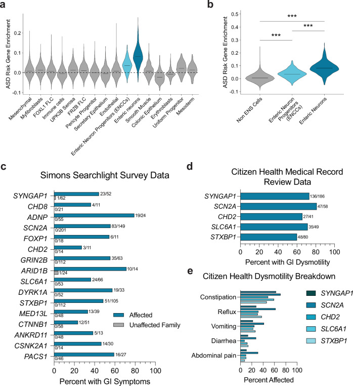
The co-occurrence of autism and gastrointestinal distress is well-established, yet the molecular underpinnings remain unknown. The identification of high-confidence, large-effect autism genes offers the opportunity to identify convergent, underlying biology by studying these genes in the context of the gastrointestinal system. Here we show that the expression of these genes is enriched in human prenatal gut neurons and their migratory progenitors, suggesting that the development and/or function of these neurons may be disrupted by autism-associated genetic variants, leading to gastrointestinal dysfunction. Here we document the prevalence of gastrointestinal issues in patients with large-effect variants in sixteen autism genes, highlighting dysmotility, consistent with potential enteric neuron dysfunction. Using Xenopus tropicalis, we individually target five of these genes (SYNGAP1, CHD8, SCN2A, CHD2, and DYRK1A) and observe disrupted enteric neuronal progenitor migration for each. Further analysis of DYRK1A reveals that perturbation causes gut dysmotility in vivo, which can be ameliorated by treatment with either of two serotonin signaling modulators, identified by in vivo drug screening. This work suggests that atypical development of enteric neurons contributes to the gastrointestinal distress commonly seen in individuals with autism and that serotonin signaling may be a productive therapeutic pathway.


Conflict of interest statement

Competing interests: E.B. is a salaried employee of Citizen Health, with vested and unvested stock options. The remaining authors do not have any competing interests.

Figures

Fig. 1
Fig. 1. hcASD gene expression is enriched in ENS cells and individuals with pathogenic variants in these genes experience GI issues.
a Enrichment of 252 hcASD genes is higher in Enteric Neuron Progenitors (ENCCs) and Enteric Neurons compared to all other cell types in single-cell RNA-sequencing data from the human prenatal gut. b 252 hcASD genes are enriched in ENCCs and Enteric Neurons compared to all other Non-ENS cells in the human prenatal gut. A one-way Kruskal-Wallis test was performed, followed by Wilcoxon rank-sum tests and Bonferroni adjustment for multiple comparisons. Non-ENS vs ENCCs padj = 5.20e−91, Non-ENS vs Enteric neurons padj = 0, ENCCs vs Enteric Neurons padj = 5.90e−81. ce The number of individuals affected and the total number of people surveyed is tallied at the end of each bar. c Simons Searchlight data documenting the percentage of affected individuals (teal bars) and their unaffected family members (gray bars) who reported GI issues in caregiver surveys. d Citizen Health medical record data showing the percentage of individuals with a SYNGAP1, SCN2A, CHD2, STXBP1 or SLC6A1 genetic variant with medical record diagnoses related to GI dysmotility. e Citizen Health medical record data by variant for dysmotility phenotypes including constipation, abdominal pain, and diarrhea.
Fig. 2
Fig. 2. hcASD gene depletion in vivo causes ENCC migration defects.
a Schematic of phox2b staining in NF stage 40 animals to mark enteric neural crest-derived cells (ENCCs, enteric neuron progenitors) in the gut, circled by a blue dashed line. phox2b also labels the hindbrain region and other migrating vagal neural crest cells. b Unilateral mutants were made by injecting the Cas9 protein and an sgRNA targeting an hcASD gene into one cell at the two-cell embryonic stage. Three days later, injected embryos were stained and ENCC area and gut area was measured on each side of the animal and compared to quantify relative gut area (CRISPR side / Control side) of migration. c Individual CRISPR mutagenesis of hcASD genes syngap1, chd8, scn2a, chd2, or dyrk1a reduce the area of ENCC migration in the gut compared to control mutagenesis of pigmentation gene slc45a2. d Area of gut migration quantification by target gene. Control in gray, hcASD genes in blue. A two-way Kruskal-Wallis test was performed followed by Wilcoxon matched-pairs signed rank test to compare the CRISPR side to the control side within each animal and a Holm-Šídák test to adjust for multiple comparisons. Control (N = 24, padj = 0.9248), syngap1 (N = 38, padj = 0.0046), chd8 (N = 25, padj = 0.001), scn2a (N = 33, padj = 0.001), chd2 (N = 31, padj = 0.0011), dyrk1a (N = 38, padj = 0.0011). All samples are independent biological replicates from the same mating pair. In the box plot, whiskers show minimum and maximum values, box represents the 25th and 75th quartiles, and the center line describes the median. Source data are provided as a Source Data file.
Fig. 3
Fig. 3. hcASD gene dyrk1a perturbation reduces ENCC migration and decreases gut motility in vivo.
a dyrk1a perturbation through CRISPR injection, morpholino (MO) injection, or Dyrk1a inhibitor (TG003) treatment all reduce the area of ENCC migration in the gut compared to control CRISPR, control MO, or vehicle control (DMSO). b Area of gut migration quantification by condition. For injection comparisons, a two-tailed Wilcoxon matched-pairs signed rank test to compare the CRISPR or morpholino side to the control side within each animal was performed. Control CRISPR (N = 18, p = 0.7987), dyrk1a CRISPR (N = 18, p = 0.0032), Control MO (N = 24, p = 0.1208), dyrk1a MO (N = 41, p < 0.0001). For small molecule treatment, a two-tailed Wilcoxon rank sum test was performed to compare DMSO (N = 18) to 5 µM TG003-treated (N = 20) embryos, p = 0.0026. All samples are independent biological replicates from the same mating pair. In the box plot whiskers show minimum and maximum values, box represents the 25th and 75th quartiles, and the center line describes the median. c Schematic of the in vivo gut motility assay. At NF stage 47, tadpoles were fed food with fluorescent beads for 2 h, then placed in a 6-well plate in 3D-printed baskets with 20 animals/well for 3 h. Tadpoles are then removed by taking out baskets and leaving excrement with fluorescent beads behind, after which the plates were imaged to count the number of beads per well. Created in BioRender. McCluskey, K. (2025) https://BioRender.com/w82f112. d Developmental inhibition of Dyrk1a (TG003-treated beginning at NF 20) results in decreased fluorescent bead excretion compared to vehicle control (DMSO). Representative images of fluorescent beads (false-colored black). e Number of fluorescent beads per well counted for DMSO (N = 3 wells, 20 tadpoles each) and TG003 (N = 3 wells, 20 tadpoles each) (Dyrk1a inhibitor) wells. A one-tailed t test with Welch’s correction was used to compare DMSO to 5 µM TG003, p = 0.0091. Data are presented as mean values +/− SEM. Source data are provided as a Source Data file.
Fig. 4
Fig. 4. Dyrk1a-associated gut dysmotility is ameliorated by acute exposure to an SSRI or 5-HTR6 agonist.
a Schematic of a serotonergic synapse labeling the presumed location of action for the drugs tested. b Acute exposure of wildtype tadpoles to escitalopram oxalate (selective serotonin reuptake inhibitor, SSRI) alone increases the average number of fluorescent beads excreted by more than one standard deviation compared to the average DMSO treatment, blue. c Acute treatment of an SSRI (10 µM escitalopram oxalate) rescued, while a 5HTR6 agonist (10 µM WAY-181187) partially rescued the decreased fluorescent bead excretion from developmentally inhibited Dyrk1a (5 µM TG003 starting at stage NF 20) animals. d Fluorescent beads per well quantified for each treatment condition. A one-way ANOVA was performed followed by unpaired one-tailed t tests with welch’s correction. Compared to TG003 treatment (N = 6 wells, 20 tadpoles each), DMSO (N = 6 wells, 20 tadpoles each p = 0.002), TG003 + SSRI (N = 3 wells, 20 tadpoles each p = 0.0423), TG003 + WAY-181187 (N = 3 wells, 20 tadpoles each p = 0.0033). Compared to DMSO, TG003 + SSRI p = 0.4264 and TG003 + WAY-181187 p = 0.0462. All samples are independent biological replicates from the same mating pair. Data are presented as mean values +/− SEM. Source data are provided as a Source Data file. e Model of how hcASD gene large-effect variants could contribute to GI dysmotility. An hcASD genetic variant leads to perturbed ENCC migration and ultimately GI dysmotility. Created in BioRender. McCluskey, K. (2025) https://BioRender.com/n91b055.

Update of

References

    1. Hughes, M. M. et al. The prevalence and characteristics of children with profound autism, 15 sites, United States, 2000–2016. Public Health Rep.138, 971–980 (2023). - PMC - PubMed
    1. McElhanon, B. O., McCracken, C., Karpen, S. & Sharp, W. G. Gastrointestinal symptoms in autism spectrum disorder: a meta-analysis. Pediatrics133, 872–883 (2014). - PubMed
    1. Wang, X. et al. The enteric nervous system deficits in autism spectrum disorder. Front. Neurosci.17, 1101071 (2023). - PMC - PubMed
    1. Wasilewska, J. & Klukowski, M. Gastrointestinal symptoms and autism spectrum disorder: links and risks – a possible new overlap syndrome. Pediatr. Health, Med. Therapeutics6, 153–166 (2015). - PMC - PubMed
    1. Fu, J. M. et al. Rare coding variation provides insight into the genetic architecture and phenotypic context of autism. Nat. Genet.54, 1320–1331 (2022). - PMC - PubMed

MeSH terms

Substances