. 2025 Mar 6;9(1):62.
doi: 10.1038/s41698-025-00849-1.

LINE-1 ORF1p expression occurs in clear cell ovarian carcinoma precursors and is a candidate blood biomarker

Affiliations

LINE-1 ORF1p expression occurs in clear cell ovarian carcinoma precursors and is a candidate blood biomarker

Pamela R de Santiago et al. NPJ Precis Oncol. .

Abstract

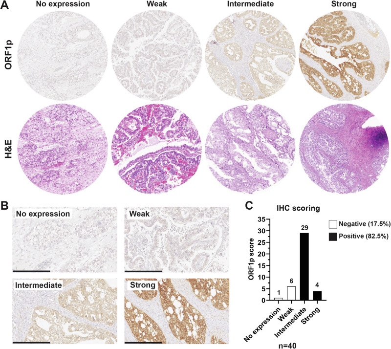
Long interspersed element 1 (LINE-1) retrotransposons are repetitive sequences that can move within the genome by an autonomous mechanism. To limit their mutagenic potential, benign cells restrict LINE-1 expression through molecular mechanisms such as DNA methylation and histone modification, but these mechanisms are usually impaired in cancer. Clear cell ovarian carcinoma (CCOC) represents 5-10% of ovarian cancers and is thought to arise from endometriosis. Women with advanced CCOC face poor prognoses, highlighting the importance of understanding early disease pathogenesis. In our study, 33 of 40 cases (over 82%) of CCOC tumors express ORF1p, a LINE-1-encoded protein. We found that LINE-1 de-repression is an early event in CCOC, as ORF1p is enhanced during the transition from typical to atypical endometriosis and persists in invasive cancer. Finally, using single-molecule array (Simoa) assays, we detected ORF1p in patient blood, suggesting it as a potential minimally invasive biomarker for this disease.


Conflict of interest statement

Competing interests: D.R.W. has a financial interest in Quanterix Corporation, a company that develops an ultra-sensitive digital immunoassay platform. He is an inventor of the Simoa technology, a founder of the company, and also serves on its Board of Directors. D.R.W.’s interests were reviewed and are managed by Brigham and Women’s Hospital and Partners Healthcare in accordance with their conflict of interest policies. A.K.G. is a co-founder of Sinochips Diagnostics, serves as a scientific advisory board member to Biovica, Clara Biotech, EXOKĒRYX, VITRAC Therapeutics, and Sinochips Diagnostics, and receives research funding from Predicine and VITRAC Therapeutics. R.D. serves on the scientific advisory board of Repare Therapeutics. All other authors declare no financial or non-financial competing interests.

Figures

Fig. 1
Fig. 1. LINE-1 is de-repressed in clear cell ovarian carcinoma.
A Formalin-fixed, paraffin-embedded human tissue from TMA. Representative H&E (bottom panel) and ORF1p IHC (upper panel) scale images used for scoring. B ORF1p IHC scale at higher magnification. Scale bar: 200 µm. C Distribution of ORF1p scoring in CCOC samples (n = 40).
Fig. 2
Fig. 2. ORF1p expression is enhanced during neoplastic transformation.
A Representative images of hematoxylin and eosin (H&E) staining and ORF1p expression (IHC) on whole-mount slides from cases of typical endometriosis (left panel) and atypical endometriosis (right panel). Scale bar: 500 µm. B ORF1p IHC scoring for typical endometriosis (n = 62) and C atypical endometriosis (n = 40).
Fig. 3
Fig. 3. LINE-1 ORF1p is expressed and released by CCOC cell lines.
A ORF1p expression (WB) in hEM3 (endometrial cell line), 12Z (endometriotic cell line), and a panel of clear cell ovarian carcinoma (CCOC) cell lines. B ORF1p expression (green) by immunofluorescence in CCOC. Scale bars: 10 µm. C ORF1p detection (WB) in conditioned media from benign and CCOC cell lines. Coomassie blue was used as loading control. D Size distribution profile of extracellular vesicles (EVs) characterized via nanoparticle tracking analysis. Bar plot indicates the average size (nm) of EVs in each cell line. Data are shown as mean ± SD. E ORF1p detection by Simoa assay in conditioned media and F sEVs. Bar plots indicate the average level of ORF1p (pg/mL) in each sample. Data are shown as mean ± SD.
Fig. 4
Fig. 4. LINE-1 ORF1p is detectable in CCOC patient blood samples.
A Circulating plasma ORF1p levels detected by two Simoa assays (34H7::Nb5-5LL left and 62H12::Ab6 right) in control (N = 50) and CCOC patients (n = 5). B Circulating serum ORF1p levels detected by two Simoa assays (34H7::Nb5-5LL left and 62H12::Ab6 right) in control (N = 10) and CCOC patients (n = 16). Patients’ cancer stages depicted when reported. LOD Limit of detection.
Fig. 5
Fig. 5. ORF1p expression regulation in CCOC and precursor cells.
A DNMT1A and ORF1p expression (WB) in hEM3 and 12Z cells after decitabine treatment for 5 or 7 days. DMSO was used as control. OVMANA lysate (+) was used as positive control for ORF1p expression. B LINE-1 methylation after decitabine treatment. STD: Standard. Data are shown as mean ± SD. C ORF1p expression in conditioned media from hEM3 and 12Z cell lines. Coomassie blue was used as loading control. D DNMT1A and E ORF1p expression (WB) in hEM3 and 12Z cells transduced with lentiviral shDNMT1A or shRNA control. Turbo-GFP (tGFP) was used as a marker for lentiviral integration. F LINE-1 ORF1p and pH2AX levels (WB) after treatment with the DNA methylation inhibitor, GSK3685032, for 1, 2, or 4 days in hEM3 and 12Z cells. G ORF1p and ARID1A detection (WB) in a panel of CCOC cell lines. H ORF1p and I ARID1A detection (WB) in hEM3 WT and KO cells. OVMANA lysate (+) was used as positive control for ORF1p expression. J ORF1p and p53 expression (WB) in a panel of CCOC cell lines. K pH2AX (marker for DNA damage) and p21 detection in CCOC cell lines after zeocin treatment (200 µg/ml for 16 h). GAPDH and Vinculin were used as loading controls. n = 3.

References

    1. Scott, E. C. & Devine, S. E. The Role of Somatic L1 Retrotransposition in Human Cancers. Viruses9, 10.3390/v9060131 (2017). - PMC - PubMed
    1. Kazazian, H. H. Jr & Moran, J. V. Mobile DNA in Health and Disease. N. Engl. J. Med.377, 361–370 (2017). - PMC - PubMed
    1. Mendez-Dorantes, C. & Burns, K. H. LINE-1 retrotransposition and its deregulation in cancers: implications for therapeutic opportunities. Genes Dev.37, 948–967 (2023). - PMC - PubMed
    1. Lander, E. S. et al. Initial sequencing and analysis of the human genome. Nature409, 860–921 (2001). - PubMed
    1. Ostertag, E. M. & Kazazian, H. H. Jr Biology of mammalian L1 retrotransposons. Annu. Rev. Genet35, 501–538 (2001). - PubMed

LinkOut - more resources