

doi: 10.1038/s42003-025-07829-y.
Genetically-encoded markers for confocal visualization of single dense core vesicles
Affiliations
- PMID: 40050695
- PMCID: PMC11885565
- DOI: 10.1038/s42003-025-07829-y
Item in Clipboard
Genetically-encoded markers for confocal visualization of single dense core vesicles
Commun Biol.
.
Abstract
Neuronal dense core vesicles (DCVs) store and release a diverse array of neuromodulators, trophic factors, and bioamines. The analysis of single DCVs has largely been possible only using electron microscopy, which makes understanding cargo segregation and DCV heterogeneity difficult. To address these limitations, we develop genetically encoded markers for DCVs that can be used in combination with standard immunohistochemistry and expansion microscopy to enable single-vesicle resolution with confocal microscopy in Drosophila.
© 2025. The Author(s).
Conflict of interest statement
Competing interests: The authors declare no competing interests.
Figures

a Schematic diagrams of Drosophila IA2 transgenes: In the UAS-IA2::mEGFP fly, mEGFP is fused to the C-terminus of IA2 (upper panel). In the UAS-trIA2::mEGFP fly, the C-terminal PTP domain is removed and replaced with mEGFP, followed by IA2’s Leu-motif (lower panel). TM: transmembrane domain, PTP: protein-tyrosine phosphatase domain. b Cartoon and representative image showing projection (dotted lines) of a trIA2::mEGFP-expressing CCAP neuron. c Sequential images showing vesicles (arrowheads) moving from head to tail (left panels) or tail to head (right panels). Scale bar: 2 µm in each panel. d Image depicts vesicle movement along the motor neuron projection over time. Right diagonals indicate representative vesicles (green arrows) moving from head to tail, while left diagonals indicate representative vesicles (magenta arrows) moving from tail to head. Vertical lines denote stationary vesicles. e Cartoon illustrating relative levels of vesicle movement. f Cartoon illustrating the approximately 4.5-fold brain size increase, with 360–900 nm DCVs. PDF and sNPF peptides are co-packaged into the same DCVs. Lower panels of g show enlarged images of outlined area in upper panels. h shows close-up of the inset in lower panels of g. Green: mEGFP, magenta: PDF, red: sNPF in g, h. Scale bar: 40 µm in upper panels of g, 2 µm in lower panels of g and 0.5 µm in h. i, cartoon of co-packaging.
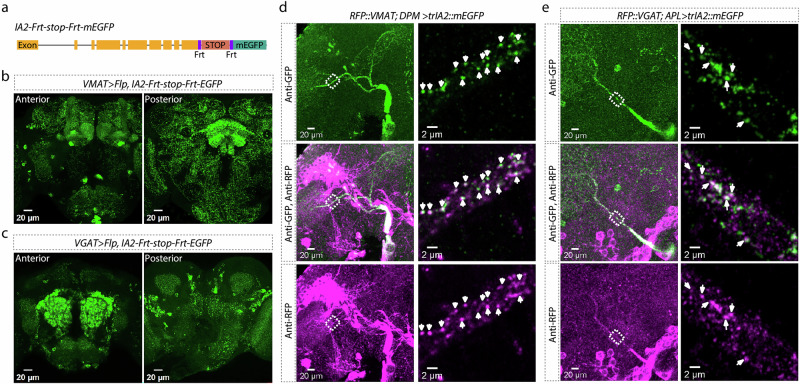
a Schematic showing CRISPR insertion of Frt-stop-Frt-mEGFP in the 3’ end of the IA2 gene. IA2 expression in VMAT-positive (b) and VGAT-positive (c) neurons. Left panels show anterior view, right panels show posterior view. Scale bar: 20 µm. d Co-localization of RFP::VMAT from endogenous VMAT locus with trIA2::EGFP. Left: DPM neuron projections in an expanded fly brain. Right: super-resolution images of the outlined area. Arrowheads indicate DCVs co-labeled by trIA2::mEGFP and RFP::VMAT. Scale bar: 20 µm on left, 2 µm on right. e Co-localization of RFP::VGAT with trIA2::EGFP. Left: APL neuron projections in an expanded fly brain. Right: super-resolution images of the outlined area. Arrowheads indicate DCVs co-labeled by trIA2::mEGFP and RFP::VGAT. Scale bar: 20 µm on left, 2 µm on right.
Update of
-
Genetically-encoded markers for confocal visualization of single dense core vesicles.bioRxiv [Preprint]. 2024 Oct 8:2024.10.07.617131. doi: 10.1101/2024.10.07.617131. bioRxiv. 2024. Update in: Commun Biol. 2025 Mar 07;8(1):383. doi: 10.1038/s42003-025-07829-y. PMID: 39416146 Free PMC article. Updated. Preprint.
-
Genetically-encoded markers for confocal visualization of single dense core vesicles.Res Sq [Preprint]. 2024 Oct 21:rs.3.rs-5021271. doi: 10.21203/rs.3.rs-5021271/v1. Res Sq. 2024. Update in: Commun Biol. 2025 Mar 07;8(1):383. doi: 10.1038/s42003-025-07829-y. PMID: 39502772 Free PMC article. Updated. Preprint.
References
-
- Edwards, R. H. Neurotransmitter release: variations on a theme. Curr. Biol.8, R883–885 (1998). - PubMed
MeSH terms
Substances
Grants and funding
LinkOut - more resources
Full Text Sources
Molecular Biology Databases
