. 2025 May;31(5):1604-1616.
doi: 10.1038/s41591-025-03574-1. Epub 2025 Mar 6.

Microglial mechanisms drive amyloid-β clearance in immunized patients with Alzheimer's disease

Affiliations

Microglial mechanisms drive amyloid-β clearance in immunized patients with Alzheimer's disease

Lynn van Olst et al. Nat Med. 2025 May.

Erratum in

Abstract

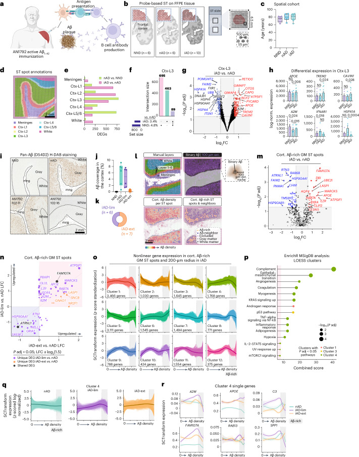
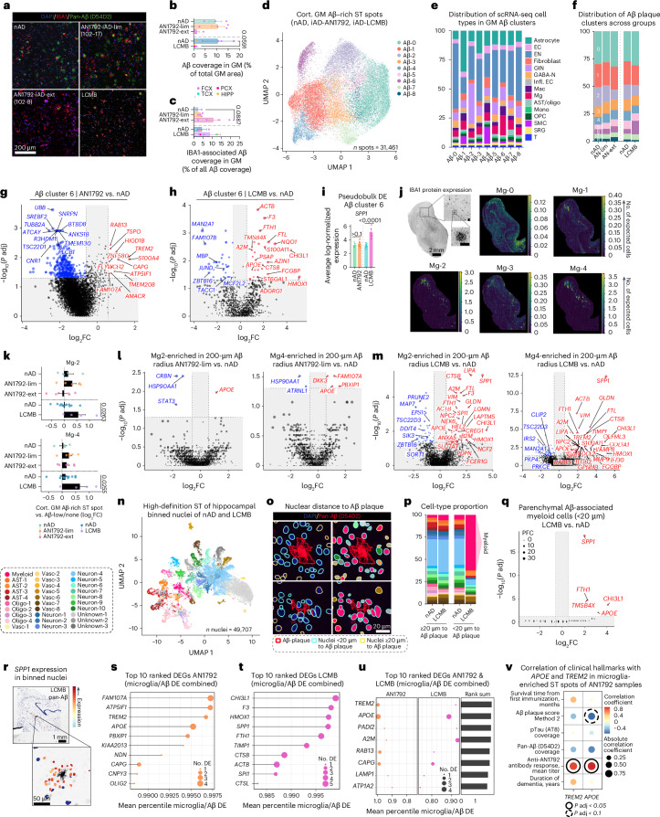
Alzheimer's disease (AD) therapies utilizing amyloid-β (Aβ) immunization have shown potential in clinical trials. Yet, the mechanisms driving Aβ clearance in the immunized AD brain remain unclear. Here, we use spatial transcriptomics to explore the effects of both active and passive Aβ immunization in the AD brain. We compare actively immunized patients with AD with nonimmunized patients with AD and neurologically healthy controls, identifying distinct microglial states associated with Aβ clearance. Using high-resolution spatial transcriptomics alongside single-cell RNA sequencing, we delve deeper into the transcriptional pathways involved in Aβ removal after lecanemab treatment. We uncover spatially distinct microglial responses that vary by brain region. Our analysis reveals upregulation of the triggering receptor expressed on myeloid cells 2 (TREM2) and apolipoprotein E (APOE) in microglia across immunization approaches, which correlate positively with antibody responses and Aβ removal. Furthermore, we show that complement signaling in brain myeloid cells contributes to Aβ clearance after immunization. These findings provide new insights into the transcriptional mechanisms orchestrating Aβ removal and shed light on the role of microglia in immune-mediated Aβ clearance. Importantly, our work uncovers potential molecular targets that could enhance Aβ-targeted immunotherapies, offering new avenues for developing more effective therapeutic strategies to combat AD.


Conflict of interest statement

Competing interests: J.A.R.N. has been a consultant/advisor relating to AD immunization programs for Elan Pharmaceuticals, GlaxoSmithKline, Novartis, Roche, Janssen, Pfizer, Biogen and Eisai. D.B. has been a consultant/advisor relating to AD immunization programs for Elan Pharmaceuticals and Biogen. D.G. has been a consultant/advisor relating to AD therapies for Merck and Novo Nordisk. They have no financial interest in relation to AD immunotherapy. The other authors declare no competing interests.

Figures

Fig. 1
Fig. 1. Active Aβ immunization sustains inflammation at the Aβ niche.
a, AN1792 active Aβ immunization. Created with BioRender.com. b, ST method and group sizes of NND, nAD and iAD FCX tissues. Created with BioRender.com. c, Study demographics indicating age of each patient. d, Manually annotated ST spots in the FCX. e, Number of DEGs for each comparison per manually annotated area. f, UpSet plot showing unique and shared DEGs across group comparisons in cortical layer III. g, DEGs in cortical layer III (iAD versus nAD). Red and blue DEGs are uniquely identified in the iAD versus nAD comparison and are not observed as DEGs in the nAD versus NND comparison. h, Pseudobulked expression for various genes in microglia-enriched gray matter ST spots. Error bars indicate the s.e.m. P values are from DESeq2. i, Representative pan-Aβ H-DAB stains for each group. j, Quantification of cortical Aβ coverage per group. k, Numbers of iAD-lim and iAD-ext patients among AN1792 actively immunized patients. l, Method of processing of Aβ IHC images. The binary Aβ signal was extended by 100 μm beyond its actual size, with a gradual decrease in signal intensity every 20 μm, allowing for detection of genes associated with Aβ density. m, DEGs from Aβ-rich gray matter ST spots (iAD versus nAD). n, LFC plots for Aβ-rich ST spots in gray matter (iAD-lim versus nAD; iAD-ext versus nAD). o, LOESS plots showing clusters of nonlinear gene expression patterns relative to Aβ density in iAD. p, Pathway enrichment analysis of genes in nonlinear expression clusters associated with Aβ density in iAD. q, LOESS plot of cluster 4 predictions in nAD, iAD-lim and iAD-ext relative to Aβ density. r, LOESS plots of select genes in LOESS cluster 4. Dark line indicating the LOESS predicted expression, and light shading represents standard error of the estimated values. c,j, Box plots are bounded by the 25th and 75th percentiles, the center line shows the median, and whiskers show the data range. o,q, LOESS plots with the dark line represent the mean LOESS predicted expression per group per cluster, and single lines indicate LOESS predicted gene expression per group per cluster. c,eh,j,k, NND = 6; nAD = 6; iAD = 13; iAD-lim = 6, iAD-ext = 7. m,n, nAD = 4; iAD = 10; iAD-lim = 6, iAD-ext = 4. or, nAD = 4; iAD = 12; iAD-lim = 6, iAD-ext = 6. DESeq2 (eh) or MAST (m and n) was used to compare expression levels. For DESeq2, covariates included sex, age, average genes detected and genomic DNA (gDNA) percentage. In MAST, manually annotated region or cortical layer, sex, age, cellular detection rate (CDR) and gDNA percentage were included as covariates, with sample ID as a random effect. All P values were false discovery rate (FDR)-adjusted using Benjamini–Hochberg correction. A2M, alpha-2-macroglobulin; APOE, apolipoprotein E; CAVIN1, caveolae-associated protein 1; Ctx, cortex; FFPE, formalin-fixed paraffin-embedded; GM, gray matter; HSPA1A, heat shock protein family A member 1A; H-DAB, hematoxylin-3,3′-diaminobenzidine; IFNAR1, interferon alpha and beta receptor subunit 1; LFC, log fold change; MSigDB, Molecular Signatures Database; P adj, adjusted P value. NS, not significant. Source data
Fig. 2
Fig. 2. Microglial phenotypes define varying degrees of Aβ clearance.
a, Reference atlas UMAP from DLPFC snRNA-seq data,. b, Spatial plots showing abundance of deconvoluted cell types. c, Spatial plots highlighting enriched ST spots for deconvoluted cell types. d, Percentages of DEGs expressed in enriched ST spots per cell type: nAD versus NND, iAD versus nAD, iAD-lim versus nAD and iAD-ext versus nAD. The number in the center of each pie chart represents the total number of DEGs. e, DEGs from microglia-enriched ST spots (iAD versus nAD). f, Top ten divergent DEGs in microglia-enriched ST spots based on PFC, comparing iAD versus nAD and nAD versus NND. g, UpSet plot showing unique and shared DEGs in microglia-enriched ST spots in gray matter across groups compared to nAD. h, LFC plots for microglia-enriched ST spots in gray matter (iAD-lim versus nAD; iAD-ext versus nAD). i, Pseudobulked expression for various genes in microglia-enriched ST gray matter spots. Error bars show the s.e.m. P values are from DESeq2. jl, Confocal images showing TMS1/ASC+IBA1+ myeloid cells (j), A2M+IBA1+ myeloid cells (k) and APOE+IBA1+ myeloid cells (l) around Aβ deposits in the FCX of iAD. m, Pathway enrichment analysis of unique and shared DEGs in microglia-enriched gray matter ST spots (iAD-lim versus nAD; iAD-ext versus nAD). n, LFC plots for microglia-enriched gray matter ST spots (NND versus nAD; iAD-ext versus nAD). o, Pseudobulked expression for various genes in microglia-enriched ST spots in gray matter. Error bars show the s.e.m. P values are from DESeq2. pq, Pathway enrichment analysis of predefined microglial states from p and q, using genes ranked by PFC in iAD-lim versus nAD and iAD-ext versus nAD. i,o, Bar plots display means ± s.e.m. di,mq, NND = 6; nAD = 6; iAD = 13; iAD-lim = 6, iAD-ext = 7. DESeq2 was used to compare expression levels, with sex, age, average genes detected and gDNA percentage included as covariates (di and no). All P values were FDR adjusted using Benjamini–Hochberg. Ast, astrocyte; CCa, cortico-cortical cluster a; CCb, cortico-cortical cluster b; CIRBP, cold-inducible RNA-binding protein; FAIM2, Fas apoptotic inhibitory molecule 2; FGFR3, fibroblast growth factor receptor 3; GSEA, gene-set enrichment analysis; IBA1, ionized calcium-binding adapter molecule 1; Int. N., interneuron; L, layer; Mg, microglia; NES, normalized enrichment score; OPC, oligodendrocyte precursor cell; Perip. Imm., peripheral immune cells; PYCARD, PYD and CARD domain containing; SMC, smooth muscle cell; SORBS3, sorbin and SH3 domain containing 3; TLR7, Toll-like receptor 7; TYROBP, TYRO protein tyrosine kinase-binding protein; UBB, ubiquitin B; UMAP, uniform manifold approximation and projection. Source data
Fig. 3
Fig. 3. Passive Aβ immunization induces distinct microglial states.
a, Lecanemab binds oligomeric and protofibrillar Aβ to promote Aβ clearance from the brain. Created with BioRender.com. b, Study participants included a 65-year-old female patient with AD who was treated with lecanemab and three matched nAD controls. Tissues analyzed included cortical areas and HIPP. Created with BioRender.com. c, Tissues were analyzed by scRNA-seq and spatial proteogenomics. Created with BioRender.com. d, Confocal images showing segmented Aβ burden and microgliosis in regions of the lecanemab-treated patient brain. e, Percentage of cortical Aβ coverage in brain regions from the lecanemab case and nAD controls. f, Percentage of cortical Aβ covered by IBA1. g, UMAP showing annotated cell types. h, Percentages of each cell type for each brain region between nAD controls and lecanemab case. i, DEGs in microglia and macrophages comparing lecanemab to nAD. j, LFC plots comparing DEGs in microglia and macrophages (lecanemab versus nAD). k, Top ten pathway enrichment analysis of DEGs in microglia and macrophages (lecanemab versus nAD). l, Pathway enrichment analysis of DEGs in microglia from FCX, TCX, PCX and HIPP (lecanemab versus nAD). m, Clustering of microglia from scRNA-seq of the lecanemab case and nAD controls. n, UMAP density plots showing microglial cluster distribution for the lecanemab case and nAD controls. o, Percentages of microglial clusters in the lecanemab case versus nAD controls. Normality tests dictated if P values were calculated using a two-tailed paired t-test or Wilcoxon test. p, Marker genes for each microglial cluster. q, Top five upregulated pathways using marker genes defining the microglial states. e,f, Bar plots display means ± s.e.m. o, Bar plots display means. e,f,o, Statistical tests, guided by Shapiro–Wilk and F tests, included t-tests, Mann–Whitney tests (e and f) and paired t-tests (o). eq, nAD = 3; LCMB = 1. i,j, MAST was used to compare expression levels, with brain region and CDR as covariates and brain region and sample ID included as a random effect. il,q, P values were FDR adjusted using Benjamini–Hochberg. Cort., cortical; GABA-N, GABAergic neuron; GIN, GABAergic interneuron; Infl. ECs, inflamed endothelial cells; LCMB, lecanemab; Mac, macrophages; mng, meninges; Oligo, oligodendrocyte; SRG, stress-responsive glia; Vasc, vascular. Source data
Fig. 4
Fig. 4. Spatial proteogenomics links the Aβ niche to microglial states.
a, Proteogenomics allowed for the simultaneous profiling of RNA and protein from lecanemab-treated and nAD controls. Created with BioRender.com. b, Manual annotations of brain regions analyzed. c, Representative images showing distinction of segmented cortical and vascular Aβ in brain regions from the lecanemab case. d, Number of DEGs for each comparison across manually annotated areas. e, DEGs from Aβ-rich gray matter ST spots (lecanemab versus nAD) in FCX, TCX, PCX and HIPP. f, Top ten pathway enrichment analysis of DEGs in Aβ-rich gray matter ST spots for each brain region (lecanemab versus nAD). g, DEPs associated with cortical Aβ ST spots from each brain region (lecanemab versus CAA control), with pink indicating shared DEGs, green indicating no shared DEGs and black indicating low expression levels not meeting DEG criteria. h, Confocal images showing CD68+IBA1+ myeloid cells surrounding Aβ deposits in the HIPP of the lecanemab-treated patient. i, LOESS plot of cluster 3 predictions in nAD (left) and lecanemab (right) relative to Aβ density. Dark line represents the mean LOESS predicted expression per group per cluster and single lines indicate LOESS predicted gene expression per group per cluster. j, LOESS plots of selected genes in LOESS cluster 3. Dark line indicates the LOESS predicted expression and light shading represents standard error of the estimated values. dg,i,j, nAD = 3; LCMB = 1. DESeq2 (d), MAST (e) or FindMarkers with a negative binomial model (g) was used to compare expression levels. For DESeq2, covariates included brain region, average genes detected and gDNA percentage. In the MAST model, manually annotated region or cortical layer, gDNA percentage and CDR were included as covariates, with brain region and sample ID as a random effect. For FindMarkers, covariates included manually annotated region or cortical layer and CDR. All P values were FDR adjusted using Benjamini–Hochberg. Source data
Fig. 5
Fig. 5. Shared microglial response drives Aβ clearance after immunization.
a, Confocal images showing pan-Aβ and IBA1 in FCX brain regions of nAD, AN1792-lim, AN1792-ext and lecanemab-treated patients. b, Percentage of cortical Aβ coverage in cortical and hippocampal regions of AN1792, nAD and the lecanemab case. c, Percentage of cortical Aβ covered by IBA1 in cortical and hippocampal regions of AN1792, nAD and the lecanemab case. d, Clustering of Aβ-rich cortical gray matter spots based on gene expression. e, C2L predictions of scRNA-seq cell types in different Aβ plaque clusters. f, Percentages of Aβ-rich clusters in AN1792, nAD and the lecanemab case. g,h, DEGs in Aβ-rich cluster 6: AN1792 versus nAD (g); lecanemab versus nAD (h). i, Pseudobulked SPP1 expression in Aβ-rich cluster 6. Error bars indicate the s.e.m. P values are from DESeq2. j, Spatial plots showing the abundance of deconvoluted scRNA-seq microglia types; scale bar, 100 μm. k, log2 fold change in predicted abundance of deconvoluted scRNA-seq microglia types in Aβ-rich ST spots versus the rest in AN1792, nAD and the lecanemab case. l,m, DEGs from Mg-2-enriched and Mg-4-enriched Aβ-associated ST spots: AN1792 versus nAD (l); lecanemab versus nAD (m). n, UMAP showing annotated binned nuclei from a high-definition ST assay. o, Spatial plots indicating the distance of nuclei to D54D2-stained Aβ plaques (left) and their annotations (right). p, Percentage of each cell type in the high-definition ST assay at ≥20 µm and <20 µm from Aβ plaques in nAD and the lecanemab case. q, DEGs from myeloid nuclei within <20 µm of Aβ plaques (lecanemab versus nAD). CDR is included as a covariate in the MAST model. r, Spatial plots showing SPP1 expression in binned nuclei around Aβ plaques in the lecanemab HIPP. s,t, Top ten upregulated response DEGs ranked by the average percentile across microglia and Aβ differential expression in AN1792 (s) and lecanemab (t). u, Top ten combined response genes to AN1792 and lecanemab by summing average percentiles of gene ranks. v, Covariate-adjusted Spearman correlation between TREM2 and APOE expression in microglia-enriched gray matter ST spots from AN1792 patients and clinical hallmarks. b,c,i,k, Bar plots display means ± s.e.m. b,c, nAD-AN1792 = 3; iAD-lim = 4; iAD-ext = 4; nAD-LCMB = 3; LCMB = 1. df, nAD-AN1792 = 4; iAD-lim = 6; iAD-ext = 4; nAD-LCMB = 3; LCMB = 1. gi, nAD-AN1792 = 4; iAD = 10; nAD-LCMB = 3; LCMB = 1. k, nAD-AN1792 = 4; iAD-lim = 6; iAD-ext = 6; nAD-LCMB = 3; LCMB = 1. l,m, nAD-AN1792 = 4; iAD-lim = 6; nAD-LCMB = 3; LCMB = 1. n,pq, nAD = 2; LCMB = 1. v, iAD = 13. g,h,l,m,q, MAST was used to compare expression levels. Covariates included sex, age, CDR and gDNA percentage with sample ID as a random effect (g and l); brain region, CDR and gDNA percentage with brain region and sample ID as a random effect (h and m); CDR (q). i, DESeq2 was used to compare expression levels. Covariates included sex, age, average genes detected, gDNA percentage (AN1792 versus nAD); average genes detected, brain region and gDNA percentage (lecanemab versus nAD). v, Covariates included sex, age, average genes detected and gDNA percentage. gi,l,m,q,v, P values were FDR adjusted using Benjamini–Hochberg. b,c,k, Statistical tests, guided by Shapiro–Wilk and F tests, included t-tests, Mann–Whitney tests, analysis of variance (ANOVA) with Tukey’s test, Welch’s ANOVA with Dunnett’s T3 test and Kruskal–Wallis with Dunn’s test. AN1792-ext, AN1792 immunized with extensive Aβ clearance; AN1792-lim, AN1792 immunized with limited Aβ clearance; DE, differential expression; ECs, endothelial cells; Mono, monocytes. Source data
Extended Data Fig. 1
Extended Data Fig. 1. Active Aβ immunization sustains inflammation at the Aβ niche.
a, Sex distribution per group. b, Number of ST spots per manually annotated area per donor for all groups. c, Average number of features (genes) per spot per manually annotated area per donor for all groups. d, Percentages of mitochondrial gene expression per spot averaged per manually annotated area per donor for all groups. e, Spatial plots showing expression of brain region-specific genes overlaid on corresponding manually annotated areas (shaded). f, Volcano plot of DEGs in cortical layer III (nAD vs. NND). g, Bar plot of the top 10 divergent DEGs in cortical layer III based on PFC, comparing iAD vs. nAD and nAD vs. NND. h, Pan-Aβ (D54D2) H-DAB staining for AN1792-immunized subjects. i, Quantification of average Aβ intensity per cortical region per donor per group. j, Representative pTau (AT8) H-DAB staining for each group. k, Quantification of cortical AT8 per group. l, Pathway enrichment analysis of unique and shared DEGs in Aβ-rich gray matter ST spots (iAD-lim vs. nAD; iAD-ext vs. nAD). m, LOESS heatmap showing non-linear gene expression patterns relative to Aβ density in iAD. n, LOESS non-linear trajectories relative to Aβ density in iAD. b-d, i, k, Bar plots display means ± SEM. a-d, f-g, NND = 6; nAD = 6; iAD = 13; iAD-lim = 6, iAD-ext = 7. i,l, nAD = 4; iAD = 10; iAD-lim = 6, iAD-ext = 4. k, iAD-lim = 5, iAD-ext = 7. l, nAD = 4; iAD-lim = 6; iAD-ext = 4. m-n, iAD = 12. f-g DESeq2 was used to compare expression levels, with sex, age, average genes detected, and gDNA percentage included as covariates. b-d, k, Statistical tests, guided by Shapiro–Wilk and F tests, included t-tests, Mann–Whitney, ANOVA with Tukey’s test, Welch ANOVA with Dunnett’s T3 test, and Kruskal–Wallis with Dunn’s test. f-g, l, P-values are FDR-adjusted using the Benjamini-Hochberg correction. Aβ, amyloid-beta; AN1792-ext, AN1792 immunized with extensive Aβ clearance; AN1792-lim, AN1792 immunized with limited Aβ clearance; DEGs, differentially expressed genes; DESeq2, Differential Expression Analysis for Sequence Count Data (version 2); FCX, frontal cortex; FDR, False Discovery Rate; gDNA, genomic DNA; GM, gray matter; H-DAB, Hematoxylin-3,3'-Diaminobenzidine; iAD, immunized Alzheimer’s disease; LCMB, lecanemab; LOESS, locally estimated scatterplot smoothing; MSigDB, Molecular Signatures Database; MT, mitochondrial; nAD, non-immunized Alzheimer’s disease; nFeatures, number of features; nSpots, number of spatial transcriptomic spots; NND, non-neurologic disease; P-adj, P-value adjusted; PFC, probabilistic fold change; pTau, phosphorylated tau; ST, spatial transcriptomics.
Extended Data Fig. 2
Extended Data Fig. 2. Microglial phenotypes define varying degrees of Aβ clearance.
a, Bubble plot heatmap showing top markers expressed by cell types in the reference atlas. b, Spatial plots displaying the abundance of deconvoluted cell types. c, Bar graphs showing the proportion of deconvoluted cell types in gray matter per donor, per group. Statistical tests, guided by Shapiro–Wilk and F tests, included ANOVA with Tukey’s test, Welch ANOVA with Dunnett’s T3 test, and Kruskal–Wallis with Dunn’s test. d, UpSet plot indicating unique and shared DEGs in deconvoluted cell types for iAD vs. nAD. e, UpSet plot showing unique and shared DEGs in deconvoluted cell types for nAD vs. NND. f-g, Volcano plot of DEGs in microglia-enriched spots in: f, iAD-lim vs. nAD; and g, iAD-ext vs. nAD. h, Bar plot of the top 10 most divergent DEGs in microglia-enriched ST spots based on PFC, comparing iAD-lim vs. nAD and iAD-ext vs. nAD. Adjusted P-values used to calculate PFC are derived from DESeq2. c, Bar plots display means ± SEM. a, 424 ROSMAP DLPFC samples. c-h, NND = 6; nAD = 6; iAD = 13; iAD-lim = 6, iAD-ext = 7. d-h, DESeq2 was used to compare expression levels, with sex, age, average genes detected, and gDNA percentage included as covariates. All P-values are FDR-adjusted using the Benjamini-Hochberg correction. Aβ, amyloid-beta; AD, Alzheimer’s disease; AN1792-ext, AN1792 immunized with extensive Aβ clearance; AN1792-lim, AN1792 immunized with limited Aβ clearance; DEGs, differentially expressed genes; DESeq2, Differential Expression Analysis for Sequence Count Data (version 2); DLPFC, dorsolateral prefrontal cortex; EN, excitatory neuron; FDR, False Discovery Rate; gDNA, genomic DNA; GM, gray matter; iAD, immunized Alzheimer’s disease; L, layer; LCMB, lecanemab; nAD, non-immunized Alzheimer’s disease; NND, non-neurologic disease; oliog., oligodendrocyte; OPC, oligodendrocyte precursor cell; P-adj, P-value adjusted; periph. imm., peripheral immune cell; PFC, probabilistic fold change; ROSMAP, Religious Orders Study and Rush Memory and Aging Project; SEM, standard error of the mean; SMC, smooth muscle cell; snRNA-seq, single-nucleus RNA sequencing; ST, spatial transcriptomics.
Extended Data Fig. 3
Extended Data Fig. 3. Passive Aβ immunization induces distinct microglial states.
a, Representative confocal images showing segmented Aβ burden and microgliosis in regions of the nAD control patient’s brain. b, SoupX contamination fraction for each scRNA-seq sample. c, Average number of features (genes) per cell per donor. d, Percentages of mitochondrial gene per cell averaged per donor. e, Integrated scRNA-seq dataset showing all analyzed cells from nAD controls and lecanemab case. f, Bubble plot heatmap of top markers expressed by cell types in the scRNA-seq dataset. g, Changes in percentages of total annotated cells for each cell type. h, Confocal images showing SPP1+IBA1+ myeloid cells surrounding Aβ deposits in the hippocampus of the lecanemab-treated patient, absent in the nAD control. i, Confocal images showing APOC1+IBA1+ myeloid cells surrounding Aβ deposits in the hippocampus of the lecanemab-treated patient. j, Volcano plot of DEGs from scRNA-seq microglia (lecanemab vs. nAD) in FCX (top left), TCX (top right), PCX (bottom left), and HIPP (bottom right). k-l, Pathway enrichment analysis of predefined microglial states from k, Sun et al., and l, Green et al., using genes ranked by PFC in scRNA-seq regional microglia (lecanemab vs. nAD). m, UMAP showing reintegrated scRNA-seq immune cells for each brain region in the lecanemab case and nAD controls. n, Marker genes for each immune cell cluster. o, Percentages of each macrophage cluster. b-d, Bar plots display means ± SEM. g, o, Bar plots display means. b-e, g, j-o, nAD = 3; LCMB = 1. j, MAST was used to compare expression levels, with CDR as a covariate and brain region * sample ID included as a random effect. c-d, g, o, Statistical tests, guided by Shapiro–Wilk and F tests, included t-tests, Mann–Whitney, ANOVA with Tukey’s test, Welch ANOVA with Dunnett’s T3 test, and Kruskal–Wallis with Dunn’s test. j-l, P-values are FDR-adjusted using the Benjamini-Hochberg correction. Aβ, amyloid-beta; AD, Alzheimer’s disease; APOC1, apolipoprotein C1; Ast, astrocyte; CCA, canonical correlation analysis; CDR, Cellular Detection Rate; DEGs, differentially expressed genes; EC, endothelial cells; ExN, excitatory neuron; FCX, frontal cortex; FDR, False Discovery Rate; GABA-N, GABAergic neuron; gDNA, genomic DNA; GIN, GABAergic interneuron; HIPP, hippocampus; IBA1, ionized calcium-binding adapter molecule 1; Infl. EC, inflamed endothelial cells; LCMB, lecanemab; Mac, macrophages; MAST, Model-based Analysis of Single-cell Transcriptomics; Mg, microglia; Mono, monocytes; MT, mitochondrial; nAD, non-immunized Alzheimer’s disease; nFeatures, number of features; Oligo, oligodendrocyte; OPC, oligodendrocyte precursor cell; P-adj, P-value adjusted; PCX, parietal cortex; PFC, probabilistic fold change; scRNA-seq, single-cell fixed RNA sequencing; SEM, standard error of the mean; SMC, smooth muscle cell; SPP1, secreted phosphoprotein 1; SRG, stress-responsive glia; ST, spatial transcriptomics; TCX, temporal cortex; UMAP, uniform manifold approximation and projection; Vasc., vascular.
Extended Data Fig. 4
Extended Data Fig. 4. Spatial proteogenomics links the Aβ niche to microglial states.
a, Manual annotations of analyzed brain regions. b, Average number of features (genes) per spot per manually annotated area per donor. c, Percentages of mitochondrial gene expression per spot averaged per manually annotated area per donor. d, Quantification of average Aβ intensity per cortical region per donor per group. e, UpSet plot indicating unique and shared DEGs in cortical Aβ-rich ST spots in FCX, TCX, PCX and HIPP for lecanemab vs. nAD. f, Volcano plot of DEGs from Aβ-rich gray matter ST spots (lecanemab vs. nAD) across all regions. g, Confocal images showing IBA1+ myeloid cells surrounding Aβ deposits that colocalize with APOE in the hippocampus of the lecanemab-treated patient, with reduced IBA1+ recruitment in the nAD control. h, Confocal images showing A2M+IBA1+ myeloid cells surrounding Aβ deposits in the hippocampus of the lecanemab-treated patient. i, Confocal images showing CD68+IBA1+ myeloid cells surrounding Aβ deposits in the hippocampus of the nAD control. j, LOESS heatmap showing non-linear gene expression patterns relative to Aβ density in lecanemab. k, LOESS non-linear trajectories relative to Aβ density in lecanemab. l, LOESS plots showing clusters of non-linear gene expression patterns relative to Aβ density in lecanemab. Dark line representing the mean LOESS predicted expression for the cluster and single lines indicating LOESS predicted gene expression per cluster. m, Pathway enrichment analysis of genes in non-linear expression clusters associated with Aβ density in lecanemab. n, LOESS plots of selected genes in LOESS cluster 3. Dark line indicating the LOESS predicted expression and light shading representing standard error of the estimated values. b-d, Bar plots display means ± SEM. nAD1, nAD2, and nAD3 each refer to separate samples. b-f, j-n, nAD = 3, LCMB = 1. e-f, MAST was used to compare expression levels. Covariates included manually annotated region or cortical layer, CDR and gDNA percentage with brain region * sample ID as a random effect (e) manually annotated region or cortical layer, CDR, gDNA percentage and brain region with brain region * sample ID as a random effect (f). b-d, Statistical tests, guided by Shapiro–Wilk and F tests, included t-tests, Mann–Whitney, ANOVA with Tukey’s test, Welch ANOVA with Dunnett’s T3 test, and Kruskal–Wallis with Dunn’s test. f, m, P-values are FDR-adjusted using the Benjamini-Hochberg correction. A2M, alpha-2-macroglobulin; Aβ, amyloid-beta; AD, Alzheimer’s disease; APO, apolipoprotein; APOC1, apolipoprotein C1; APOE, apolipoprotein E; CDR, Cellular Detection Rate; CD68, cluster of differentiation 68; Cort., cortical; Ctx, cortex; CTSB, cathepsin B; DEGs, differentially expressed genes; DESeq2, Differential Expression Analysis for Sequence Count Data (version 2); FCGBP, Fc fragment of IgG binding protein; FDR, False Discovery Rate; gDNA, genomic DNA; GM, gray matter; IBA1, ionized calcium-binding adapter molecule 1; ITGAX, integrin subunit alpha X; L, layer; LCMB, lecanemab; LOESS, locally estimated scatterplot smoothing; MAST, Model-based Analysis of Single-cell Transcriptomics; MSigDB, Molecular Signatures Database; MT, mitochondrial; nAD, non-immunized Alzheimer’s disease; nFeatures, number of features; P-adj, P-value adjusted; SPP1, secreted phosphoprotein 1; ST, spatial transcriptomics.
Extended Data Fig. 5
Extended Data Fig. 5. Shared microglial response drives Aβ clearance after immunization.
a, UMAP showing cortical Aβ-rich ST spots based on gene and protein expression, colored by brain region and donor. b, UMAP density plots for each group. c, Top two marker genes for each cortical Aβ-rich cluster. d, Bar plots showing C2L predictions of scRNA-seq cell types proportionally in the gray matter per group. e, Volcano plot showing DEGs distinguishing cortical Aβ-rich cluster 6 from all other cortical Aβ-rich clusters. f, Bar plots showing C2L predictions of scRNA-seq microglia and macrophage subtypes proportionally in Aβ-rich cluster 6 per group. g, Bar graphs showing pseudobulked TREM2, TSPO, S100A4, APOE, A2M, TMSB4X, RAB13, FCGBP, CTSB, CHI3L1, and FAM107A expression in Aβ-rich cluster 6. Error bars indicate SEM. P-values are from DESeq2. h, Bar plots showing log2 fold-change in predicted abundance of deconvoluted scRNA-seq microglia types in Aβ-rich ST spots versus the rest in AN1792, nAD, and the lecanemab case. i, Bar plots showing log2 fold-change in pseudobulked expression of A2M, APOE, FAM107A, LIPA, SPP1, and TREM2 in Aβ-associated Mg2-enriched and Mg4-enriched ST spots compared to the nAD control group for AN1792 and the lecanemab case. j, Chord plots showing inferred CellChat cell-cell communication of APOE, complement, and SPP1 signaling pathways among different scRNA-seq cell types. The width of the chords reflects the strength of interaction or communication probability, with thicker chords indicating stronger signaling. k, Visium HD ST method. Created using BioRender.com. l, UMAP showing annotated binned nuclei from the high-definition ST assay, colored by donor. m, Number of features (genes) per binned nuclei in high-definition ST data per donor. n, Percentage of mitochondrial genes per binned nuclei in high-definition ST data per donor. o, Top three marker genes for overarching cell types annotated in the high-definition ST data. p-q, Top 10 upregulated response DEGs in microglia or Aβ DE ranked by their average percentile in Aβ (Y-axis) and microglia (X-axis) DE: o, in AN1792; r, in lecanemab. g, h-i, Bar plots display means ± SEM. m-n, Violin plots showing the data range and median. Points represent individual cells. nAD3 and nAD4 refer to separate samples. a-c, e-f, nAD-AN1792 = 4, iAD-lim = 6; iAD-ext = 4; nAD-LCMB = 3; LCMB = 1. d, nAD-AN1792 = 6, iAD-lim = 6; iAD-ext = 7; nAD-LCMB = 3; LCMB = 1. g, nAD-AN1792 = 4; iAD = 10; nAD-LCMB = 3; LCMB = 1. h, nAD-AN1792 = 4; iAD-lim = 6; iAD-ext = 6; nAD-LCMB = 3; LCMB = 1. i, iAD = 9 (Mg-2), 10 (Mg-4); LCMB = 1. j, iAD = 5; nAD = 3; LCMB = 1. l-o, nAD = 2; LCMB = 1. g, DESeq2 was used to compare expression levels. Covariates included sex, age, average genes detected, gDNA percentage (AN1792 vs. nAD); average genes detected, brain region, gDNA percentage (LCMB vs. nAD). h. Statistical tests, guided by Shapiro–Wilk and F tests, included t-tests, Mann–Whitney, ANOVA with Tukey’s test, Welch ANOVA with Dunnett’s T3 test, and Kruskal–Wallis with Dunn’s test. All P-values are FDR-adjusted using the Benjamini-Hochberg correction. A2M, alpha-2-macroglobulin; Aβ, amyloid-beta; AD, Alzheimer’s disease; APOC1, apolipoprotein C1; APOE, apolipoprotein E; CD68, cluster of differentiation 68; Cort., cortical; Ctx, cortex; CTSB, cathepsin B; DEGs, differentially expressed genes; DESeq2, Differential Expression Analysis for Sequence Count Data (version 2); FDR, False Discovery Rate; FCGBP, Fc fragment of IgG binding protein; GM, gray matter; HD, high-definition; IBA1, ionized calcium-binding adapter molecule 1; ITGAX, integrin subunit alpha X; L, layer; LCMB, lecanemab; LOESS, locally estimated scatterplot smoothing; MSigDB, Molecular Signatures Database; MT, mitochondrial; nAD, non-immunized Alzheimer’s disease; nFeatures, number of features; P-adj, P-value adjusted; SPP1, secreted phosphoprotein 1; ST, spatial transcriptomics.

References

    1. Karran, E. & Hardy, J. Antiamyloid therapy for Alzheimer’s disease–are we on the right road? N. Engl. J. Med.370, 377–378 (2014). - PubMed
    1. Brody, D. L. & Holtzman, D. M. Active and passive immunotherapy for neurodegenerative disorders. Annu. Rev. Neurosci.31, 175–193 (2008). - PMC - PubMed
    1. Holmes, C. et al. Long-term effects of Aβ42 immunisation in Alzheimer’s disease: follow-up of a randomised, placebo-controlled phase I trial. Lancet372, 216–223 (2008). - PubMed
    1. Nicoll, J. A. et al. Neuropathology of human Alzheimer disease after immunization with amyloid-beta peptide: a case report. Nat. Med.9, 448–452 (2003). - PubMed
    1. Nicoll, J. A. et al. Aβ species removal after Aβ42 immunization. J. Neuropathol. Exp. Neurol.65, 1040–1048 (2006). - PubMed

MeSH terms