. 2025 Mar 6;23(1):71.
doi: 10.1186/s12915-025-02162-6.

Another tail of two sites: activation of the Notch ligand Delta by Mindbomb1

Affiliations

Another tail of two sites: activation of the Notch ligand Delta by Mindbomb1

Nicole Vüllings et al. BMC Biol. .

Abstract

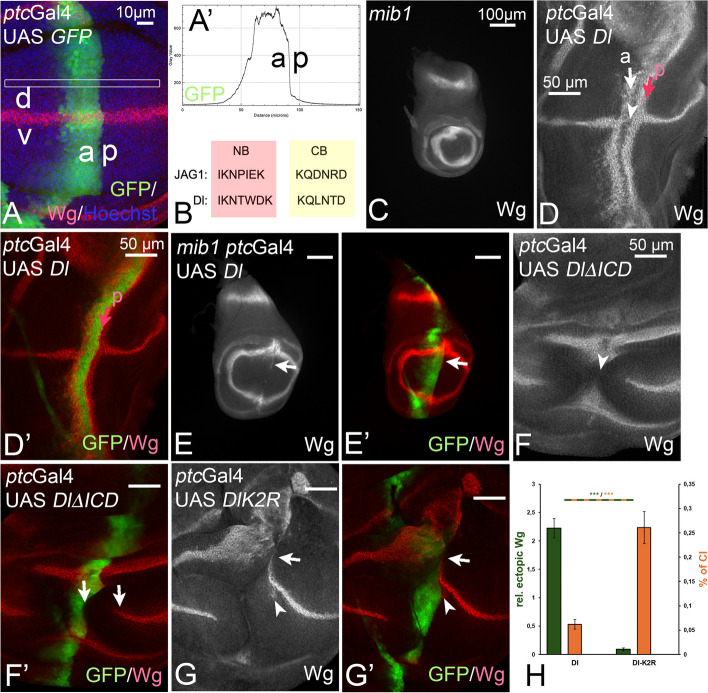
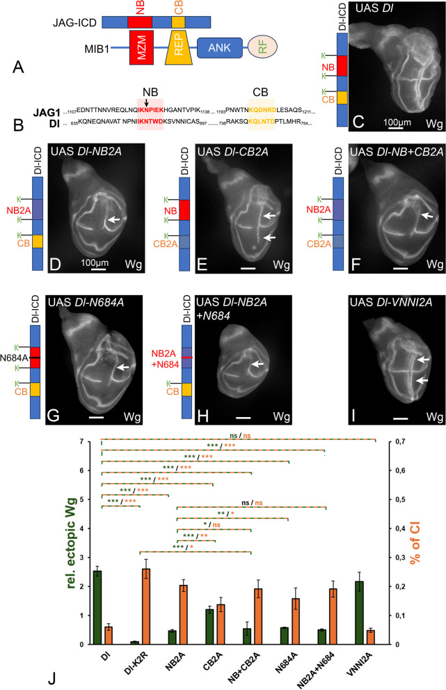
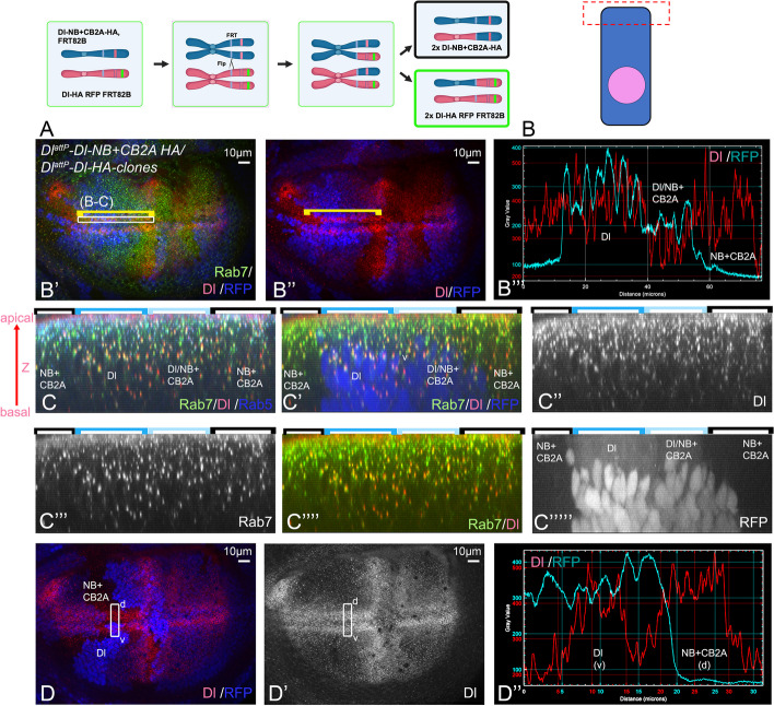
Background: Notch signalling plays a crucial role in many developmental, homoeostatic and pathological processes in metazoans. The pathway is activated by binding of the ligand to the Notch receptor, which changes the conformation of the receptor by exerting a pulling force. The pulling force is generated by the endocytosis of the interacting ligand into the signal-sending cell. Endocytosis of ligands requires the action of the E3 ligases Mindbomb1 (Mib1) and Neuralized (Neur) that ubiquitylate lysines (Ks) of their intracellular domains. It has been shown that human MIB1 binds JAGGED1 (JAG1) via a bipartite binding motif in its ICD. This interaction is required for the activation of JAG1. However, it is not known whether this bipartite binding mode is of general importance. It is also not rigorously tested whether it occurs in vivo. Moreover, it is not known whether Mib1 ubiquitylates specific Ks in the ICD of ligands, or is rather non-selective.

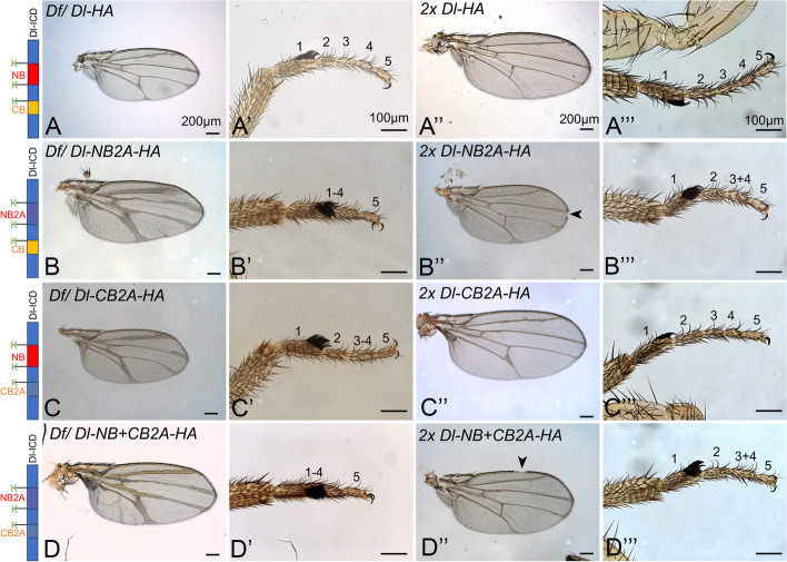
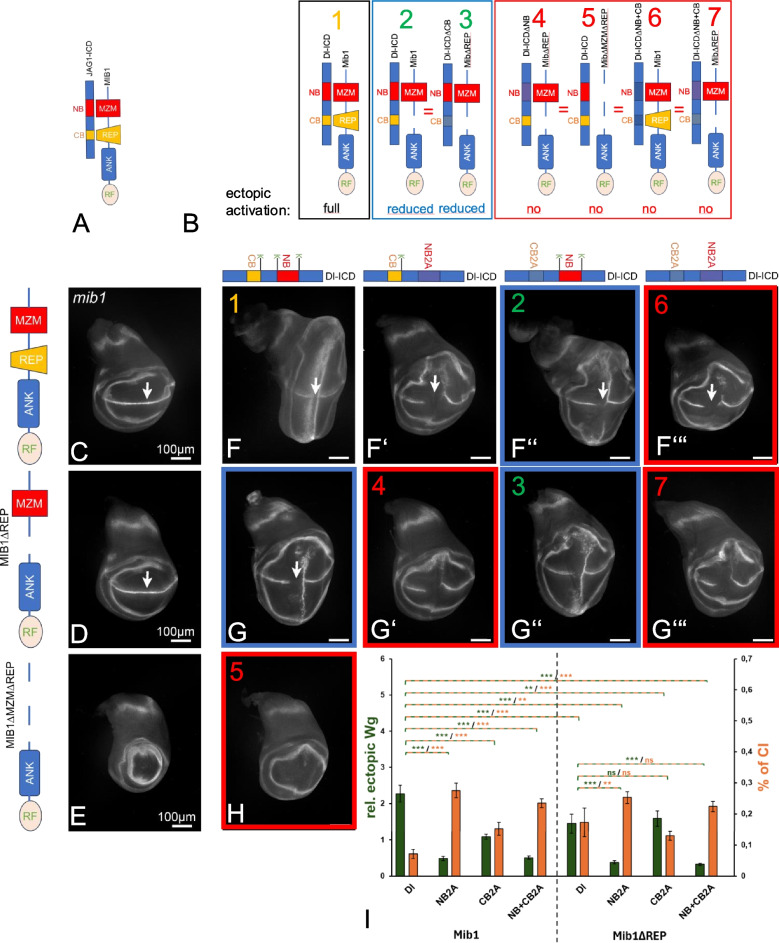
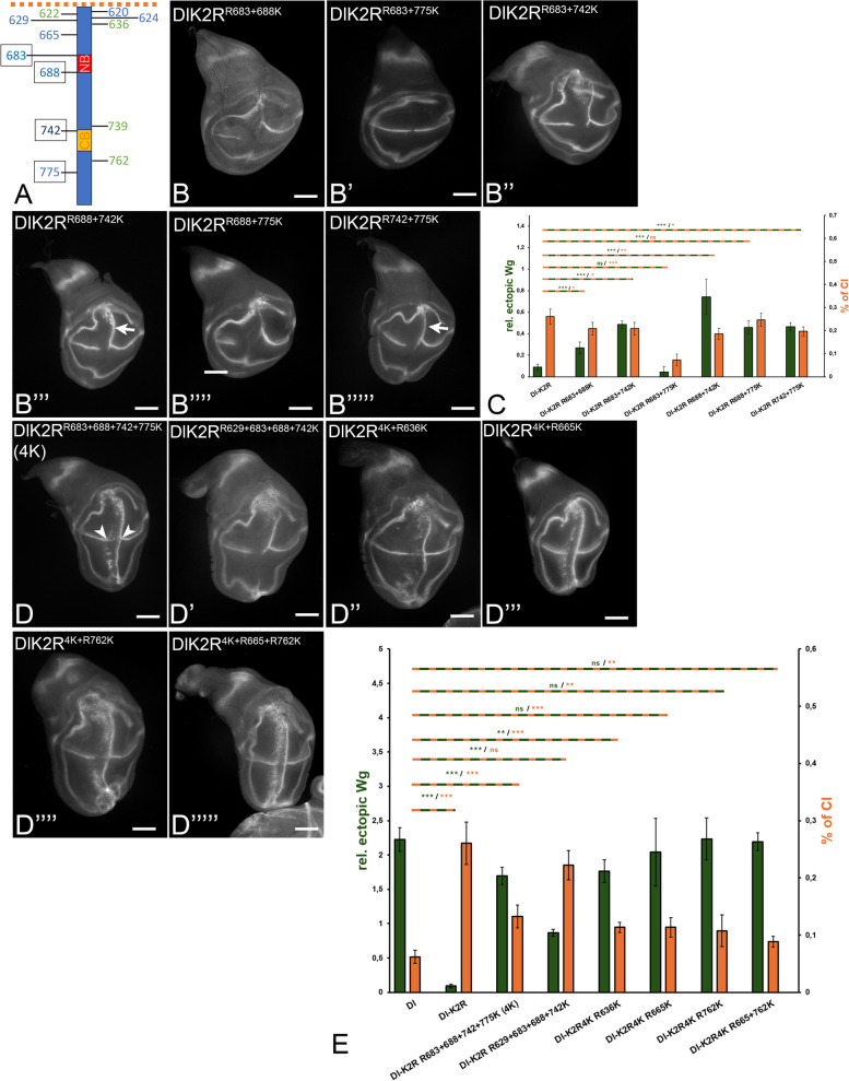
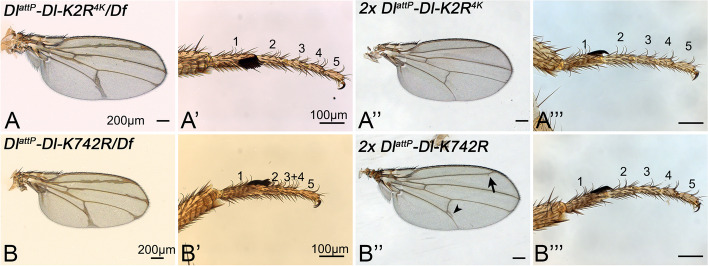
Results: We therefore investigated how Mib1 interacts with the Notch ligand Delta of Drosophila in an in vivo trans-activation assay and determined the Ks which are required for signalling. We show that the activation of Dl by Mib1 follows similar rules as has been found for mammalian MIB1 and JAG1. We present evidence that a combination of six Ks of the ICD is required for the full signalling activity of Dl by Mib1, with K742 being the most important one.

Conclusions: Altogether, our analysis further reveals the rules of Mib1-mediated DSL-ligand-dependent Notch-signalling.

Keywords: Cis-inhibition; DSL-ligands; Delta; Endocytosis; Mindbomb1; Notch-pathway; Ubiquitylation.


Conflict of interest statement

Declarations. Ethics approval and consent to participate: Not applicable. Consent for publication: All the authors of this manuscript consent to its publication as a research article in BMC Biology. Competing interests: The authors declare no competing interests.

Figures

Fig. 1
Fig. 1
Effects of expression of DlΔICD and DlK2R during wing development. A The constructs are expressed with ptcGal4, which drives expression in a stripe along the anterior side (a) of the A/P-boundary of the wing imaginal disc. Expression in the wing disc is perpendicular to the D/V-boundary (d, v) along which the Notch target gene wg is expressed. A’ Expression is graded within the ptcGal4 domain, increasing from anterior to the A/P-boundary. The gradient is measured by the pixel intensity of the GFP-signal in the region boxed in A. B The sequence comparison of the NB and CB of JAG1 with Dl. C Expression of Wg in mib1 mutant discs. The expression along the D/V-boundary is lost and the wing area is dramatically reduced. D, D’ Ectopic expression of Dl with ptcGal4 in wildtype discs results in the induction of two stripes of ectopic Wg expression running perpendicular to the endogenous expression along the D/V-boundary. One anterior broader stripe (arrow, a) and a thinner stripe located in the posterior boundary cells (red arrow, p). At the intersection with the D/V-boundary, the endogenous expression of Wg is cell-autonomously suppressed in the domain of high expression due to CI (arrowhead). The cell-autonomy of CI is revealed by the expression of wg directly adjacent to the posterior boundary of the ptcGal4 domain (red arrow). E, E’ Expression of Dl in a mib1-mutant wing disc results in a very weak activation of Wg in the dorsal compartment of the wing area (arrow), confirming that Dl can signal weakly in the absence of Mib1 function, but requires Mib1 function for its full activity (compare with C, D, D’). F, F’ Expression of DlΔICD causes a large gap in the endogenous expression of Wg (arrowhead in F). A gap can be observed between the posterior expression boundary of the ptcGal4 domain and the endogenous expression of Wg (arrows in F’). This non-cell autonomous suppression of Wg expression indicates that DlΔICD acts in a dominant-negative manner. No ectopic expression of Wg is induced, indicating the activity of Dl is abolished if the ICD is deleted. G, G’ Expression of DlK2R results in a suppression of the endogenous expression of Wg. Note that this effect is restricted to the domain of expression and accompanied by weak ectopic activation of Wg in posterior boundary cells in the dorsal compartment (arrow and arrowhead). This indicates that DlK2R is weakly active and possess stronger cis-inhibitory abilities than Dl, indicated by the larger gap in the endogenous expression domain of Wg (arrow and arrowhead) [8]. H Quantification of the signalling activity and CI of Dl and DlK2R (see Fig. 2 and Methods for more details). It confirms that the loss of the Ks results in a dramatic reduction in signalling, accompanied by a strong increase in CI
Fig. 2
Fig. 2
The importance of the NB and CB for the activity of Dl. A Cartoon of the bipartite interaction mode between JAG1 and MIB1. The NB and CB of JAG1 interact with the MZM and REP domain of MIB1, respectively. B Sequence comparison of sections of the ICDs of JAG1 and Dl that include the NB/ICD2 (shaded in red) and the predicted CB/ICD3 (shaded in yellow). The arrow points to the conserved asparagine (N) in the NB. C Expression of Dl by ptcGal4 for comparison. D Expression of Dl-NB2A results in a weaker ectopic induction of Wg expression, largely restricted to the dorsal compartment (arrow). The anterior stripe is missing due to increased CI. E Likewise, expression of Dl-CB2A also induces weaker ectopic expression of Wg than Dl, indicating that it is required for the full activity of Dl. Note that expression of Dl-CB2A leads to a stronger Wg activation than Dl-NB2A (longer stripe of ectopic expression), indicating that the CB is less important. F The mutation of both boxes results in an ectopic induction of Wg expression comparable to Dl-NB2A (compare with D), indicating that the NB is more important for Dl function. G Dl-N684 induced ectopic expression of Wg comparable to Dl-NB2A, indicating its importance for the function of the NB. H Expression of Dl-NB2A with re-introduced N684 causes a phenotype that is similar to that caused by Dl-NB2A (compare with D). I The mutation of the NN-motif in the ICD (Dl-DNNI2A) has no effect on the activity of Dl (arrows, compare with B). J Quantification of the CI and transactivation abilities of the Dl-variants shown in CI. CI was measured by dividing the length of the gap of endogenous Wg expression by the total length of the endogenous Wg expression along the D/V boundary. Ectopic activation of the Notch pathway was determined by measuring the length of the anterior and/or posterior ectopic Wg expression and dividing it by the endogenous wg expression along the D/V boundary. ‘relative ectopic Wg’ displays the extend of ectopic Wg expression, while ‘% of CI’ shows the % of interruption of the endogenous Wg expression domain. Ten discs were measured for each genotype. p < 0.05 = *; p < 0.01 = **; p < 0.001 = ***
Fig. 3
Fig. 3
Adult phenotypes of DlattP-knock-in alleles encoding Dl-variants with NB, CB or NB + CB mutated over the deficiency Df(3R) DlBSC850 (Df) and in homozygosity. All variants contain the Ks flanking the boxes, to exclude effects caused by the absence of crucial ubi acceptors. A, A’ Dl-HA/Df results in a haplo-insufficient phenotype with strong broadening of wing veins. The tarsal region consisting of 5 segments has a wildtype appearance. A’’, A’’’ Two copies of Dl-HA restore the WT phenotype of the wing and leg. B, B’ The phenotype of one copy of Dl-NB2A/Df flies. The wing veins are broadened (B) and the tarsal segments 1–4 are fused (B’). B’’, B’’’ Phenotype of homozygous Dl-NB2A flies. The wing vein broadening and tarsal segment fusion phenotypes are less severe. The arrowhead points to a small Notch in the wing margin. C, C’ The mutation of the CB causes a milder vein and leg phenotype. C’’, C’’’ In homozygosity of Dl-CB2A, only weak wing and leg defects. D, D’ Simultaneous mutation of the NB and CB causes developmental defects similar to Dl-NB2A (compare with BB’’’). The arrowhead points to a small notch in the wing margin
Fig. 4
Fig. 4
Comparison of the endocytic behaviour of Dl-NB + CB2A-HA with DlK2R-HA. A Cartoon describing the generation of the adjacently located homozygous clones by Flp/FRT-mediated clonal analysis (mitotic recombination). The mitotic recombination in G2 results in the free segregation of the replicated mutant alleles because of their localisation on different centromeres. The result is homozygous founder cells for each genotype in 25% of the cases. BB’’ Comparison of the apical membrane localisation of the Dl-variants in cells of clones homozygous for DlattP-Dl-NB + CB2A-HA (absence of RFP) and DlattP-Dl-HA (homozygous for RFP) detected with a Dl antibody directed against the extracellular domain. B’’’ Pixel intensity plot of the RFP channel which identifies the clones and the Dl channel. The yellow bracket highlight the region of measurement adjacent to the D/V-boundary where Dl is expressed in a continuous stripe from anterior to posterior at the same level in wildtype discs. It includes regions homozygous for both Dl alleles. No significant difference is observed in the apical levels of Dl in DlattP-Dl-NB + CB2A-HA and DlattP-Dl-HA homozygous cells, indicating that both Dl variant are present in the apical membrane at comparable levels. CC’’’’ Z section of the region of Dl expression highlight in B’ with the yellow bracket. It reveals that intracellular Dl and Dl-NB + CB2A are located in Rab7- and Rab5-positive endosomes. DD’’ Another example of the correct location of and levels of Dl-NB + CB2A in the apical membrane. Here the Dl-homozygous clone is located in the ventral stripe of Dl and the Dl-NB + CB2A-homozygous clone in the dorsal stripe. The pixel density plot shown in D’’ confirms the similarity of the levels of the two Dl-variants in the apical membrane domain of the cells
Fig. 5
Fig. 5
The MZM and REP domains of Mib1 interact with the NB and CB of Dl, respectively. A Reported interaction between JAG1 and MIB1. The NB and CB of JAG1 bind to the MZM and REP domains of MIB1, respectively. B Prediction for the ectopic activation of Notch by combinations of Dl- and Mib1-variants in mib1 mutant discs. Note that the combination of Dl-NB2A with Mib1ΔREP is predicted not to function, since the interaction of both the NB with the MZM and the CB with the REP should be compromised. It should therefore cause a similar phenotype as the other combinations in the red frame. However, the combination of Mib1ΔREP with Dl-CB2A should lead to the reduction of ectopic activation in a similar manner like the combination of Dl with Mib1ΔREP (combinations in the blue frame. CE Rescue of mib1 mutant flies with Mib1-variants expressed under control of tubP. Rescue is performed with one copy of the constructs. C tubP-mib1 completely rescued mib1 mutants and the expression of Wg along the D/V boundary is re-installed (arrow, compare with Fig. 1E). D Likewise, tubP-mib1ΔREP resulted in a re-instalment of Wg expression along the D/V-boundary (arrow). E In contrast, tubP-mib1ΔMZM + REP failed to rescue mib1 mutants. The disc resembled that of mib1 mutant discs (compare with Fig. 1E). FH’’’ Combinations of Mib1- and Dl-variants expressed in mib1 mutant discs. The Mib1 variants are expressed under control of tubP; the Dl-variants are expressed with ptcGal4. The coloured frames highlight the results, which confirmed the predictions outlined in B. I Quantification of the signalling activities and CI of the combinations
Fig. 6
Fig. 6
Identification of Ks in the ICD of Dl relevant for signalling. See also Fig. 6S1 for a summary of all variants tested. A Location of the 12 Ks in the ICD of Ser of Drosophila. B, C Consequences of expression of Dl and DlK2R for comparison. D Quantification of the expression of Dl and DlK2R. EE’’’’ Consequences of the re-introduction of individual core Ks into DlK2R. Quantification in F. G, G’ Consequences of the exchange of individual core Ks to R into Dl. Quantification in I. H, H’ Exchange of all 4 core Ks (H) or the 4 core Ks and K665 and K762 to R. Quantification in I. For further information, see text
Fig. 7
Fig. 7
Identification of combinations of Ks in the ICD of Dl relevant for signalling. See also Fig. 6S1 for a summary of all variants tested. A Location of the 12 Ks in the ICD of Ser of Drosophila. B, C Consequences of expression of Dl and DlK2R for comparison. BB’’’’’ The phenotype of expression of DlK2R-variants where combinations of two core Ks were re-introduced. C Quantification of the activity of the Dl-variants shown in BB’’’’’. DD’’’’’ Identification of the combination of Ks required for dl signalling. E Quantification of the activity of the Dl-variants shown in DD’’’’’. It reveals the re-introduction of a combination of six Ks, including the core Ks, re-establishes the full activity of Dl, but is still insufficient to completely normalise CI. For further information, see text
Fig. 8
Fig. 8
The importance of the four core Ks of the ICD off Dl. AA’’’ DlattP-DlK2R4K is a functional Dl allele. The phenotype of DlattP-DlK2R4K over the deficiency resembles that of DlattP-Dl (compare with Fig. 3A, A). A, A’ Heterozygous DlattP-DlK2R4K flies display the phenotype typical for Dl heterozygosity (compare with Fig. 3A). A’’, A’’’ Homozygosity of DlattP-DlK2R4K results in a wildtype phenotype. B, B’ Phenotype of DlattP-Dl-K742R is stronger than that of Dl-heterozygous flies, indicated by the fusion of the tarsal segments 3 + 4. B’’, B’’’ This phenotype is abolished by the addition of a second copy of DlattP-Dl-K742R. Nevertheless, a small amount of extra vein material is still observed, indicating that even two copies cannot restore the wildtype phenotype completely (arrow and arrowhead). For further information, see text

References

    1. Siebel C, Lendahl U. Notch signaling in development, tissue homeostasis, and disease. Physiol Rev. 2017;97:1235–94. - PubMed
    1. Seib E, Klein T. The role of ligand endocytosis in notch signalling. Biol Cell. 2021. 10.1111/boc.202100009. - PubMed
    1. Gordon WR, Arnett KL, Blacklow SC. The molecular logic of Notch signaling–a structural and biochemical perspective. J Cell Sci. 2008;121:3109–19. - PMC - PubMed
    1. Schnute B, Troost T, Klein T. Endocytic trafficking of the Notch receptor. Adv Exp Med Biol. 2018;1066:99–122. 10.1007/978-3-319-89512-3_6. - PubMed
    1. Le Borgne R, Bardin A, Schweisguth F. The roles of receptor and ligand endocytosis in regulating Notch signaling. Development. 2005;132:1751–62. - PubMed

LinkOut - more resources