. 2025 Mar 6;25(1):79.
doi: 10.1186/s12935-025-03712-2.

Repurposing pitavastatin and atorvastatin to overcome chemoresistance of metastatic colorectal cancer under high glucose conditions

Affiliations

Repurposing pitavastatin and atorvastatin to overcome chemoresistance of metastatic colorectal cancer under high glucose conditions

Wei-Ming Cheng et al. Cancer Cell Int. .

Abstract

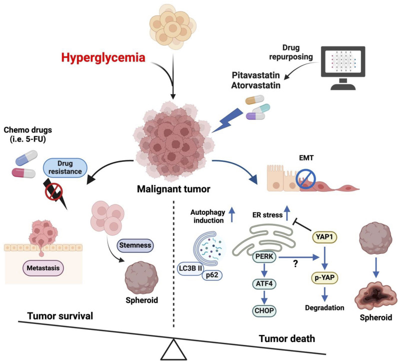
Background: Colorectal cancer (CRC) poses a significant clinical challenge because of drug resistance, which can adversely impact patient outcomes. Recent research has shown that abnormalities within the tumor microenvironment, especially hyperglycemia, play a crucial role in promoting metastasis and chemoresistance, and thereby determine the overall prognosis of patients with advanced CRC.

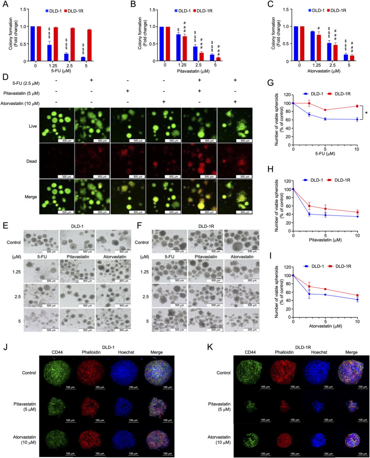
Methods: This study employs data mining and consensus molecular subtype (CMS) techniques to identify pitavastatin and atorvastatin as potential agents for targeting high glucose-induced drug resistance in advanced CRC cells. CRC cells maintained under either low or high glucose conditions were established and utilized to assess the cytotoxic effects of pitavastatin and atorvastatin, both with and without 5-fluorouracil (5-FU). CRC 3D spheroids cultured were also included to demonstrate the anti-drug resistance of pitavastatin and atorvastatin.

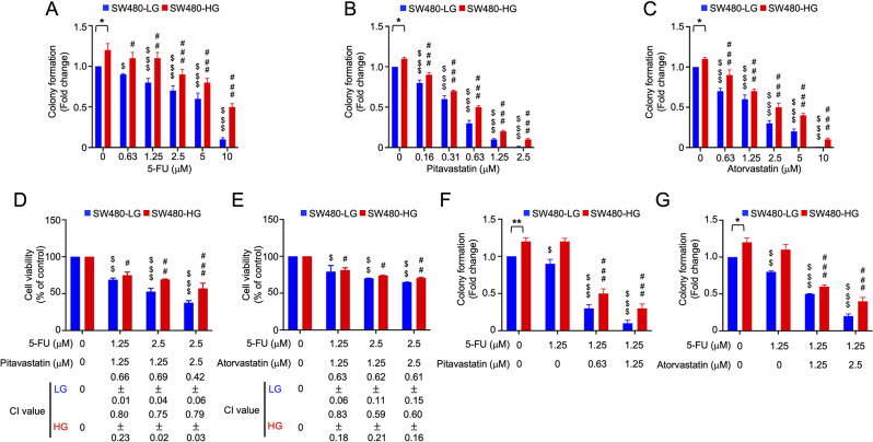
Results: A bioinformatics analysis identified pitavastatin and atorvastatin as promising drug candidates. The CMS4 CRC cell line SW480 (SW480-HG) was established and cultured under high glucose conditions to simulate hyperglycemia-induced drug resistance and metastasis in CRC patients. Pitavastatin and atorvastatin could inhibit cell proliferation and 3D spheroid formation of CMS4 CRC cells under high glucose conditions. In addition, both pitavastatin and atorvastatin can synergistically promote the 5-FU-mediated cytotoxic effect and inhibit the growth of 5-FU-resistant CRC cells. Mechanistically, pitavastatin and atorvastatin can induce apoptosis and synergistically promote the 5-FU-mediated cytotoxic effect by activating autophagy, as well as the PERK/ATF4/CHOP signaling pathway while decreasing YAP expression.

Conclusion: This study highlights the biomarker-guided precision medicine strategy for drug repurposing. Pitavastatin and atorvastatin could be used to assist in the treatment of advanced CRC, particularly with CMS4 subtype CRC patients who also suffer from hyperglycemia. Pitavastatin, with an achievable dosage used for clinical interventions, is highly recommended for a novel CRC therapeutic strategy.

Keywords: Atorvastatin; Colorectal cancer; Consensus molecular subtype; Drug resistance; Hyperglycemia; Pitavastatin.


Conflict of interest statement

Declarations. Ethics approval and consent to participate: Not applicable. Consent for publication: All authors have participated in the study and consented to publication. Competing interests: The authors declare no competing interests.

Figures

Fig. 1
Fig. 1
Pitavastatin and atorvastatin are identified as putative treatments for metastatic CMS4 CRC. (A) PRISM screening, a high-throughput DNA-barcoding technique, is used to analyze the cell viability of 35 CRC cells treated with 4,686 compounds. PRISM drug sensitivity represents the log2-fold change in cell proliferation rate following drug treatment compared with an untreated group. Mann-Whitney U tests (p < 0.05) were used to identify specifically sensitive drugs in metastatic CRC cells and CMS-specific groups. (B-C) The numbers of compounds and on-market drugs at the intersection of drug candidates representing five groups (i.e., CMS1-4 specific drugs and metastasis-specific drugs). The horizontal bar represents the set size (i.e., the total number of drugs identified as candidates from each of the five groups). In contrast, the vertical bar represents the size of the intersection (i.e., the number of drugs included in each intersecting set). (D-E) Illustrations of the specific sensitivity of pitavastatin, atorvastatin, lovastatin, simvastatin, and mevastatin against metastatic (*Metastatic cells compared to primary cells) and CMS4 cells (* CMS4 cells compared to other CMS cells). (F) Analysis of the PRISM database used to compare the sensitivity of CRC cells to pitavastatin, atorvastatin, and 5-FU. There was no pitavastatin sensitivity data for SW837 cells found in the database. The cut-off value of drug sensitivity was smaller than 0.3. One symbol represents p < 0.05; two symbols indicate p < 0.01
Fig. 2
Fig. 2
Pitavastatin and atorvastatin can overcome high glucose-induced drug resistance and synergistically promote 5-FU-mediated cytotoxicity. (A-C) Colony formation assay of SW480-LG and SW480-HG following treatment with 5-FU, pitavastatin, or atorvastatin for nine days. (D-E) The combination index (CI) of SW480-LG and SW480-HG cells treated with 5-FU in combination with pitavastatin or atorvastatin. CI defines synergism (CI < 1), an additive effect (CI = 1), and antagonism (CI > 1). (F-G) Colony formation assay of SW480-LG and SW480-HG following treatment with a combination of 5-FU and pitavastatin or atorvastatin. Data represent the mean ± SEM (N = 3). $: Denotes a comparison with the SW480-LG control; #: Indicates a comparison with the SW480-HG control; The number of symbols corresponds to the significance level: one symbol indicates p < 0.05; two symbols indicate p < 0.01; and three symbols indicates p < 0.001. LG: Low glucose; HG: High glucose
Fig. 3
Fig. 3
Pitavastatin and atorvastatin can inhibit the migration ability and spheroid formation stimulated by high glucose. (A) A transwell assay shows decreased cell migration following treatment with pitavastatin or atorvastatin for 24 h in SW480-LG and SW480-HG cells. Representative images are shown. An untreated SW480-LG condition serves as a baseline for comparison. (B) Following a 48-hour treatment with pitavastatin and atorvastatin, both statins demonstrated an ability to influence the expression of ZO-1 and Snail proteins. (C-D) SW480-LG and SW480-HG cells were cultured using an ACD 3D culture system at a density of 1000 cells per well. Images were acquired using an Olympus IX83 inverted microscope with a 10X objective. The scale bar represents 500 μm. (E-G) Effects of 5-FU, pitavastatin, and atorvastatin on spheroid formation by SW480-LG and SW480-HG cells. Data represent mean ± SEM (N = 3). $: Indicates a comparison with the SW480-LG control; #: Indicates a comparison with the SW480-HG control; The number of symbols corresponds to the significance level: one symbol represents p < 0.05; two symbols indicate p < 0.01; and three symbols denote p < 0.001. LG: Low glucose; HG: High glucose
Fig. 4
Fig. 4
Pitavastatin and atorvastatin affect intrinsic 5-FU resistance in CRC cells. (A-C) Colony formation assay of DLD-1 and DLD-1R cells following treatment with 5-FU, pitavastatin, or atorvastatin for nine days. (D) The Cyto3D™ dead-live assay following cotreatment with 5-FU and pitavastatin or atorvastatin in DLD-1 spheroids. Live-dead imaging of DLD-1 spheroids was conducted using the Cyto3D Live-Dead assay. Bright-field and fluorescence images were acquired using an Olympus IX83 inverted microscope with a 10X objective. The scale bar represents 500 μm. (E-F) DLD-1 and DLD-1R were seeded onto R3CE plates at 1000 cells per well density. 5-FU, pitavastatin, and atorvastatin were added to the culture medium at the indicated concentrations for 7 days. Images were acquired using an Olympus IX83 inverted microscope with a 10X objective. The scale bar represents 500 μm. (G-I) Effects of 5-FU, pitavastatin, and atorvastatin on spheroid formation by DLD-1 and DLD-1R cells. (J-K) Drug-treated DLD-1 and DLD-1R spheroids were stained with CD44 (labeled with Alexa Fluor™ 488), Alexa Fluor™ 568 Phalloidin, and/or Hoechst 33,342. IF staining images were acquired using a Zeiss LSM900 confocal microscope with a 20X objective. The scale bar represents 100 μm. Data represent mean ± SEM (N = 3). DLD-1R: DLD-1 5-FU resistance
Fig. 5
Fig. 5
Underlying mechanisms driving the effects of pitavastatin and atorvastatin on CRC cells. The network illustrates the connections between pathways associated with pitavastatin (A) and atorvastatin (B). The size of each dot denotes a pathway gene set, while lines between two dots represent connections between two pathways. Finally, the line width represents the strength of the relationship. (C-D) Western blotting data indicates that treatment with pitavastatin and atorvastatin for 48 h significantly influences the expression of markers associated with autophagy, ER, and apoptosis in both SW480-LG and SW480-HG cells. LG: Low glucose; HG: High glucose
Fig. 6
Fig. 6
Pitavastatin and atorvastatin induce apoptosis partly by stimulating autophagy and inducing ER stress. Western blotting data show the impact of various inhibitors on the expression of markers associated with autophagy, ER, and apoptosis. (A) SW480-LG and SW480-HG cells were co-treated with 3-MA and pitavastatin or atorvastatin. (B) Both SW480-vehicle and SW480-ATG5 KO cells were treated with pitavastatin and atorvastatin. (C) GSK2606414 was used in a cotreatment with pitavastatin or atorvastatin on SW480-LG and SW480-HG cells. LG: Low glucose; HG: High glucose
Fig. 7
Fig. 7
Schematic representation of the mechanism of action of pitavastatin and atorvastatin. This illustration was created using BioRender.com

References

    1. Bray F, Ferlay J, Soerjomataram I, Siegel RL, Torre LA, Jemal A. Global cancer statistics 2018: GLOBOCAN estimates of incidence and mortality worldwide for 36 cancers in 185 countries. CA Cancer J Clin. 2018;68(6):394–424. - PubMed
    1. Bailey CE, Hu CY, You YN, Bednarski BK, Rodriguez-Bigas MA, Skibber JM, Cantor SB, Chang GJ. Increasing disparities in the age-related incidences of colon and rectal cancers in the united States, 1975–2010. JAMA Surg. 2015;150(1):17–22. - PMC - PubMed
    1. Kasi PM, Shahjehan F, Cochuyt JJ, Li Z, Colibaseanu DT, Merchea A. Rising proportion of young individuals with rectal and Colon cancer. Clin Colorectal Cancer. 2019;18(1):e87–95. - PubMed
    1. Benson AB, Venook AP, Al-Hawary MM, Arain MA, Chen YJ, Ciombor KK, Cohen S, Cooper HS, Deming D, Farkas L, et al. Colon cancer, version 2.2021, NCCN clinical practice guidelines in oncology. J Natl Compr Canc Netw. 2021;19(3):329–59. - PubMed
    1. Miller KD, Nogueira L, Mariotto AB, Rowland JH, Yabroff KR, Alfano CM, Jemal A, Kramer JL, Siegel RL. Cancer treatment and survivorship statistics, 2019. CA Cancer J Clin. 2019;69(5):363–85. - PubMed

LinkOut - more resources