. 2025 Mar 6;24(1):67.
doi: 10.1186/s12943-025-02272-3.

Circular RNA circCLASP2 promotes nasopharyngeal carcinoma progression through binding to DHX9 to enhance PCMT1 translation

Affiliations

Circular RNA circCLASP2 promotes nasopharyngeal carcinoma progression through binding to DHX9 to enhance PCMT1 translation

Miao Peng et al. Mol Cancer. .

Abstract

Background: Circular RNAs (circRNAs), characterized by their covalently closed-loop structures, constitute a distinct class of non-coding RNAs. They play pivotal regulatory roles within cells and are intricately associated with the progression of malignant tumors. However, their roles and the underlying mechanisms in nasopharyngeal carcinoma (NPC) progression have yet to be fully uncovered and comprehensively understood.

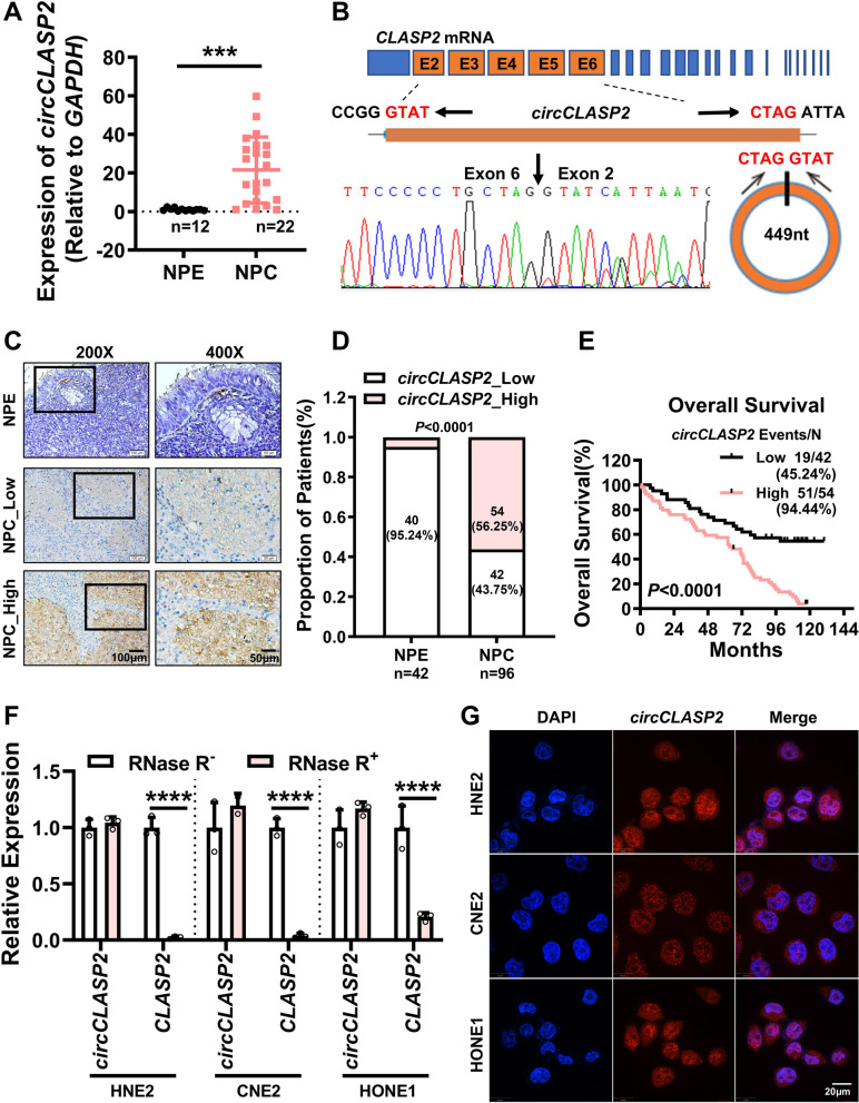
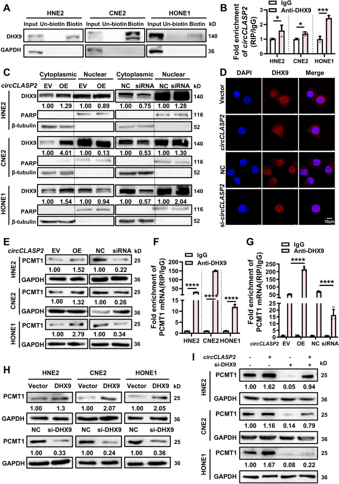
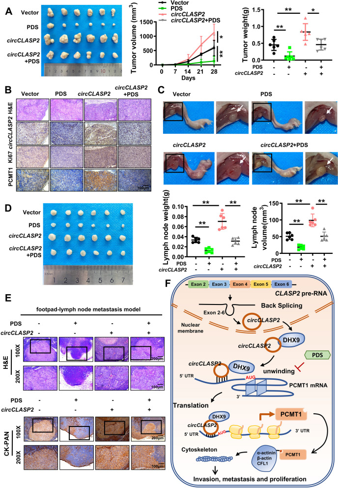
Methods: Employing RNA sequencing technology, high-abundance circular RNAs in NPC were identified. Expression analysis of circCLASP2 in NPC tissues was conducted using quantitative real-time polymerase chain reaction (qRT-PCR) and in situ hybridization experiments. Through in vitro and in vivo functional assays, the influence of circCLASP2 on the proliferation and metastasis of NPC was investigated. LC-MS/MS technology analyzed the binding partners of circCLASP2, its differentially regulated targets, and the associated proteins of PCMT1. Interactions among circCLASP2, DHX9 protein, and PCMT1 mRNA were elucidated through RNA immunoprecipitation and RNA pull-down techniques. The effects of circCLASP2 and DHX9 on RNA G-quadruplex (rG4) structures and PCMT1 mRNA translation were explored through immunofluorescence (IF), ribosomal gradient separation, and dual-luciferase reporter assays. Immunoprecipitation (IP) revealed the downstream effector of the circCLASP2-DHX9-PCMT1 regulatory axis and Phalloidin staining confirmed its ultimate effect on the cytoskeleton. PDS treatment was applied for interventions in NPC, demonstrating potential therapeutic avenues.

Results: Our research revealed that circCLASP2, a novel circRNA that has not been reported in tumors, is upregulated in NPC and fosters cell proliferation and metastasis both in vitro and in vivo. Mechanistically, circCLASP2 acts as a molecular scaffold, facilitating the approximation of DHX9 to PCMT1 mRNA. DHX9 unwinds the inhibitory rG4 structure near the translation initiation site on PCMT1 mRNA, increasing PCMT1 expression. PCMT1 binds to and upregulates cytoskeleton-associated proteins, modulating cytoskeleton strength and dynamics and ultimately driving NPC cell proliferation and metastasis. In both in vitro and in vivo experiments, PDS significantly inhibits NPC growth and metastasis, showcasing promising therapeutic potential.

Conclusions: Our investigation pinpointed a circular RNA, circCLASP2, which is upregulated in NPC and augments cytoskeletal functions via the DHX9-PCMT1 axis, contributing to the malignancy progression of NPC. This pathway holds promise as a potential therapeutic target for NPC. Furthermore, these molecules could also serve as biomarkers for adjunct diagnosis and prognosis assessment in NPC.

Keywords: CircCLASP2; Cytoskeleton; DHX9; Nasopharyngeal Carcinoma; PCMT1.


Conflict of interest statement

Declarations. Ethics approval and consent to participate: The present study was approved by the Ethics Committee of Central South University. Consent for publication: Not applicable. Competing interests: The authors declare no competing interests.

Figures

Fig. 1
Fig. 1
CircCLASP2 is highly expressed in NPC and is associated with poor prognosis in patients. A The differential expression of circCLASP2 in 12 chronic rhinitis epithelial tissues (NPE) and 22 NPC tissues was detected by qRT-PCR, and statistically analyzed by Student's t test (two-tailed). B Schematic representation of circCLASP2. Sanger sequencing confirmed that exons 2–6 of CLASP2 mRNA were back spliced to form circCLASP2. Black arrows indicate the primer locations and orientations for detecting the circCLASP2 splice junction. C-E ISH assays revealed elevated expression of circCLASP2 in 96 NPC paraffin-embedded tissue sections compared to that in 42 NPEs and was associated with adverse prognosis in NPC patients. The ISH results were graded based on the depth of staining and the positivity rate. A comprehensive score greater than or equal to 5 was considered as high circCLASP2 expression, while a score less than 5 was categorized as low circCLASP2 expression. C Representative ISH images, left: scale bar = 100 μm; right: scale bar = 50 μm. D Statistical analysis of ISH scores. Statistical analysis was performed using the unpaired t test. E Kaplan–Meier survival analysis showed significantly reduced overall survival rates in NPC patients with high circCLASP2 expression compared to those with low circCLASP2 expression. F Relative expression levels of circCLASP2 and CLASP2 mRNA in NPC cells after treatment with RNase R. G FISH assays for detecting subcellular localization of circCLASP2 in NPC cells. Nuclei were stained with DAPI (blue), and circCLASP2 was identified with probes (red). Scale bar = 20 μm. Data were presented as mean ± SD. ***, P < 0.001; ****, P < 0.0001
Fig. 2
Fig. 2
CircCLASP2 promotes proliferation and metastasis of NPC cells in vitro and in vivo. A MTT assay determining the proliferation capability of HNE2 cells with overexpression or knockdown of circCLASP2. B Transwell invasion assay assessing the invasion capability of HNE2 cells with overexpression or knockdown of circCLASP2. Scale bar = 100 μm. The right panel shows statistical analysis. C Wound healing assay measuring the migration ability of HNE2 cells with overexpression or knockdown of circCLASP2, with images taken at the same field of view at 0 h and 24 h. Scale bar = 200 μm. The right panel shows statistical analysis. D-H CNE2 cells transfected with empty vector, circCLASP2 overexpression plasmid, siRNA negative control (NC), or si-circCLASP2 were injected subcutaneously or into the footpad of BALB/c mice (n = 6 per group), with each mouse receiving an injection of 2 × 106 cells. D Subcutaneous tumors in mice from each group (left). Growth curve of volumes (middle) and statistical analysis of the weights (right) of subcutaneous tumors in nude mice. E Representative images of H&E, ISH, and IHC staining of subcutaneous tumor sections. Scale bar = 100 μm. F Representative images of the "footpad-lymph node" metastasis model showing the injection site in the foot and ipsilateral popliteal lymph node. G Popliteal lymph nodes from the "footpad-lymph node" metastasis model (left). Statistical analysis of volumes (middle) and weights (right) of lymph nodes in nude mice. H Representative images of H&E and CK-PAN staining of popliteal lymph node sections from the "footpad-lymph node" metastasis model, showing metastatic tumor cells. Top image scale bar = 200 μm; Bottom image scale bar = 100 μm. Data were presented as mean ± SD. *, P < 0.05; **, P < 0.01; ***, P < 0.001; ****, P < 0.0001
Fig. 3
Fig. 3
CircCLASP2 promotes PCMT1 expression via facilitating the interaction between DHX9 and PCMT1 mRNA in the cytoplasm. A Using a biotin-labeled circCLASP2 probe, RNA pull-down assays enriched circCLASP2 binding proteins in NPC cell lines HNE2, CNE2 and HONE1, with Western blotting showing the interaction between circCLASP2 and DHX9. GAPDH served as a negative control. B RIP assays using DHX9 antibody to detect the interaction between DHX9 and circCLASP2 in HNE2, CNE2, and HONE1. IgG was used as a negative control. C Western blotting assessing the abundance of DHX9 protein in the cytoplasm and nucleus of HNE2, CNE2, and HONE1 cells following overexpression or knockdown of circCLASP2. PARP and β-tubulin served as nuclear and cytoplasmic markers, respectively. D IF experiments examining the effect of circCLASP2 on the subcellular localization of DHX9. E Western blotting measuring PCMT1 protein levels in HNE2, CNE2, and HONE1 following overexpression or knockdown of circCLASP2. F RIP assays were conducted to detect the interaction between DHX9 and PCMT1 mRNA in HNE2, CNE2, and HONE1, using DHX9 antibody. IgG served as a negative control. G RIP assays using DHX9 antibody to assess the effects of overexpressing or knocking down circCLASP2 on the interaction between DHX9 and PCMT1 mRNA. H Western blotting measuring PCMT1 protein levels in HNE2, CNE2, and HONE1 following overexpression or knockdown of DHX9. I Western blotting assessing PCMT1 protein levels in HNE2, CNE2, and HONE1 after simultaneous overexpression of circCLASP2 and knockdown of DHX9. Data were presented as mean ± SD. *, P < 0.05; ***, P < 0.001; ****, P < 0.0001
Fig. 4
Fig. 4
PCMT1 is associated with the progression of NPC and involved in the circCLASP2-regulated pathway of NPC malignancy. A-B ISH and IHC analysis of 34 NPC and 7 adjacent non-tumor NPE tissues demonstrated the correlation between the expressions of circCLASP2, DHX9 and PCMT1. A The expression levels of circCLASP2, DHX9 and PCMT1 in NPC and NPE tissues, detected by ISH and IHC. Left: scale bar = 100 μm; right: scale bar = 50 μm. B Correlation analysis of circCLASP2 ISH and PCMT1 IHC scores. C Western blotting detected circCLASP2’s role in rescuing PCMT1 protein expression after knockdown in NPC cells. MTT assay (D), Transwell invasion assay (E), and wound healing assay (F) assessing the impact of circCLASP2-PCMT1 axis on the migration, invasion, and proliferation of NPC cells. Data were presented as mean ± SD. **, P < 0.01; ***, P < 0.001; ****, P < 0.0001
Fig. 5
Fig. 5
DHX9 enhances PCMT1 mRNA translation by unwinding the G4 structure. A FISH-IF experiments showing the presence of rG4 structure on PCMT1 mRNA in HNE2 cells. Blue: DAPI; Red: Sequences flanking the region on PCMT1 mRNA that potentially form rG4 structures; Green: BG4; Yellow: colocalization of red and green fluorescence. Scale bar = 10 μm. B Dual-luciferase reporter assay system measuring luciferase activity in HNE2 cells after overexpressing circCLASP2 and/or knocking down DHX9. C Dual-luciferase reporter assay system measuring luciferase activity in HNE2 cells after overexpressing circCLASP2 and/or PDS treatment. D Ribosome separation with sucrose density gradient assessing the effects of different treatments on the translation efficiency of PCMT1 mRNA in NPC cells. Treatment conditions included knockdown of circCLASP2, knockdown of DHX9, overexpression of circCLASP2 with concurrent knockdown of DHX9, and overexpression of circCLASP2 with PDS treatment. RNA from various ribosomal components was extracted for qRT-PCR to quantify the enrichment of PCMT1 mRNA on monosomes and polysomes. GAPDH mRNA served as a control. E Western blotting detecting the impact of overexpressing circCLASP2 or DHX9 with concurrent PDS treatment on PCMT1 expression in HNE2, CNE2, and HONE1 cells. Data were presented as mean ± SD. *, P < 0.05; **, P < 0.01; ***, P < 0.001; ****, P < 0.0001
Fig. 6
Fig. 6
PCMT1 modulates cytoskeletal polymerization through regulating cytoskeleton-associated proteins. A PCMT1 binding proteins were captured by IP, which were then separated by SDS-PAGE and visualized using silver staining. B IP detecting the interactions between PCMT1 and α-actinin, β-actin, or CFL1 in NPC cells. Specific primary antibodies were used for enrichment, followed by detection via Western blotting. C Western blotting assessing the impact of overexpressing or knocking down PCMT1 on the protein levels of α-actinin, β-actin, and CFL1. D Phalloidin staining in NPC cells showing actin filament intensity and aggregation after overexpressing or knocking down circCLASP2, DHX9 and PCMT1. Scale bar = 10 μm
Fig. 7
Fig. 7
G4 stabilizer PDS inhibits the proliferation and metastasis of NPC cells both in vitro and in vivo. A-E CNE2 cells (2 × 10.6) transfected with empty vectors or circCLASP2 overexpression vectors were injected subcutaneously or into the footpads of BALB/c mice. PDS or saline was administered via the tail vein. A Tumor tissues from in situ xenograft mice models (n = 6 per group) (left). Measurements of tumor volume (middle) and weight (right) in the in situ xenograft model. B Representative images of H&E, ISH, and IHC staining of subcutaneous tumor sections from the xenograft model. Scale bar = 100 μm. C Representative images of the footpad and ipsilateral popliteal lymph node from the "footpad-lymph node" metastasis model. D Images of the popliteal lymph nodes in the "footpad-lymph node" metastasis model (n = 6 per group) (left). Statistical analysis of the weight (middle) and volume (right) of popliteal lymph nodes in the "footpad-lymph node" metastasis model. E Representative images of H&E and CK-PAN staining of popliteal lymph node sections from the "footpad-lymph node" metastasis model, showing metastatic tumor tissue. Top: scale bar = 200 μm; Bottom: scale bar = 100 μm. F Schematic diagram illustrating the circCLASP2-DHX9-PCMT1 axis driving malignant progression of NPC. Data were presented as mean ± SD. *, P < 0.05; **, P < 0.01

References

    1. Wang Y, Yan Q, Fan C, Mo Y, Wang Y, Li X, et al. Overview and countermeasures of cancer burden in China. Sci China Life Sci. 2023;66:2515–26. - PMC - PubMed
    1. Fan C, Tang Y, Wang J, Xiong F, Guo C, Wang Y, et al. The emerging role of Epstein-Barr virus encoded microRNAs in nasopharyngeal carcinoma. J Cancer. 2018;9:2852–64. - PMC - PubMed
    1. Wang D, Zuo S, Ge J, Qu H, Wu J, Yi N, et al. CircTP63-N suppresses the proliferation and metastasis of nasopharyngeal carcinoma via engaging with HSP90AB1 to modulate the YAP1/Hippo signaling pathway. Sci China Life Sci. 2024. 10.1007/s11427-023-2737-2. - PubMed
    1. Tu C, Zeng Z, Qi P, Li X, Guo C, Xiong F, et al. Identification of genomic alterations in nasopharyngeal carcinoma and nasopharyngeal carcinoma-derived Epstein-Barr virus by whole-genome sequencing. Carcinogenesis. 2018;39:1517–28. - PubMed
    1. Qu H, Wang Y, Yan Q, Fan C, Zhang X, Wang D, et al. CircCDYL2 bolsters radiotherapy resistance in nasopharyngeal carcinoma by promoting RAD51 translation initiation for enhanced homologous recombination repair. J Exp Clin Cancer Res. 2024;43:122. - PMC - PubMed

MeSH terms

LinkOut - more resources