. 2025 Mar;34(3):e70071.
doi: 10.1111/exd.70071.

Do Melanocytes Have a Role in Controlling Epidermal Bacterial Colonisation and the Skin Microbiome?

Affiliations

Do Melanocytes Have a Role in Controlling Epidermal Bacterial Colonisation and the Skin Microbiome?

Omera Bi et al. Exp Dermatol. 2025 Mar.

Abstract

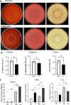
In addition to producing melanin to protect epidermal keratinocytes against DNA damage, melanocytes may have important roles in strengthening innate immunity against pathogens. We have developed a functional, pigmented, human full-thickness 3D skin equivalent to determine whether the presence of melanocytes impacts epidermal bacterial growth and regulates the expression of genes involved in the immune response. We introduced primary epidermal melanocytes to construct a 3-cell full-thickness skin equivalent with primary dermal fibroblasts and epidermal keratinocytes. Immunohistochemistry verified the appropriate ratio and spatial organisation of melanocytes. Alpha-MSH induced melanogenesis, confirming an appropriate physiological response. We compared this 3-cell skin equivalent with the 2-cell version without melanocytes in response to inoculation with 3 species of bacteria: Staphylococcus epidermidis, Corynebacterium striatum, and Cutibacterium acnes. There was a significant decrease in the colonisation of bacteria in the skin equivalents containing functional melanocytes. There was increased expression of immune-response genes (S100A9, DEFB4A, IL-4R) following microorganism exposure; however, there were marked differences between the unpigmented and pigmented skin equivalents. This physiologically relevant human 3D-skin equivalent opens up new avenues for studying complex skin pigmentation disorders, melanoma, and UV damage, as well as the rapidly evolving field of the skin microbiome and the balance between commensal and pathogenic species.

Keywords: bacteria; full thickness skin equivalent; immune response; melanocytes; skin microbiome.


Conflict of interest statement

The authors declare no conflicts of interest.

Figures

FIGURE 1
FIGURE 1
Schematic illustration of the optimised methodology to develop a pigmented 3‐cell human full‐thickness skin equivalent. (A) Unpigmented 2‐cell skin equivalent: (Step 1) A dermal equivalent (DE) was generated with low passage dermal fibroblasts (DF), fibrinogen, and collagen. (Step 2) Once established, early passage epidermal keratinocytes (EK) in keratinocyte media (KC) were seeded on top. (Step 3) Growth media was added to the surface of the full‐thickness skin equivalents (FTSE) and to the bottom of the well every 2 days until the construct was moved to the air– liquid interface (ALI) at day 12. (Step 4) FTSE were transferred into deep wells and growth media was added to the bottom of the well for a further 12 days. (B) Pigmented 3‐cell skin equivalent: The same procedure as described in (A) was followed except at (Step 2) low passage epidermal melanocytes (MC) in melanocyte media were added to the cell suspension of EK in a ratio of 1:10 MC:EK. (C) Inoculation with bacterial species: (Step 1) Pigmented and unpigmented skin equivalents were inoculated with a mixture of S. epidermidis, C. striatum , and C. acnes with a final concentration of each of 1 × 10⁴ CFU cm2 before incubating at 37°C with 5% CO2 for 24 h. (Step 2) Each FTSE was divided in half and 5 × 8 mm punch biopsies were taken, diluted in 1:100 Dey‐Engley Neutralising Broth (DEB), vortexed, and then plated onto Mannitol Salt Agar (MSA) and Aerobic Coryneform Agar (ACA) for aerobic culture at 37°C for 24 h, and Reinforced Clostridial Agar (RCAF) for anaerobic culture at 37°C for 72 h. (Step 3) Bacterial colonies were counted on each agar type to determine CFU/cm2. (Step 2a) Punch biopsies were taken from the 2nd half of each FTSE and stored in RNA later. (Step 4) RT‐qPCR was performed to quantify the expression of S100A9, DEFB4A, and IL‐4A, with gene expression normalised to GAPDH and β‐actin.
FIGURE 2
FIGURE 2
Upregulation of melanogenesis via α‐MSH in a pigmented 3‐cell human full‐thickness skin equivalent. Pigmented 3‐cell full‐thickness skin equivalents were incubated for 12 days in the presence or absence of α‐MSH (10−5 or 10−6 M), then snap frozen, cut into 6 μM sections and melanocytes localised by immunohistochemistry (peroxidase method) (A–C) or immunofluorescence (D–E). Expression of TRP1 is shown by brown staining (A‐C) or red fluorescence (D–E); basement membrane localised by staining for laminin (green), and nuclei by DAPI (blue). (G) Expression of TRP1 is shown by green fluorescence in human skin in vivo, and nuclei by DAPI (blue). (H) Total cell corrected fluorescence; data are presented as mean +/− SEM of 3 biological repeats (6 images per condition) of the FTSEs. (I) Gene expression of TRP1 in the FTSEs quantitated by RT‐qPCR. Data are presented as mean +/− SEM of 3 biological repeats performed in triplicate. Statistical analysis was performed using one‐way Anova followed by Šídák multiple comparison test (H) and one‐way ANOVA followed by Dunn's multiple comparisons test (I), (ns non‐significant, *p ≤ 0.05).
FIGURE 3
FIGURE 3
Presence of melanocytes in a human full‐thickness skin equivalent modulates the growth of epidermal microflora and immune‐response genes. A combination of S. epidermis, C. striatum , or C. acnes was grown on the epidermis of unpigmented or pigmented human full‐thickness skin equivalents for 24 h each at a concentration of 1 × 104 CFU cm2. Bacterial colonies were counted after plating onto relevant agar to quantitate CFU cm2 (A and B). Unpigmented (UN‐P), pigmented (P). Data are presented as mean ± SEM of 5 technical repeats. Statistical analysis was performed using an unpaired two‐tailed t‐test to compare unpigmented and pigmented groups. (C) A combination of S. epidermis, C. striatum , and C. acnes was grown on the epidermis of unpigmented or pigmented human full‐thickness skin equivalents, each at a concentration of 1 × 104 CFU cm2 for 24 h. Expression of anti‐microbial genes S100A9, DEFB4A, and IL‐4A was quantitated by RT‐qPCR following homogenisation of colonised skin equivalents. Gene expression was normalised with GAPDH and β‐actin. Data are representative of three individual skin equivalents and three technical repeats. One‐way ANOVA followed by Dunnett's multiple comparison test was used to assess differences among groups. ns non‐significant, p > 0.05, *p ≤ 0.05, **p ≤ 0.01, *** p ≤ 0.001, **** p ≤ 0.0001. (one‐way ANOVA).

References

    1. Speeckaert R., Belpaire A., Speeckaert M., and van Geel N., “The Delicate Relation Between Melanocytes and Skin Immunity: A Game of Hide and Seek,” Pigment Cell & Melanoma Research 35, no. 4 (2022): 392–407. - PubMed
    1. Koike S. and Yamasaki K., “Melanogenesis Connection With Innate Immunity and Toll‐Like Receptors,” International Journal of Molecular Sciences 21, no. 24 (2020): 9769, 10.3390/ijms21249769. - DOI - PMC - PubMed
    1. Tapia C. V., Falconer M., Tempio F., et al., “Melanocytes and Melanin Represent a First Line of Innate Immunity Against Candida albicans ,” Medical Mycology 52, no. 5 (2014): 445–454. - PubMed
    1. Grice E. A. and Segre J. A., “The Skin Microbiome,” Nature Reviews Microbiology 9, no. 4 (2011): 244–253. - PMC - PubMed
    1. Cario M. and Taieb A., “Isolation and Culture of Epidermal Melanocytes,” Methods in Molecular Biology 1993 (2019): 33–46 New York, NY, Springer New York. - PubMed

Supplementary concepts

Grants and funding

LinkOut - more resources