. 2025 Apr;14(11):e2405238.
doi: 10.1002/adhm.202405238. Epub 2025 Mar 6.

A Dual-Layer Hydrogel Barrier Integrating Bio-Adhesive and Anti-Adhesive Properties Prevents Postoperative Abdominal Adhesions

Affiliations

A Dual-Layer Hydrogel Barrier Integrating Bio-Adhesive and Anti-Adhesive Properties Prevents Postoperative Abdominal Adhesions

Mena Asha Krishnan et al. Adv Healthc Mater. 2025 Apr.

Abstract

Postoperative abdominal adhesions are a common and painful complication after surgery, leading to high healthcare costs and diminished quality of life. This report presents a novel bilayer hydrogel barrier featuring an inner adhesive layer and an outer antiadhesive layer. The inner adhesive layer hydrogel (PT) is prepared by mixing polyethyleneimine (PEI) and thioctic acid (TA). The outer layer (HP) hydrogel is fabricated by the conjugation reaction of thermoresponsive zwitterionic hyaluronic acid, phenylboronic acid, and epigallocatechin gallate complex and polyvinyl alcohol based on dynamic boronic ester bond. The PEI/TA layer enhances attachment to moist tissue surfaces in vivo, and the anti-adhesive layer HP hydrogel promotes biocompatibility and anti-inflammation while minimizing protein adsorption and improving mechanical stability. The bilayer hydrogel (HPPT) exhibited rapid gelation, robust adhesion in dynamic and moist environments, superior viscoelastic properties and cellular biocompatibility. A mouse-cecum abdominal wall adhesion model is utilized to evaluate efficacy, and the HPPT hydrogel shows local retention, anti-inflammatory effect, and inhibits fibrin deposition while minimizing adhesion formation. These findings highlight the innovative structural and functional properties of the HPPT hydrogel, positioning it as a promising therapeutic barrier in peritoneal surgery aimed at reducing postoperative adhesions and enhancing surgical outcomes.

Keywords: anti‐adhesion barrier; anti‐fouling; bilayer hydrogel; zwitterionic hydrogel.


Conflict of interest statement

The authors declare no conflict of interest.

Figures

Figure 1
Figure 1
Schematic illustration for fabrication of bilayer HPPT hydrogel barrier. In A), The inner layer was synthesized by coaercevate formation of TA and PEI resulting in intramolecular hydrophobic and intermolecular H‐bonding. B) The bioadhesive when applied on injured site repels water. The outer layer is formed by C) EGCG‐PBA complex which was conjugated with D) HA‐SPDA polymer and amine terminal pNiPAM, 4‐(4,6‐dimethoxy‐1,3,5‐triazin‐2‐yl)‐4‐methylmorpholinium chloride (DMTMM) at RT for 3 days to form HSPE‐pNiPAM microgel, and was mixed with PVA to form HP hydrogel within 10s. E) The anti‐adhesive, zwitterionic HP hydrogel outer layer was applied, to prevent adhesion. This schematic illustration was developed using BioRender.com, in compliance with the terms of an authorized license.
Figure 2
Figure 2
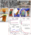
Characterization of structure, morphology, ex vivo stability and rheological properties. A) SEM images of porous structure of freeze‐dried HP hydrogel. Scale bar = 200 µm and 1µm. B) The ex vivo stability of the HPPT hydrogel was examined over a 24‐hour period on inverted rat abdominal tissues maintained at 37 °C in a 5% CO2 environment. C) The adhesion of rat tissue to 1st adhesive PEI/TA layer was assessed against gravitational pull. D) The introduction of a second HP anti‐adhesive layer completely prevented tissue adherence. Stability assessments of the HPPT bilayer on rat abdomen tissue were performed in various states: E) original, F) twisted, G) bent, and H) held vertically. Rheological studies of HP hydrogel and HPPT gel were conducted, providing insights through I) time‐sweep curves (0‐5 min duration, 1 Hz frequency, 10% strain, maintained at 37 °C), J) frequency‐sweep curves (0.1‐100 rad s−1 frequency range, under 10% strain, at 37 °C), K) step‐strain testing t high strains (600%) versus low strains (10%) at 1 Hz and 37 °C, and (L) shear‐rate sweep curves extending from 0.1 to 100 s⁻¹.
Figure 3
Figure 3
Cytotoxicity evaluation of the 5% PEI/TA adhesive layer and HPPT bilayer was conducted on A) MeT5a and B) L929 cell lines, utilizing hydrogel extracts over a time course of day 1, day 3, and day 7 (n = 4; ns = no significant). C) Confocal laser scanning microscopy (CLSM) images illustrated cell attachment on HP and HPPT hydrogels, assessed using Live/Dead staining after an 18‐hour incubation period. A semi‐quantitative analysis revealed the area occupied by live D) L929 cells (***p < 0.001) and E) MeT5a cells (***p < 0.001) (n = 4). Additionally, F) the adsorption profile of bovine serum albumin (BSA) on the surfaces of HP and HPPT hydrogels was evaluated (n = 4; ***p < 0.001).
Figure 4
Figure 4
In vivo evaluation of anti‐adhesion efficacy. A) Schematic detailing the procedural steps involved in abrading the peritoneal surfaces of the cecum and the abdominal wall, followed by the application of a PEI/TA adhesive layer (highlighted in yellow), and subsequently layering an anti‐adhesive HP hydrogel (highlighted in purple). B) Photographic documentation taken during the surgical establishment of a mouse cecum‐abdominal wall defect model, showcasing the application of a bilayer HPPT hydrogel. C) Gross observations highlighted typical morphologies of the cecum and abdominal wall among the various experimental groups: Sham, HP, HPPT, and the controls which included Injury and Seprafilm at a 14‐day post‐surgery. The black circles denote areas of adhesions, and the black arrows indicate regions devoid of adhesion. D) A detailed adhesion scoring system was employed to categorize the severity of adhesions at predetermined time points (n = 8). E) Statistical comparisons of adhesion scores among the experimental groups were conducted to determine significance (n = 8, ***p<0.001, **p<0.01). Histopathological analyses of tissue samples from the Sham, Injury, HP, HPPT, and Seprafilm groups were performed at 14 days post‐surgery, utilizing Hematoxylin and Eosin (H&E) staining F) and G) Masson's trichrome staining for enhanced visualization. The cecum and abdominal wall regions are denoted as CE and AW, respectively, with black arrows marking the adhesion sites. Magnification for histological images was set at 20×; scale bars represent 300 µm.
Figure 5
Figure 5
In vivo retention of HP and HPPT hydrogels. A) IVIS imaging was conducted over a 14‐day period for both HP and HPPT treatment groups, utilizing Cy7.5 dye for labelling. B) The quantification of average radiance for the HP and HPPT groups illustrates the in vivo retention of the gel at various time points (n = 3). C) The average area of radiance was determined via contour plots, effectively quantifying the fluorescence area for the HP and HPPT groups throughout the designated time points (n = 3).
Figure 6
Figure 6
A) CLSM images of immunofluorescence staining after day 14 post‐surgery. Tissue sections were stained with DAPI (blue) alongside t‐PA and PAI‐1 (both in green). Scale bars indicate 100 µm. A semi‐quantitative analysis of the immunofluorescence images was performed to assess the percentage area of B) PAI‐1 and C) tPA expression relative to the DAPI‐stained regions across the tissue samples (n = 3). Statistical significance was denoted as ***p<0.001, and **p<0.01. Additionally, the relative mRNA expression levels of D) PAI‐1 and E) tPA were analyzed in excised tissues obtained 14 days post‐operatively (n = 3).
Figure 7
Figure 7
Analysis of tissue sections at 14 days post‐surgery to assess the inflammatory response. A) CLSM immunofluorescence staining demonstrated expressions of CD68 (red) and CD206 (green) in the excised tissue samples from the cecum, abdomen, and adhesion sites across all experimental groups. Semi‐quantitative evaluation of the CLSM‐stained sections was performed, revealing the percentage area of B) CD68 and C) CD206 relative to DAPI‐stained nuclei (***p<0.001, **p<0.01, *p<0.05. Additionally, the expression levels of mRNA corresponding to D) TNF‐α, E) TGF‐β1, and F) IL‐6 were quantified from isolated tissues (n = 3) (**p<0.001, *p<0.05).
Figure 8
Figure 8
Analysis of tissue sections at 14 days post‐surgery to assess the inflammatory response. A) CLSM immunofluorescence staining demonstrated expressions of iNOS (green) in the excised tissue samples from the cecum in Sham, HP and HPPT groups, and adhesion sites in Injury and Seprafilm groups. Semi‐quantitative evaluation of the CLSM‐stained sections was performed, revealing the percentage area of B) iNOS relative to DAPI‐stained nuclei (***p<0.001). Additionally, the expression levels of mRNA corresponding to C) iNOS were quantified from isolated tissues (n = 3) (*p<0.05).

Similar articles

References

    1. a) Tabibian N., Swehli E., Boyd A., Umbreen A., Tabibian J. H., Ann Med Surg. 2017, 15, 9; - PMC - PubMed
    2. b) van Goor H., Colorectal Dis. 2007, 9 Suppl 2, 25. - PubMed
    1. Prystowsky J. B., Stryker S. J., Ujiki G. T., Poticha S. M., Arch. Surg. 1988, 123, 855. - PubMed
    1. ten Broek R. P., Issa Y., van Santbrink E. J., Bouvy N. D., Kruitwagen R. F., Jeekel J., Bakkum E. A., Rovers M. M., van Goor H., BMJ. 2013, 347, f5588. - PMC - PubMed
    1. a) Ivarsson M. L., Bergström M., Eriksson E., Risberg B., Holmdahl L., Br. J. Surg. 1998, 85, 1549; - PubMed
    2. b) Holmdahl L., Eriksson E., Eriksson B. I., Risberg B., Surgery. 1998, 123, 539. - PubMed
    1. a) Chegini N., Kotseos K., Zhao Y., Bennett B., McLean F. W., Diamond M. P., Holmdahl L., Burns J., Hum Reprod. 2001, 16, 1291; - PubMed
    2. b) Holmdahl L., Kotseos K., Bergström M., Falk P., Ivarsson M. L., Chegini N., Surgery. 2001, 129, 626. - PubMed

MeSH terms