. 2025 Apr 2;33(4):1434-1448.
doi: 10.1016/j.ymthe.2025.02.046. Epub 2025 Mar 5.

Gene therapy rescues brain edema and motor function in a mouse model of megalencephalic leukoencephalopathy with subcortical cysts

Affiliations

Gene therapy rescues brain edema and motor function in a mouse model of megalencephalic leukoencephalopathy with subcortical cysts

Alejandro Brao et al. Mol Ther. .

Abstract

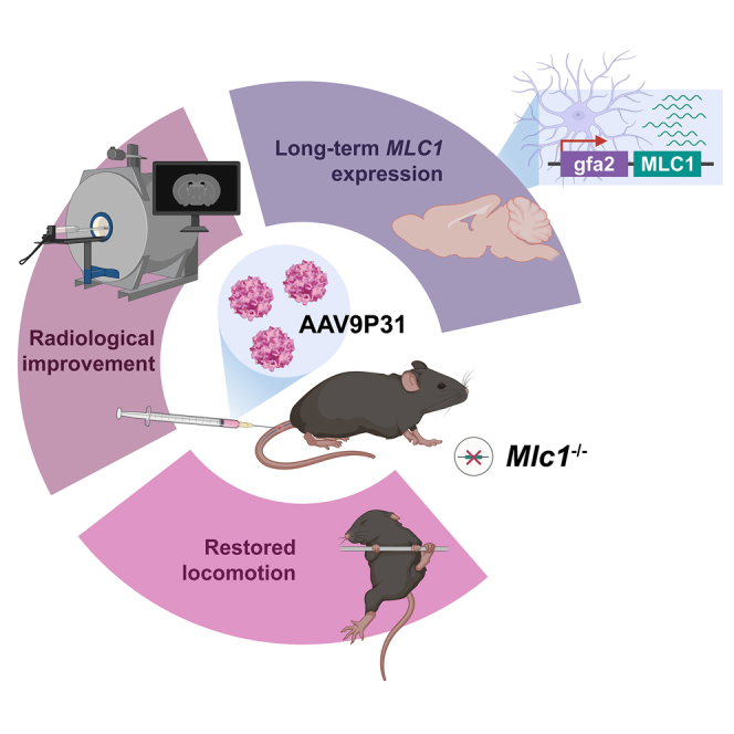
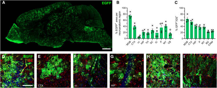
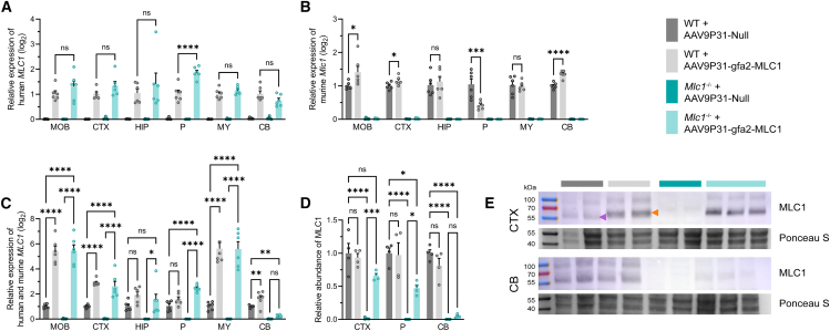
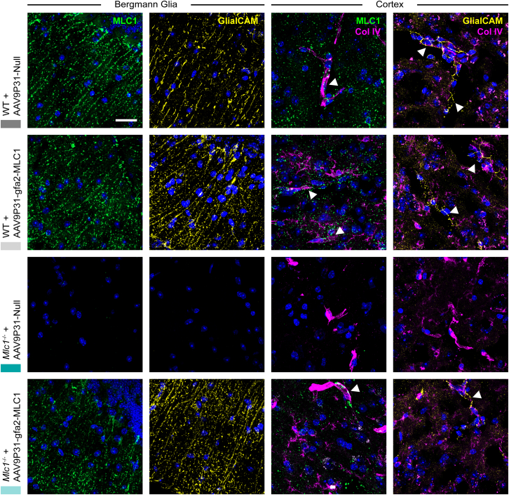
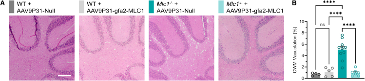
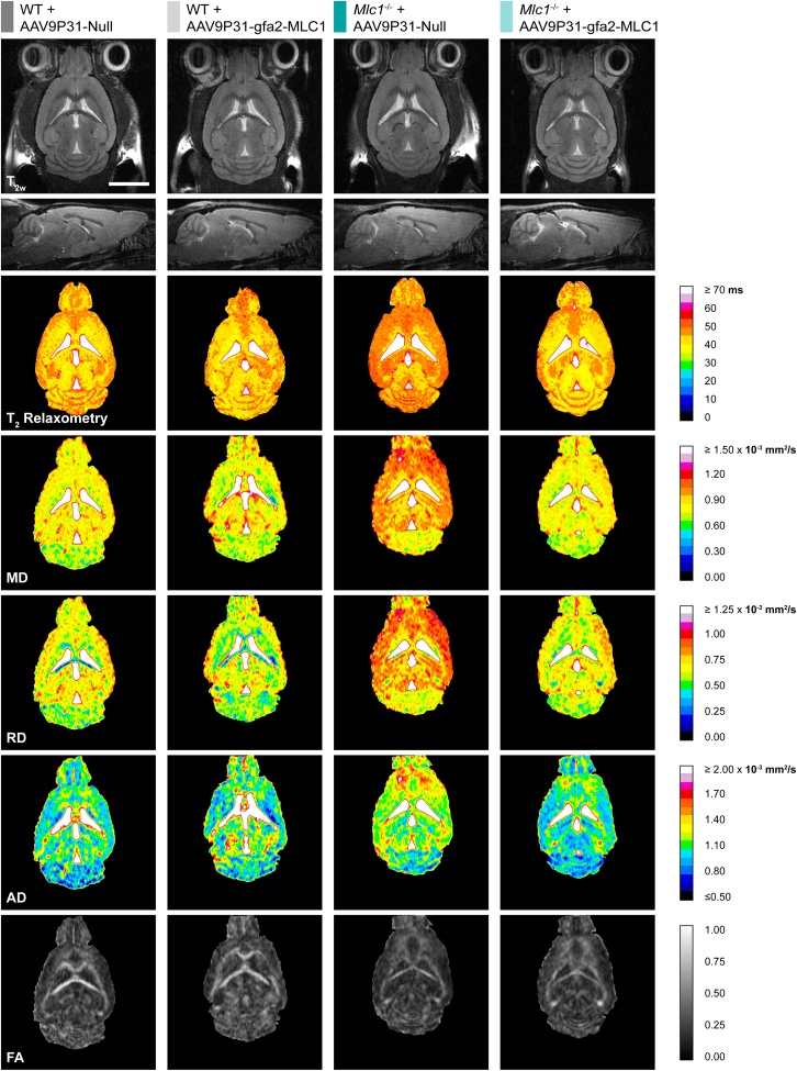
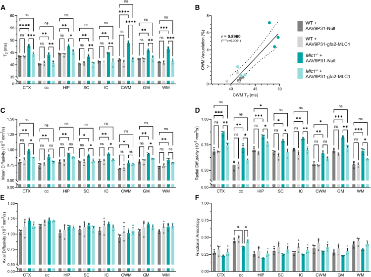
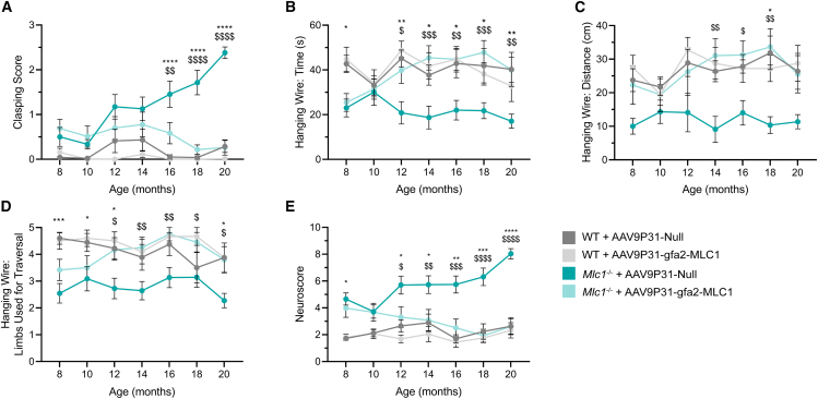
Megalencephalic leukoencephalopathy with subcortical cysts (MLC) is an ultrarare, infantile-onset leukodystrophy characterized by white matter edema for which there is no treatment. More than 75% of diagnosed cases result from biallelic loss-of-function mutations in the astrocyte-specific gene MLC1, leading to early-onset macrocephaly, cerebellar ataxia, epilepsy, and mild cognitive decline. To develop a gene therapy for MLC, we administered an adeno-associated viral vector capable of crossing the murine blood-brain barrier, delivering the human MLC1 cDNA under the control of a human astrocyte-specific promoter, to 10-month-old Mlc1-/- mice. We observed long-term astrocyte-driven expression of MLC1 up to 1 year after viral vector administration in all brain areas analyzed. Despite the late-stage intervention, in vivo magnetic resonance imaging revealed normalization of water accumulation. Notably, our therapy successfully reversed locomotor deficits in Mlc1-/- mice, as evidenced by improved performance in motor tests assessing cerebellar ataxia-like behaviors. Collectively, these findings not only demonstrate the sustained efficacy of our gene therapy but also highlight the reversibility of vacuolation and motor impairments in Mlc1-/- mice, suggesting that MLC patients could benefit from treatment even after symptom onset.

Keywords: AAV; astrocytes; cerebellum; gene therapy; leukodystrophy; megalencephalic leukoencephalopathy with subcortical cysts; myelin; neuroimaging; rare disease; white matter.


Conflict of interest statement

Declaration of interests The authors declare no competing interests.

Figures

None
Graphical abstract
Figure 1
Figure 1
Astrocyte transduction using AAV9P31 vectors (A) Immunoreactive EGFP (green) biodistribution in a sagittal mouse brain slice 4 weeks after intravenous administration of AAV9P31-gfa2-EGFP (3.2 × 1013 vg/kg). Scale bar corresponds to 1 mm. EGFP biodistribution quantified as (B) percentage of EGFP+ area relative to that of different neuroanatomic regions (MOB, main olfactory bulb; CTX, cortex; cc, corpus callosum; HIP, hippocampal formation; TH, thalamus; SC, superior colliculi; IC, inferior colliculi; P, pons; MY, medulla oblongata; CB, cerebellum; BG, Bergmann glia; CWM, cerebellar white matter) and as (C) percentage of EGFP+ GS+ cells relative to total GS+ cells (n = 4). High magnification micrographs illustrating EGFP and GS (red) colocalization in (D) the olfactory bulb, (E) cortex, (F) corpus callosum, (G) hippocampus, (H) pons, and (I) molecular layer of the cerebellum. Nuclei were counterstained with Hoechst 33342 (blue). Scale bar corresponds to 100 μm.
Figure 2
Figure 2
Analysis of MLC1 mRNA and protein levels after astrocyte transduction using AAV9P31 vectors Relative expression of (A) human MLC1, (B) murine Mlc1, and (C) total human and murine MLC1 mRNA in selected neuroanatomic regions (MOB, olfactory bulb; CTX, cortex; HIP, hippocampal formation; P, pons; MY, medulla oblongata; CB, cerebellum) determined by the Pfaffl method. Expression ratios were log2-transformed with an offset of +1 (log2 (Pfaffl ratio +1)) to normalize the data and prevent undefined values for ratios equal to zero, particularly in the Mlc1−/− + AAV9P31-Null experimental group (see statistical analysis in the materials and methods section for further details). (D) Relative abundance of MLC1 protein in cortex, pons, and cerebellum, quantified by western blot. (E) Detection of MLC1 dimers in western blot of membrane protein-enriched fractions from the cortex and cerebellum, alongside with Ponceau S staining. Note the distinct electrophoretic mobility of murine MLC1 dimers (purple arrowhead) and human MLC1 dimers (orange arrowhead), with the latter exhibiting a higher apparent molecular weight due to the HA tag at its N terminus. Due to the higher affinity of the antibody for human MLC1 compared with murine MLC1, raw band intensities overestimate human MLC1 abundance. After adjusting for antibody affinity (see protein extraction and western blot in the materials and methods section for further details), MLC1 protein levels in treated Mlc1−/− mice are below the physiological range (n = 6 per group in RT-qPCR and 4 in western blot; ∗p ≤ 0.05, ∗∗p ≤ 0.01, ∗∗p ≤ 0.001, ∗∗∗∗p ≤ 0.0001).
Figure 3
Figure 3
Immunostaining of MLC1 and GlialCAM Gene therapy provided a sustained expression of MLC1 (green) in the Bergmann glia of treated Mlc1−/− mice and rescued GlialCAM (yellow) membrane targeting. MLC1 and GlialCAM were also detected in the cortical region, most notably surrounding and close to blood vessels, as validated by collagen IV (magenta) immunostaining (indicated with arrowheads). Nuclei (blue) were counterstained with Hoechst 33342. Scale bar corresponds to 25 μm.
Figure 4
Figure 4
Histopathological analysis of white matter vacuolation (A) Cerebellum in H&E-stained brain sections. Scale bar corresponds to 250 μm. (B) Quantification of CWM vacuolation in H&E-stained sections (n = 4–10 per group, ∗∗∗∗p ≤ 0.0001).
Figure 5
Figure 5
Magnetic resonance imaging Representative sections or maps of (from top to bottom) T2-weighted images (T2w; axial and sagittal view), T2 relaxometry, mean diffusivity (MD), radial diffusivity (RD), axial diffusivity (AD), and fractional anisotropy (FA) of WT and Mlc1−/− mice administered with null or gfa2-MLC1 AAV vectors. Scale bar corresponds to 5 mm.
Figure 6
Figure 6
Quantification of T2 relaxometry and diffusion parameters (A) T2 time of selected neuroanatomic regions (CTX, cortex; cc, corpus callosum; HIP, hippocampal formation; SC, superior colliculi; IC, inferior colliculi; CWM, cerebellar white matter) and global gray (including CTX, HIP, SC, IC) and white (including cc, CWM) matter. Note the rise in T2 in CWM in Mlc1−/− mice is associated with the development of vacuoles (see Figure 4), but not in other brain areas. (B) Correlation between CWM vacuolation and in its in vivo T2 time. (C) MD, (D) RD, (E) AD, and (F) FA of selected neuroanatomic regions and global gray and white matter. While no changes in axial diffusivity were detected between WT and untreated Mlc1−/− mice, statistically significant differences in the other diffusion metrics were partially or completely corrected upon treatment (n = 3 per group, ∗p ≤ 0.05, ∗∗p ≤ 0.01, ∗∗∗p ≤ 0.001, ∗∗∗∗p ≤ 0.0001).
Figure 7
Figure 7
Motor function is rescued after gene therapy in Mlc1−/− mice (A) Score in the hindlimb clasping test. (B) Latency to fall from the wire in the hanging wire test (up to 60 s). (C) Distance traveled along the wire in the hanging wire test (up to 60 s). (D) Number of limbs (forelimbs, hindlimbs, and/or tail) used for traversal on the wire in the hanging wire test. (E) Neuroscore or phenotypic index (n = 10 in WT + AAV9P31-Null group, 10 in WT + AAV9P31-gfa2-MLC1 group, 15 in Mlc1−/− + AAV9P31-Null group, and 13 in Mlc1−/− + AAV9P31-gfa2-MLC1; p value of “WT + AAV9P31-Null vs. Mlc1−/− + AAV9P31-Null” and “Mlc1−/− + AAV9P31-Null vs. Mlc1−/− + AAV9P31-gfa2-MLC1” comparisons plotted as “∗” and “$,” respectively; ∗,$p ≤ 0.05, ∗∗,$$p ≤ 0.01, ∗∗∗,$$$p ≤ 0.001, ∗∗∗∗,$$$$p ≤ 0.0001).

Similar articles

Cited by

References

    1. van der Knaap M.S., Barth P.G., Stroink H., van Nieuwenhuizen O., Arts W.F., Hoogenraad F., Valk J. Leukoencephalopathy with swelling and a discrepantly mild clinical course in eight children. Ann. Neurol. 1995;37:324–334. doi: 10.1002/ana.410370308. - DOI - PubMed
    1. Leegwater P.A.J., Boor P.K.I., Yuan B.Q., Van Der Steen J., Visser A., Könst A.A.M., Oudejans C.B.M., Schutgens R.B.H., Pronk J.C., Van Der Knaap M.S. Identification of novel mutations in MLC1 responsible for megalencephalic leukoencephalopathy with subcortical cysts. Hum. Genet. 2002;110:279–283. doi: 10.1007/s00439-002-0682-x. - DOI - PubMed
    1. Ben-Zeev B., Levy-Nissenbaum E., Lahat H., Anikster Y., Shinar Y., Brand N., Gross-Tzur V., MacGregor D., Sidi R., Kleta R., et al. Megalencephalic leukoencephalopathy with subcortical cysts; a founder effect in Israeli patients and a higher than expected carrier rate among Libyan Jews. Hum. Genet. 2002;111:214–218. doi: 10.1007/s00439-002-0770-y. - DOI - PubMed
    1. Yüzbaşioğlu A., Topçu M., Kocaefe Y.Ç., Ozgüç M. Novel mutations of the MLC1 gene in Turkish patients. Eur. J. Med. Genet. 2011;54:281–283. doi: 10.1016/j.ejmg.2010.11.014. - DOI - PubMed
    1. Abdel-Salam G.M.H., Abdel-Hamid M.S., Ismail S.I., Hosny H., Omar T., Effat L., Aglan M.S., Temtamy S.A., Zaki M.S. Megalencephalic leukoencephalopathy with cysts in twelve Egyptian patients: novel mutations in MLC1 and HEPACAM and a founder effect. Metab. Brain Dis. 2016;31:1171–1179. doi: 10.1007/s11011-016-9861-7. - DOI - PubMed

MeSH terms

Supplementary concepts

LinkOut - more resources