

Chylothorax After Thoracic Surgery: How We Manage It
- PMID: 40051248
- PMCID: PMC11885794
- DOI: 10.1111/1759-7714.70036
Chylothorax After Thoracic Surgery: How We Manage It
Abstract
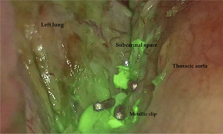
Chylothorax is a rare but insidious condition, characterized by the accumulation of chyle in the pleural space, which is particularly common after cardiothoracic surgeries. It presents significant challenges in both diagnosis and treatment. In this technical report, we present our experience in managing four cases of postsurgical chylothorax, each one treated with a different approach. The first and second cases were successfully managed with Lipiodol lymphangiography, which allowed for the visualization and occlusion of the injured lymphatic duct, leading to the resolution of the chylothorax. The third case involved thoracic duct embolization, a procedure that resulted in the closure of the duct responsible for the chylous effusion. The last case involved a patient who developed left-sided chylothorax following a pulmonary resection. The patient experienced chylous leakage early in the postoperative period and underwent a revision thoracoscopy for hemostasis and thoracic duct ligation. During the procedure, indocyanine green (ICG) fluorescence was used to effectively identify and ligate the injured chylous duct. This case series highlights the variety of therapeutic strategies available for the management of chylothorax, emphasizing the importance of a structured, stepwise approach tailored to the specific needs of each patient.
Keywords: chylothorax; indocyanine green fluorescence; lymphangiography; lymphatic embolization; thoracic surgery.
© 2025 The Author(s). Thoracic Cancer published by John Wiley & Sons Australia, Ltd.
Conflict of interest statement
The authors declare no conflicts of interest.
Figures


References
-
- McGrath E. E., Blades Z., and Anderson P. B., “Chylothorax: Aetiology, Diagnosis and Therapeutic Options,” Respiratory Medicine 104 (2010): 1–8. - PubMed
-
- Agrawal A., Chaddha U., Kaul V., Desai A., Gillaspie E., and Maldonado F., “Multidisciplinary Management of Chylothorax,” Chest 162 (2022): 1402–1412. - PubMed
-
- Martucci N., Tracey M., and Rocco G., “Postoperative Chylothorax,” Thoracic Surgery Clinics 25 (2015): 523–528. - PubMed
-
- Nair S. K., Petko M., and Hayward M. P., “Aetiology and Management of Chylothorax in Adults,” European Journal of Cardio‐Thoracic Surgery 32 (2007): 362–369. - PubMed
Publication types
MeSH terms
LinkOut - more resources
Full Text Sources
Medical
