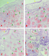
Angiomatous Nasal Polyps in a 43-Year-Old Female From North-Central Nigeria: A Case Report
- PMID: 40051951
- PMCID: PMC11884828
- DOI: 10.7759/cureus.78506
Angiomatous Nasal Polyps in a 43-Year-Old Female From North-Central Nigeria: A Case Report
Abstract
Angiomatous nasal polyps (ANPs) are uncommon variants of nasal polyps, which, like their more common counterparts, are a significant and common cause of morbidity for patients, regardless of demographic variations. As a group, nasal polyps are non-neoplastic and commonly arise within the maxillary antrum, sphenoid, and ethmoid sinuses, protruding via the ostia into the nasal cavity. Their precise cause is unknown, but several factors related to genetics, local infections, family history, and environment have been implicated in their etiopathogenesis. ANPs are commonly diagnosed in the younger age group and are particularly significant because they may mimic benign and malignant vascular tumors occurring within the nasopharynx. We report a rare case of angiomatous nasal polyp in a 43-year-old Nigerian female. ANPs should be included in the differential diagnosis of mass lesions of the nasal cavity in adult patients (male or female) who present with obstructive symptoms and should be distinguished from both benign and malignant neoplastic mimics.
Keywords: angiomatous nasal polyp; chronic rhinosinusitis; nasal congestion; nasal polyps; rhinorrhea.
Copyright © 2025, Ezike et al.
Conflict of interest statement
Human subjects: Consent for treatment and open access publication was obtained or waived by all participants in this study. Conflicts of interest: In compliance with the ICMJE uniform disclosure form, all authors declare the following: Payment/services info: All authors have declared that no financial support was received from any organization for the submitted work. Financial relationships: All authors have declared that they have no financial relationships at present or within the previous three years with any organizations that might have an interest in the submitted work. Other relationships: All authors have declared that there are no other relationships or activities that could appear to have influenced the submitted work.
Figures


References
-
- Histopathology. Hellquist HB. Allergy Asthma Proc. 1996;17:237–242. - PubMed
-
- Nasal polyps: still more questions than answers. Bateman ND, Fahy C, Woolford TJ. J Laryngol Otol. 2003;117:1–9. - PubMed
-
- Pathogenesis of nasal polyps: an update. Pawliczak R, Lewandowska-Polak A, Kowalski ML. Curr Allergy Asthma Rep. 2005;5:463–471. - PubMed
-
- Update on the molecular biology of nasal polyposis. Bernstein JM. Otolaryngol Clin North Am. 2005;38:1243–1255. - PubMed
Publication types
LinkOut - more resources
Full Text Sources
Research Materials
