

Extracellular Vesicles From Follicular Fluid in Infertile Women: Size, Morphology and miRNA Content Analysis
- PMID: 40052100
- PMCID: PMC11881956
- DOI: 10.1002/jex2.70041
Extracellular Vesicles From Follicular Fluid in Infertile Women: Size, Morphology and miRNA Content Analysis
Abstract
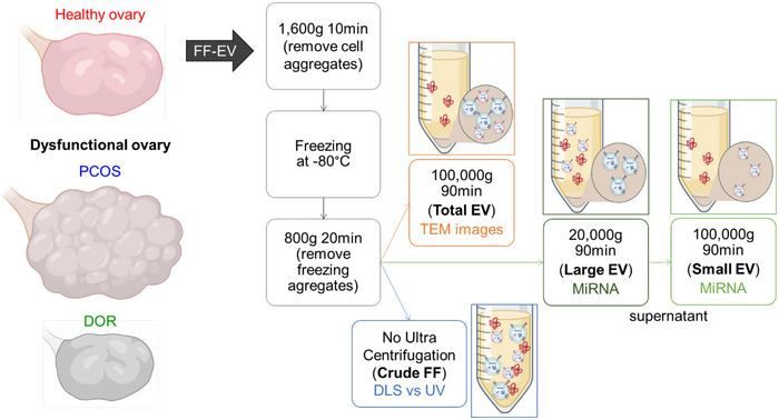
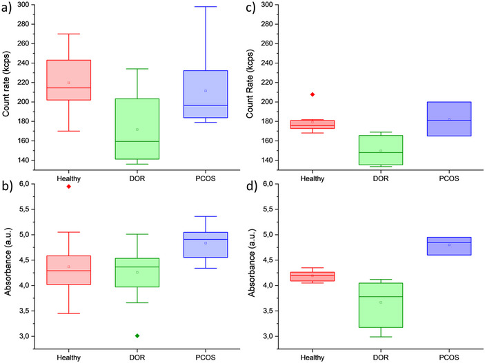
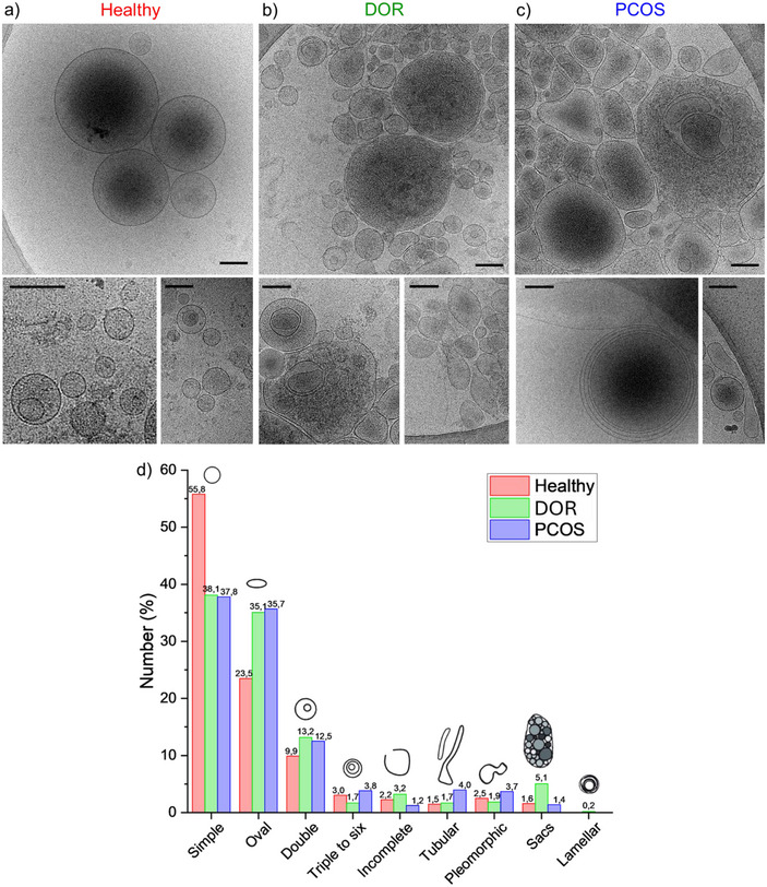
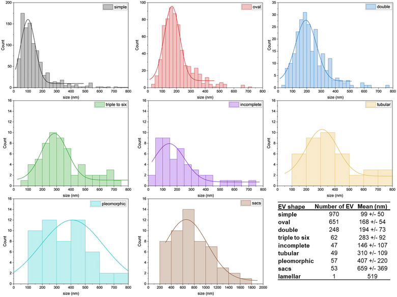
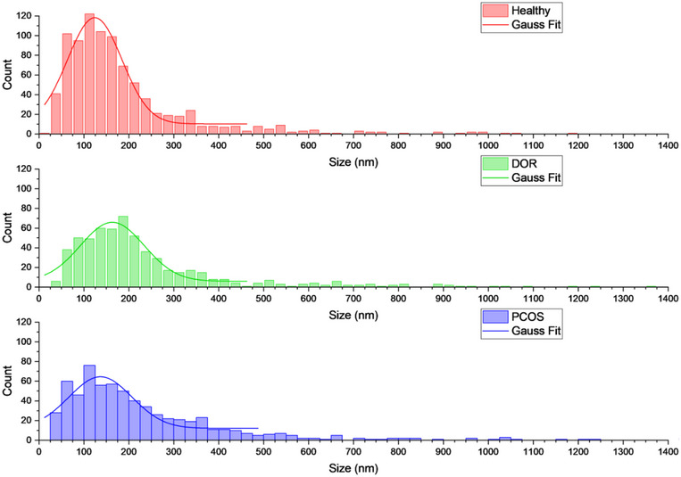
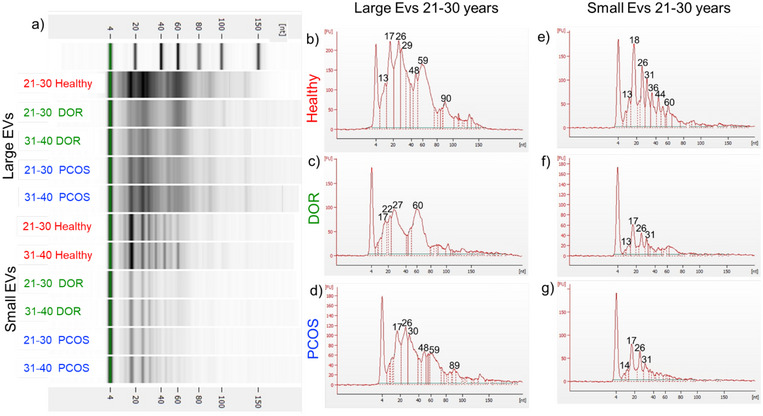
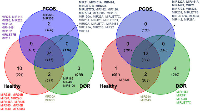
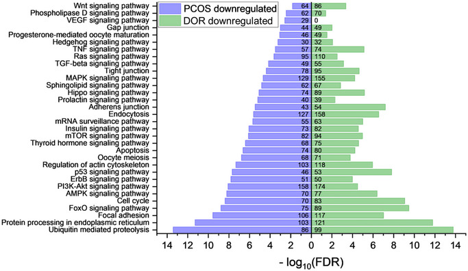
The declining birth rates and fertility challenges in Europe have intensified global concerns over rising infertility, particularly among women. This study decisively investigates follicular fluid-related extracellular vesicles (FF-EVs) from infertile patients with polycystic ovary syndrome (PCOS) or diminished ovarian reserve (DOR) undergoing in vitro fertilization (IVF), comparing them to a healthy control group. We have identified significant variations in protein content and polydispersity in crude follicular fluid using UV-Vis absorption and dynamic light scattering (DLS) techniques. Furthermore, the morphology of the extracellular vesicles (EVs) and the patterns of non-coding RNA content, including miRNAs, reveal distinct differences in infertile patients. These findings offer critical insights into the molecular signatures associated with these conditions. This study plays a vital role in advancing reproductive healthcare by pinpointing potential targets that can enhance diagnosis and deepen our understanding of ovarian disorders.
Keywords: Cryo‐TEM; diminished ovarian reserve (DOR); extracellular vesicles (EVs); follicular fluid (FF); miRNA; morphology; polycystic ovarian syndrome (PCOS).
© 2025 The Author(s). Journal of Extracellular Biology published by Wiley Periodicals, LLC on behalf of the International Society for Extracellular Vesicles.
Conflict of interest statement
The authors declare no conflicts of interest.
Figures
Similar articles
-
Prolidase activity in women with polycystic ovarian syndrome undergoing assisted conception.J Obstet Gynaecol. 2024 Dec;44(1):2346228. doi: 10.1080/01443615.2024.2346228. Epub 2024 Apr 26. J Obstet Gynaecol. 2024. PMID: 38973654
-
The follicular fluid metabolome in infertile individuals between polycystic ovary syndrome and diminished ovarian reserve.Arch Biochem Biophys. 2022 Dec 15;732:109453. doi: 10.1016/j.abb.2022.109453. Epub 2022 Nov 5. Arch Biochem Biophys. 2022. PMID: 36347279
-
UHPLC-MS-MS analysis of oxylipins metabolomics components of follicular fluid in infertile individuals with diminished ovarian reserve.Reprod Biol Endocrinol. 2021 Sep 14;19(1):143. doi: 10.1186/s12958-021-00825-x. Reprod Biol Endocrinol. 2021. PMID: 34521427 Free PMC article.
-
Extracellular vesicles and their content in the context of polycystic ovarian syndrome and endometriosis: a review.J Ovarian Res. 2024 Aug 5;17(1):160. doi: 10.1186/s13048-024-01480-7. J Ovarian Res. 2024. PMID: 39103867 Free PMC article. Review.
-
Unraveling the complexity of follicular fluid: insights into its composition, function, and clinical implications.J Ovarian Res. 2024 Nov 26;17(1):237. doi: 10.1186/s13048-024-01551-9. J Ovarian Res. 2024. PMID: 39593094 Free PMC article. Review.
Cited by
-
Exosomal Communication Between Cumulus-Oocyte Complexes and Granulosa Cells: A New Molecular Axis for Oocyte Competence in Human-Assisted Reproduction.Int J Mol Sci. 2025 Jun 3;26(11):5363. doi: 10.3390/ijms26115363. Int J Mol Sci. 2025. PMID: 40508172 Free PMC article. Review.
References
LinkOut - more resources
Full Text Sources