

Interstitial Capillary Congestion and Diffuse Alveolar Damage: Incidental or Factual Findings in the Lungs of Autopsy Cases Positive for Coronavirus Disease-19
- PMID: 40053438
- PMCID: PMC12103138
- DOI: 10.4103/aam.aam_263_24
Interstitial Capillary Congestion and Diffuse Alveolar Damage: Incidental or Factual Findings in the Lungs of Autopsy Cases Positive for Coronavirus Disease-19
Abstract
Background: Interstitial capillary congestion and diffuse alveolar damage (DAD) were frequently observed in patients who died from Coronavirus disease-19 (COVID-19). The research question pertains to observing these findings in COVID-19-positive patients lacking pulmonary symptoms. The histological examination of lung samples from COVID-19-positive patients who do not succumb to COVID-19-related pulmonary complications can provide an answer. This study analyzed postmortem lung autopsy samples from individuals who did not succumb to COVID-19-related pulmonary complications. The research article aimed to examine the morphological variations in postmortem lung samples of COVID-19 patients who did not succumb to the disease, and to compare these changes with those observed in cases of COVID-19-related deaths, utilizing existing English literature.
Methodology: This prospective study included subjects who died without complications from COVID-19-related injuries, had positive real-time polymerase chain reaction throat swabs, and exhibited no pulmonary manifestation of COVID-19 disease. A comprehensive histomorphological analysis of the lung samples was conducted.
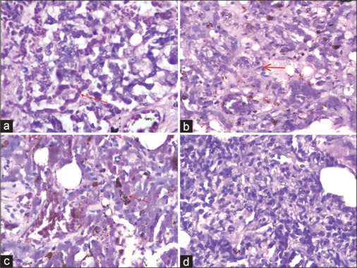
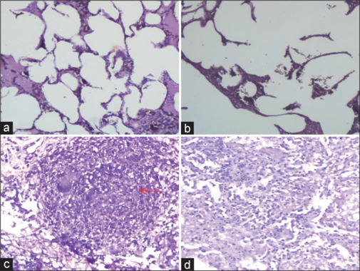
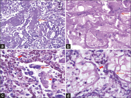
Results: A total of 20 subjects were enrolled in this study. Capillary congestion was the most prevalent histomorphological change observed in lung autopsies, seen in 90% (18/20) of cases, followed by emphysema in 75% (15/20) and the acute phase of DAD in 25% (5/20) of cases. Acute bronchopneumonia and fibrotic nodules were identified in 20% (4/20) and 10% (2/20) of the study population, respectively.
Conclusion: In postmortem lung autopsy of COVID-19-positive patients lacking symptomatic pulmonary issues, capillary congestion, diffuse alveolar destruction, and emphysema were prevalent. The findings indicate that COVID-19 exhibits varied responses to damage and inflammation that do not correlate with mortality. This study enhances the understanding of pathophysiological lung tissue variations in COVID-19 patients who have non-COVID-19-related deaths, potentially educating forensic pathologists and supporting future research endeavors.
Résumé Contexte:Une congestion capillaire interstitielle et des lésions alvéolaires diffuses ont été fréquemment observées chez les patients décédés du COVID-19. La question de recherche concerne l’observation de ces résultats chez des patients positifs au COVID-19 dépourvus de symptômes pulmonaires. L’examen histologique d’échantillons pulmonaires de patients positifs au COVID-19 qui ne succombent pas aux complications pulmonaires liées au COVID-19 peut apporter une réponse. Cette étude a analysé des échantillons d’autopsie pulmonaire post-mortem provenant d’individus qui n’ont pas succombé aux complications pulmonaires liées au COVID-19. L’article de recherche visait à examiner les variations morphologiques dans des échantillons pulmonaires post-mortem de patients atteints du COVID-19 qui n’ont pas succombé à la maladie et à comparer ces changements avec ceux observés dans les cas de décès liés au COVID-19, en utilisant la littérature anglaise existante.Méthodologie:Cette étude prospective a inclus des sujets décédés sans complications des suites de blessures liées au COVID-19, ayant eu des prélèvements de gorge RT-PCR positifs et n’ayant présenté aucune manifestation pulmonaire de la maladie COVID-19. Une analyse histomorphologique complète des échantillons de poumons a été réalisée.Résultats:Au total, 20 sujets ont été inscrits dans cette étude. La congestion capillaire était le changement histomorphologique le plus répandu observé dans les autopsies pulmonaires, observé dans 90% (18/20) des cas, suivi de l’emphysème dans 75% (15/20) et de la phase aiguë de lésions alvéolaires diffuses dans 25% (5/20) de cas. Une bronchopneumonie aiguë et des nodules fibrotiques ont été identifiés respectivement chez 20% (4/20) et 10% (2/20) de la population étudiée.Conclusion:Lors de l’autopsie pulmonaire post-mortem de patients positifs au COVID-19 dépourvus de problèmes pulmonaires symptomatiques, la congestion capillaire, la destruction alvéolaire diffuse et l’emphysème étaient répandus. Les résultats indiquent que le COVID-19 présente des réponses variées aux dommages et à l’inflammation qui ne sont pas corrélées à la mortalité. Cette étude améliore la compréhension des variations physiopathologiques du tissu pulmonaire chez les patients atteints de la COVID-19 dont les décès ne sont pas liés à la COVID-19, ce qui pourrait potentiellement former les médecins légistes et soutenir les futurs efforts de recherche.
Contexte:: Une congestion capillaire interstitielle et des lésions alvéolaires diffuses ont été fréquemment observées chez les patients décédés du COVID-19. La question de recherche concerne l’observation de ces résultats chez des patients positifs au COVID-19 dépourvus de symptômes pulmonaires. L’examen histologique d’échantillons pulmonaires de patients positifs au COVID-19 qui ne succombent pas aux complications pulmonaires liées au COVID-19 peut apporter une réponse. Cette étude a analysé des échantillons d’autopsie pulmonaire post-mortem provenant d’individus qui n’ont pas succombé aux complications pulmonaires liées au COVID-19. L’article de recherche visait à examiner les variations morphologiques dans des échantillons pulmonaires post-mortem de patients atteints du COVID-19 qui n’ont pas succombé à la maladie et à comparer ces changements avec ceux observés dans les cas de décès liés au COVID-19, en utilisant la littérature anglaise existante.
Méthodologie:: Cette étude prospective a inclus des sujets décédés sans complications des suites de blessures liées au COVID-19, ayant eu des prélèvements de gorge RT-PCR positifs et n’ayant présenté aucune manifestation pulmonaire de la maladie COVID-19. Une analyse histomorphologique complète des échantillons de poumons a été réalisée.
Résultats:: Au total, 20 sujets ont été inscrits dans cette étude. La congestion capillaire était le changement histomorphologique le plus répandu observé dans les autopsies pulmonaires, observé dans 90% (18/20) des cas, suivi de l’emphysème dans 75% (15/20) et de la phase aiguë de lésions alvéolaires diffuses dans 25% (5/20) de cas. Une bronchopneumonie aiguë et des nodules fibrotiques ont été identifiés respectivement chez 20% (4/20) et 10% (2/20) de la population étudiée.
Conclusion:: Lors de l’autopsie pulmonaire post-mortem de patients positifs au COVID-19 dépourvus de problèmes pulmonaires symptomatiques, la congestion capillaire, la destruction alvéolaire diffuse et l’emphysème étaient répandus. Les résultats indiquent que le COVID-19 présente des réponses variées aux dommages et à l’inflammation qui ne sont pas corrélées à la mortalité. Cette étude améliore la compréhension des variations physiopathologiques du tissu pulmonaire chez les patients atteints de la COVID-19 dont les décès ne sont pas liés à la COVID-19, ce qui pourrait potentiellement former les médecins légistes et soutenir les futurs efforts de recherche.
Keywords: Autopsie COVID-19; COVID-19; Coronavirus disease-19; congestion capillaire interstitielle; coronavirus disease-19 autopsy; diffuse alveolar damage; emphysema; emphysème; interstitial capillary congestion; lung morphology; lésions alvéolaires diffuses; morphologie pulmonaire.
Copyright © 2025 Annals of African Medicine.
Conflict of interest statement
There are no conflicts of interest.
Figures
References
-
- Malik YA. Properties of coronavirus and SARS-CoV-2. Malays J Pathol. 2020;42:3–11. - PubMed
MeSH terms
LinkOut - more resources
Full Text Sources
Medical
