. 2025 Mar 7;11(10):eadq1047.
doi: 10.1126/sciadv.adq1047. Epub 2025 Mar 7.

TBK1 and IKKε prevent premature cell death by limiting the activity of both RIPK1 and NLRP3 death pathways

Affiliations

TBK1 and IKKε prevent premature cell death by limiting the activity of both RIPK1 and NLRP3 death pathways

Fabian A Fischer et al. Sci Adv. .

Abstract

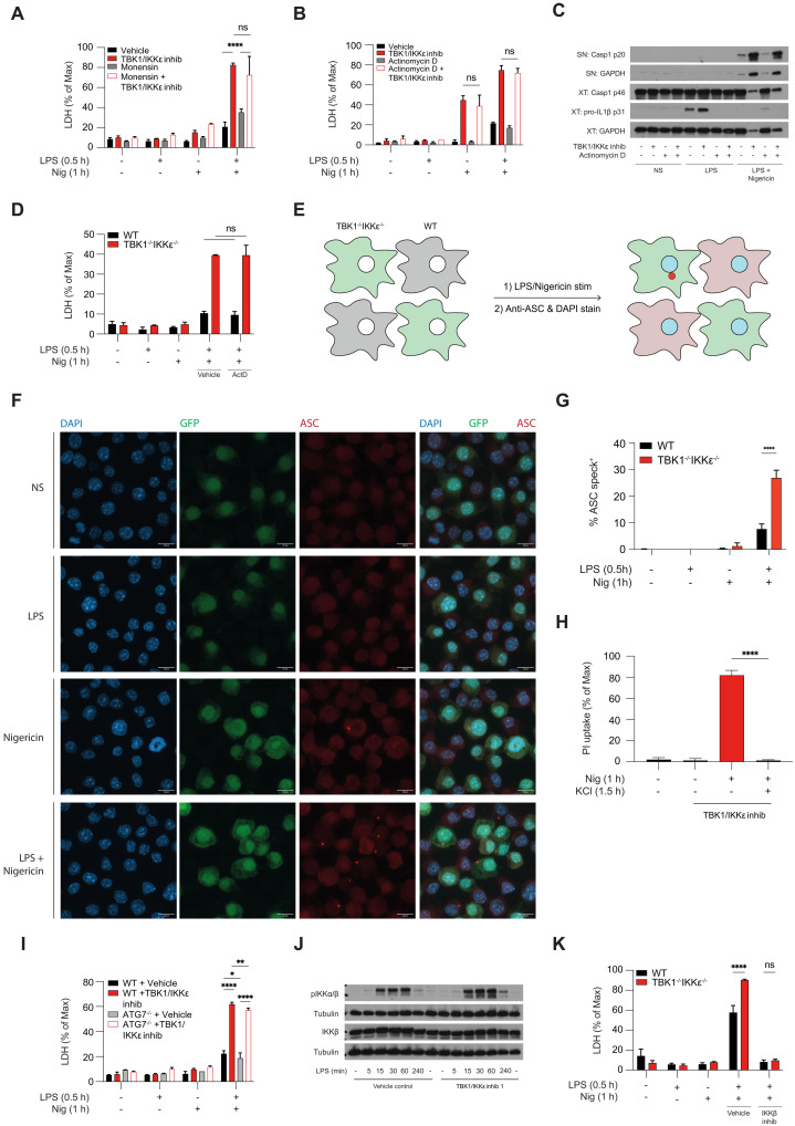
The loss of TBK1, or both TBK1 and the related kinase IKKε, results in uncontrolled cell death-driven inflammation. Here, we show that the pathway leading to cell death depends on the nature of the activating signal. Previous models suggest that in steady state, TBK1/IKKε-deficient cells die slowly and spontaneously predominantly by uncontrolled tumor necrosis factor-RIPK1-driven death. However, upon infection of cells that express the NLRP3 inflammasome, (e.g., macrophages), with pathogens that activate this pathway (e.g., Listeria monocytogenes), TBK1/IKKε-deficient cells die rapidly, prematurely, and exclusively by enhanced NLRP3-driven pyroptosis. Even infection with the RIPK1-activating pathogen, Yersinia pseudotuberculosis, results in enhanced RIPK1-caspase-8 activation and enhanced secondary NLRP3 activation. Mechanistically, TBK1/IKKε control endosomal traffic, and their loss disrupts endosomal homeostasis, thereby signaling cell stress. This results in premature NLRP3 activation even upon sensing "signal 2" alone, without the obligatory "signal 1." Collectively, TBK1/IKKε emerge as a central brake in limiting death-induced inflammation by both RIPK1 and NLRP3 death-inducing pathways.


Figures

Fig. 1.
Fig. 1.. TBK1/IKKε limit NLRP3-driven cell death independently of TNF-RIPK1 signaling.
(A to C) WT, TBK1 KO (Tbk1−/−), IKKε KO (Ikkε−/−), or dual TBK1 (Tbk1−/−Ikkε−/−) iBMDMs were primed with LPS (1 μg/ml) for 30 min followed by stimulation with 10 to 15 μM nigericin for 1 hour (h). (D to I) WT, TNFR1 KO (Tnfr1−/−), or RIPK1 kinase–dead (Ripk1D138N) BMDMs were primed with LPS (100 ng/ml) for 4 hours followed by stimulation with 7.5 μM nigericin. The cells were treated 30 min before nigericin with 3 μM (target-specific concentration) of the TBK1/IKKε inhibitor MRT68601. (J and K) WT, RIPK1D138N, or TNFR1 KO (Tnfr1−/−) BMDMs were incubated for 24 hours with 3 μM MRT68601 in the presence of 10 μM MCC950 (NLRP3 inhib) or 50 μM Nec-1 (RIPK1 inhib). [(A), (D), (G), (J), and (K)] Cell viability was measured using LDH release. [(B), (E), and (H)] Cytokine secretion was measured using enzyme-linked immunosorbent assay. [(C), (F), and (I)] Caspase-1 cleavage in supernatants (SNs) and cell extracts (XTs) was measured using immunoblot. Data are shown as means + SD of duplicates [(A) and (B)] or triplicates [(D), (E), (G), (H), (J), and (K)] from one representative experiment of three independent experiments and one representative immunoblot of three (C) or two shown [(F) and (I)]. ns, not significant.
Fig. 2.
Fig. 2.. TBK1/IKKε limit the activity of both RIPK1 and NLRP3 death pathways in macrophages.
BMDMs were primed with LPS (100 ng/ml) or TNF (100 ng/ml) for 30 min, followed by stimulation with 7.5 μM nigericin for 6 hours. Cells were treated with 3 μM MRT68601, 10 μM MCC950, and 50 μM Nec-1 at the time of priming. After nigericin stimulation, SNs were harvested and cells were lysed for immunoblot analysis. (A to L) Cell viability was measured every 60 min by measuring PI uptake signals. Data are shown as means + SD of duplicates from one representative of three independent experiments.
Fig. 3.
Fig. 3.. Cell death in response to infections is augmented when TBK and IKKε control is lost.
(A) Experimental scheme: Overnight cultures of L.m. were spun, reconstituted in PBS, and used to acutely infect WT or Tbk1−/−Ikkε−/− iBMDMs with titrations of L.m. for 1 hour, with or without 30-min preincubation with 10 μM MCC950 in (D and E). [B and (D)] Cell viability was measured using LDH release. [C and (E)] Caspase-1 cleavage was measured in iBMDM SNs and cell XTs using immunoblotting. (F to I) Primary BMDMs were infected with Y. pseudotuberculosis for 90 min or for 2 hours for the P2X7 inhibitor experiments, and mixed SN and cell XTs were analyzed by immunoblotting. Cell death was measured by LDH release assay. Where indicated, BMDMs were treated with 50 μM Nec-1s or 10 μM MRT67307 or 10 μM P2X7 inhibitor for 30 min before infection. Data are shown as means + SD of infection triplicates (B) or duplicates (D) from one representative experiment of three (B) or two (E) independent experiments and one representative immunoblot in (C) and (E), three experiments in (F) and (G), two of which with MOI 7.5, 5, and 2.5, and one with MOI 5 and 2.5 and two experiments in (H) and (I) with MOI 7.5.
Fig. 4.
Fig. 4.. Deletion of TBK1/IKKε allows for priming-independent NLRP3 activation.
(A to C) WT or Tbk1−/−Ikkε−/− iBMDMs were primed with LPS (1 μg/ml) for 30 min followed by stimulation with 10 to 15 μM nigericin for 3.5 hours in the presence of 10 μM of the NLRP3 inhibitor MCC950 in (C). [(A) to (C)] Cell viability was measured using LDH release. (B) Caspase-1 cleavage was measured in SN and lysates using immunoblotting. Data are shown as means + SD of triplicates (A) or duplicates (C) from one representative experiment of three independent experiments and one representative immunoblot in (B).
Fig. 5.
Fig. 5.. TBK1/IKKε regulate NLRP3 activation via a cell-intrinsic mechanism.
(A, B, C, H, and I) BMDMs were primed with LPS (100 ng/ml) for 30 min followed by stimulation with 7.5 μM nigericin for 1 hour. (J) BMDMs were primed with LPS (100 ng/ml) in a time course from 5 min to 4 hours. [(D) and K] WT or Tbk1−/−Ikkε−/− iBMDMs were primed with LPS (1 μg/ml) for 30 min followed by stimulation with 10 to 15 μM nigericin for 1 hour. The cells were pretreated with 3 μM MRT68601 (TBK1/IKKε inhib) for 30 min before LPS treatment, actinomycin D (5 μg/ml) or 2 μM monensin for 45 min before LPS treatment, 5 μM BI605906 (IKKβ inhib) for 1 hour before LPS treatment. Cell viability was measured using LDH release [(A), (B), (D), (I), and (K)] or PI uptake (H). [(C) and (J)] Caspase-1 cleavage was measured in SNs and lysates using immunoblotting. [(E) to (G)] WT or Tbk1−/−Ikkε−/− iBMDMs were cocultured in the same dish for 24 hours before being primed with LPS (1 μg/ml) for 30 min followed by stimulation with 15 μM nigericin for 1 hour in the presence of 10 μM of the caspase-1 inhibitor VX-765 to prevent lytic cell death. ASC specks were visualized by confocal imaging using an anti-ASC antibody and 4′,6-diamidino-2-phenylindole (DAPI) stain or WT (GFP) and Tbk1−/−Ikkε−/− (GFP+) iBMDMs. Data are shown as collapsed Z-stacks of representative images and quantification showing % cells forming ASC specks from multiple fields of view from three independent experiments is on the right. Data are shown as means + SD of triplicates [(A), (B), (H), and (I)] or duplicates [(D) and (K)] from one representative experiment of two [(A), (I), and (K)] or three [(B), (D), and (H)] independent experiments, as means + SEM from three independent experiments (G) and one representative immunoblot in (C) and (J).
Fig. 6.
Fig. 6.. Loss of TBK1/IKKε disrupts the endosomal homeostasis and lowers the threshold for NLRP3 activation.
(A) HEK293T cells expressing NLRP3-FLAG were generated using lentiviral transduction. (B and C) HEK293T-NLRP3-FLAG cells pretreated with 3 μM MRT68601 (TBK1/IKKε inhib) were stimulated with 10 μM nigericin for 1 hour. The cells were then fixed and immuno-labeled for TGN46 and FLAG. (D) HMDMs were pretreated with 3 μM MRT68601 and 10 μM of the caspase-1 inhibitor VX-765 to prevent lytic cell death followed by stimulation with 7.5 μM nigericin for 30 min. The cells were subsequently fixed and immuno-labeled for TGN46. Data are shown as collapsed Z-stacks of representative images from multiple fields of view from two [(B) and (C)] or three independent experiments for (D). (E) Quantification of images from three experiments showing the fraction of cells forming compact, versus elongated versus fully dispersed TGN46 endosomal network (representative shapes shown on the right).
Fig. 7.
Fig. 7.. Loss of endosomal homeostasis by Rab5 deletion also lowers the threshold for NLRP3 activation.
(A) HMDMs were primed with LPS (100 ng/ml) in a time course and pretreated with 3 μM MRT68601 (TBK1/IKKε inhib) for 30 min before LPS treatment. Rab7 activation was assessed by immunoblot for phospho-serine72 levels compared to total Rab7. (B to E) BMDMs were subjected to siRNA-mediated knockdown of Rab7 [(B) and (D)] or Rab5 [(C) and (E)] for 2 days before replating and stimulation on the third day. BMDMs were primed with LPS (100 ng/ml) and treated with 3 μM MRT68601 for 30 min followed by stimulation with 7.5 μM nigericin for 2 hours. [(B) and (C)] Knockdown efficiency was confirmed using immunoblotting. [(D) and (E)] Cell viability was measured using PI uptake every 30 min over 2 hours. Data are shown as means + SD of duplicates from one representative experiment of three [(D) and (E)] independent experiments and one representative immunoblot for (A) to (C).

References

    1. Balka K. R., Louis C., Saunders T. L., Smith A. M., Calleja D. J., D’Silva D. B., Moghaddas F., Tailler M., Lawlor K. E., Zhan Y., Burns C. J., Wicks I. P., Miner J. J., Kile B. T., Masters S. L., De Nardo D., TBK1 and IKKε act redundantly to mediate STING-induced NF-κB responses in myeloid cells. Cell Rep. 31, 107492 (2020). - PubMed
    1. Tan Y., Kagan J. C., Innate immune signaling organelles display natural and programmable signaling flexibility. Cell 177, 384–398.e11 (2019). - PMC - PubMed
    1. Xu D., Jin T., Zhu H., Chen H., Ofengeim D., Zou C., Mifflin L., Pan L., Amin P., Li W., Shan B., Naito M. G., Meng H., Li Y., Pan H., Aron L., Adiconis X., Levin J. Z., Yankner B. A., Yuan J., TBK1 suppresses RIPK1-driven apoptosis and inflammation during development and in aging. Cell 174, 1477–1491.e19 (2018). - PMC - PubMed
    1. Lafont E., Draber P., Rieser E., Reichert M., Kupka S., de Miguel D., Draberova H., von Massenhausen A., Bhamra A., Henderson S., Wojdyla K., Chalk A., Surinova S., Linkermann A., Walczak H., TBK1 and IKKε prevent TNF-induced cell death by RIPK1 phosphorylation. Nat. Cell Biol. 20, 1389–1399 (2018). - PMC - PubMed
    1. Eren R. O., Kaya G. G., Schwarzer R., Pasparakis M., IKKε and TBK1 prevent RIPK1 dependent and independent inflammation. Nat. Commun. 15, 130 (2024). - PMC - PubMed

MeSH terms

LinkOut - more resources