. 2025 Mar 7;11(10):eado1648.
doi: 10.1126/sciadv.ado1648. Epub 2025 Mar 7.

Hyperammonemia induces programmed liver cell death

Affiliations

Hyperammonemia induces programmed liver cell death

Annarein J C Kerbert et al. Sci Adv. .

Abstract

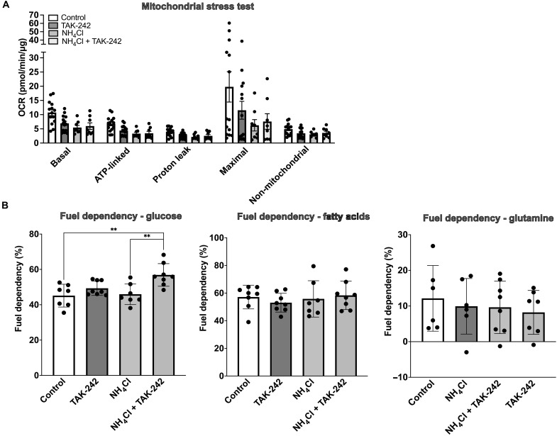
Hyperammonemia is common in liver cirrhosis and causally associated with hepatic encephalopathy development. Little is known about its hepatotoxic effects, which we aimed to characterize in this study. In a mouse model of chronic hyperammonemia without preexisting liver disease, we observed development of liver fibrogenesis and necroptotic cell death. Hyperammonemia also induced dysregulation of its main metabolic pathway, the urea cycle, as reflected by down-regulation of urea cycle enzyme protein expression and accumulation of its metabolites. Inhibition of receptor-interacting serine/threonine-protein kinase 1 (RIPK1) and its upstream inducer Toll-like receptor 4 (TLR4) protected against liver injury and further hyperammonemia. In clinically relevant rodent models of hyperammonemia (genetic ornithine transcarbamylase deficiency and bile duct ligation-induced cirrhosis), TLR4 inhibition reduced circulating ammonia. In conclusion, hyperammonemia induces liver fibrogenesis and RIPK1-mediated cell death, which is associated with urea cycle dysfunction. Inhibition of RIPK1 and TLR4 protects against hyperammonemia-induced liver injury and are potential therapeutic targets for hyperammonemia and chronic liver disease progression.


Figures

Fig. 1.
Fig. 1.. Hyperammonemia induces liver injury, which is prevented by TLR4 and RIPK1 inhibition.
(A) Experimental design. (B) Bar graphs representing circulating ammonia, BUN, and ALT levels. OP, TAK-242, and RIPA-56 treatment significantly reduced circulating ammonia levels. The TLR4KO genotype protected against severe hyperammonemia development. Hyperammonemia did not lead to a significant increase in plasma BUN in WT-AA, as opposed to OP- and TAK-242–treated mice. Hyperammonemia was associated with a significant increase in plasma ALT, which was prevented by OP, TAK-242, and RIPA-56 treatment and by the TLR4KO genotype. (C) Bar graphs showing quantification of Sirius Red and TUNEL staining. (D) Microscopy imaging of Sirius Red and TUNEL staining. Hyperammonemia was associated with liver fibrogenesis (Sirius Red) and liver cell death (TUNEL). No fibrogenesis or cell death was observed in OP-, TAK-242–, and RIPA-56–treated mice. Data are presented as means ± SD or median ± interquartile range. Groups are compared by ordinary one-way ANOVA with Tukey post hoc test or Kruskal Wallis test with post hoc Dunn’s test. *P < 0.05; **P < 0.01; ***P < 0.001; ****P < 0.0001.
Fig. 2.
Fig. 2.. Hyperammonemia induces up-regulation of hepatic apoptotic and necroptotic proteins, which is prevented by TLR4 and RIPK1 inhibition.
(A) Western blots and quantification of bax, bcl-2, diablo, and cleaved caspase-3 proteins in liver tissue of WT and TLR4KO mice fed with the NP or AA diet with or without treatment with OP, TAK-242, or RIPA-56. Proteins were selected based on a Proteome Profiler Mouse Apoptosis Array (fig. S1) and normalized to glyceraldehyde-3-phosphate dehydrogenase (GAPDH). Hyperammonemia induced up-regulation of bax, bcl2, and cleaved caspase-3. This was reduced in mice treated with OP, TAK-242, or RIPA-56. Up-regulation of these proteins was less pronounced in TLR4KO mice. (B) Western blots and quantification of liver RIPK1 and RIPK3 proteins. Hyperammonemia induced up-regulation of both RIPK1 and RIPK3. The up-regulation of RIPK1 was prevented by OP and the up-regulation of RIPK3 by OP, TAK-242, and RIPA-56 treatment. In TLR4KO mice, no changes in RIPK1/3 expression were observed with the AA diet. (C) Liver multiplex immunofluorescence analysis of RIPK3 and cleaved caspase-3. The results validate the up-regulation of both proteins in livers of mice fed with the AA diet but not in those treated with OP, TAK-242, or RIPA-56. Western blot analysis was performed using pooled samples (n = 4 to 6 animals per group), and therefore, no SDs or statistical comparisons between groups are shown. For multiplex immunofluorescence quantification, groups were compared by ordinary one-way ANOVA with Tukey post hoc test or Kruskal Wallis test with post hoc Dunn’s test. Data are presented as means ± SD or median ± interquartile range. *P < 0.05
Fig. 3.
Fig. 3.. Hyperammonemia induces accumulation of urea cycle metabolites, which is prevented by OP and TLR4 inhibition.
(A) PCA of the liver metabolomics dataset. (B) Heatmaps showing the top 50 differential metabolites in the liver metabolomics analysis, according to ANOVA P values < 0.05 between WT-AA and WT-NP. (C) Individual peak areas of urea cycle intermediates and related metabolites that were among the top 50 differential metabolites presented in (B). Hyperammonemia in WT-AA was associated with increased peak areas of urea cycle metabolites, primarily ornithine and aspartic acid. In WT-AA, significantly increased peak areas were also observed for glutamic acid and NAG, the allosteric activator of CPS1. These changes were not observed in mice treated with TAK-242 or OP and in TLR4KO mice. Data in (C) are presented as means ± SD. Groups are compared by ordinary one-way ANOVA with Tukey post hoc test. *P < 0.05; **P < 0.01; ***P < 0.001; ****P < 0.0001.
Fig. 4.
Fig. 4.. Hyperammonemia induces down-regulation of mitochondrial urea cycle enzymes (UCEs), which is prevented by TLR4 inhibition.
(A) Liver multiplex immunofluorescence images and quantification of mitochondrial UCEs CPS1, OTC, and GLUD1. Significant weaker signals were seen for all three enzymes in WT-AA mice. A protective effect of OP and TAK-242 treatment was observed. (B) Liver multiplex immunofluorescence images and quantification of GS and the mitochondrial marker TOM20. A trend toward a weaker signal of TOM20 was seen in WT-AA as compared to WT-NP, which was increased by OP and TAK-242 treatment. Similar trends in expression of CPS1, OTC, and GLUD1 were observed when they were normalized to TOM20, suggesting that the changes in these enzymes are possibly linked with changes in this mitochondrial marker. Data are presented as means ± SD. Groups are compared by ordinary one-way ANOVA with Tukey post hoc test. *P < 0.05; **P < 0.01; ***P < 0.001.
Fig. 5.
Fig. 5.. TLR4 inhibition reduces circulating ammonia levels and preserves gene expression of urea cycle enzymes (UCEs).
(A) Study design. (B) A stepwise increase in plasma ammonia was observed throughout the sham, BDL, and BDL-LPS group. Prophylactic TAK-242 treatment resulted in significantly lower ammonia levels compared to BDL-LPS. (C) Bar graphs representing hepatic gene expression (2−ΔΔCt) of the key UCEs CPS1 and OTC. In BDL and BDL-LPS groups, significantly lower gene expression levels of CPS1 and OTC were observed as compared to sham. Prophylactic TAK-242 treatment restored gene expression levels of CPS1 but not of OTC. (D) Western blots and quantification of liver CPS1 and OTC. No changes in CPS1 protein expression levels were observed between groups. A significant decrease in OTC protein expression was observed in the BDL group as compared with sham. No impact of LPS on OTC protein expression was observed. (E) In line with Western blot analysis, OTC enzyme activity was significantly reduced in BDL animals. No effect of LPS on OTC enzyme activity was observed. Data are presented as means ± SD. For (B), (C), and (E), groups are compared by ordinary one-way ANOVA with Tukey post hoc test. Western blot analysis was performed using pooled samples (n = 4 to 6 animals per group), and therefore, no SDs or statistical comparisons between groups are shown. *P < 0.05; **P < 0.01.
Fig. 6.
Fig. 6.. TLR4 inhibition reduces circulating ammonia levels in rodent models of genetic urea cycle disorder.
(A) Study design. (B) Bar graphs representing circulating ammonia, BUN, and ALT levels in the OTCspf-ash mouse model. Data show a significant increase in blood ammonia in OTC-HPD mice compared with WT-CD, which was prevented by TAK-242 treatment but not by SP. No significant changes in plasma ALT and BUN were observed among the groups. (C) Microscopy and quantification of Sirius Red and TUNEL staining. No fibrogenesis or cell death was observed in this model. Data are presented as means with SD or median with interquartile range. Groups are compared by ordinary one-way ANOVA with Tukey post hoc test or Kruskal Wallis test with post hoc Dunn’s test. *P < 0.05; ****P < 0.0001.
Fig. 7.
Fig. 7.. Ammonium chloride reduces the oxygen consumption rate (OCR) in primary mouse hepatocytes (PMHs).
(A) Bar chart showing mitochondrial respiration changes measured by the Seahorse flux analyzer in PMHs exposed to 5 mM NH4Cl with or without 0.2 μM TAK-242 or TAK-242 alone. The OCR was analyzed for basal respiration, ATP-linked respiration, proton leak respiration, maximal respiration, and nonmitochondrial oxygen consumption. The results show a nonsignificant decrease in OCR of PMHs upon exposure to NH4Cl. TAK-242 treatment led to a trend toward increased OCR for basal and maximal respiration. (B) Bar charts representing the results of the Seahorse XF Mito Fuel Flex test, showing an increase in glucose-dependent ATP generation in PMHs exposed to NH4Cl + TAK-242. No changes in fatty acids or glutamine-dependent ATP generation were observed among the groups. Groups are compared by ordinary one-way ANOVA with Tukey post hoc test. **P < 0.01.

References

    1. Haussinger D., Regulation of hepatic ammonia metabolism: The intercellular glutamine cycle. Adv. Enzyme Regul. 25, 159–180 (1986). - PubMed
    1. Rudman D., DiFulco J. T., Galambos J. T., Smith R. B. D., Salam A. A., Warren W. D., Maximal rates of excretion and synthesis of urea in normal and cirrhotic subjects. J. Clin. Invest. 52, 2241–2249 (1973). - PMC - PubMed
    1. Kaiser S., Gerok W., Haussinger D., Ammonia and glutamine metabolism in human liver slices: New aspects on the pathogenesis of hyperammonaemia in chronic liver disease. Eur. J. Clin. Invest. 18, 535–542 (1988). - PubMed
    1. Fabbri A., Marchesini G., Bianchi G., Bugianesi E., Bortoluzzi L., Zoli M., Pisi E., Unresponsiveness of hepatic nitrogen metabolism to glucagon infusion in patients with cirrhosis: Dependence on liver cell failure. Hepatology 18, 28–35 (1993). - PubMed
    1. Shangraw R. E., Jahoor F., Effect of liver disease and transplantation on urea synthesis in humans: Relationship to to acid-base status. Am. J. Physiol. 276, G1145-52 (1999). - PubMed

MeSH terms

LinkOut - more resources