. 2025 Apr 8;20(4):102425.
doi: 10.1016/j.stemcr.2025.102425. Epub 2025 Mar 6.

Robust generation of photoreceptor-dominant retinal organoids from porcine induced pluripotent stem cells

Affiliations

Robust generation of photoreceptor-dominant retinal organoids from porcine induced pluripotent stem cells

Kimberly L Edwards et al. Stem Cell Reports. .

Abstract

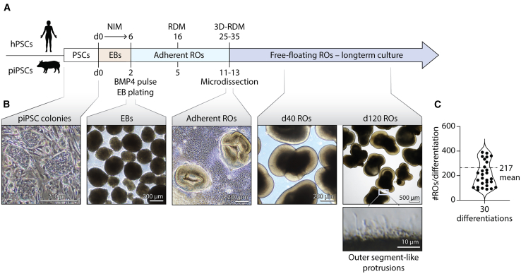
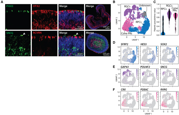
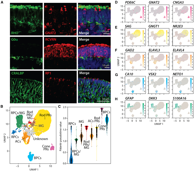
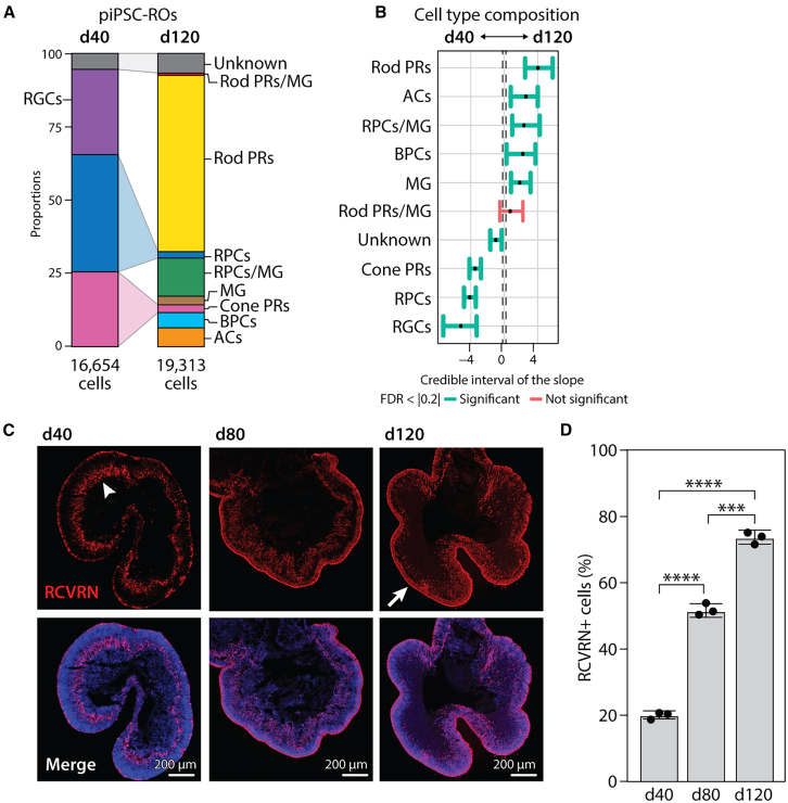
Outer retinal degenerative diseases (RDDs) and injuries leading to photoreceptor (PR) loss are prevailing causes of blindness worldwide. While significant progress has been made in the manufacture of human pluripotent stem cell (hPSC)-derived PRs, robust production of pluripotent stem cell (PSC)-PRs from swine, a popular preclinical large animal model, would provide an avenue to collect conspecific functional and safety data to complement human xenograft studies. Toward this goal, we describe the highly efficient generation of PR-dominant porcine induced PSC (piPSC)-derived retinal organoids (ROs) using modifications of our established hPSC-RO differentiation protocol. Porcine iPSC-ROs were characterized using immunocytochemistry (ICC) and single-cell RNA sequencing (scRNA-seq), which revealed the presence and maturation of major neural retina cell types, including PRs and retinal ganglion cells, which possess molecular signatures akin to those found in hPSC-ROs. In late piPSC-ROs, a highly organized outer neuroepithelium was observed with rods and cones possessing outer segments and axon terminals expressing pre-synaptic markers adjacent to dendritic terminals of bipolar cells. The existence of piPSC lines and protocols that support reproducible, scalable production of female and male ROs will facilitate transplant studies in porcine models of retinal injury and RDDs unconfounded by immunological and evolutionary incompatibilities inherent to human xenografts.

Keywords: allograft; cell transplantation; iPSC-derived retinal cells; neural retina; photoreceptors; porcine iPSCs; retinal differentiation; retinal organoids; scRNA-seq.


Conflict of interest statement

Declaration of interests The authors declare no competing interests.

Figures

Figure 1
Figure 1
Production and differentiation of ROs from piPSCs (A) Schematic showing the timing of key steps in the piPSC-RO differentiation protocol used for this study compared to that of an established hPSC-RO differentiation protocol (Capowski et al., 2019). (B) Representative light microscopic live images of (from left to right) piPSC colonies grown on an optimal density MEF feeder layer, d2 EBs in suspension, d12 adherent RO colonies prior to dissection, and free-floating d40 and d120 ROs in 3D culture after dissection. A higher-magnification image of the surface of a d120 piPSC-RO shows outer segment-like protrusions. (C) Plot showing the number of piPSC-ROs obtained from each of 30 consecutive differentiations (n = 30 differentiations, mean = 217 ± 19 ROs/differentiation), as indicated by the presence of a compact, uniform, phase-bright outer neuroepithelial rim 2 days after microdissection of adherent cultures. Four wells of a 6-well plate containing 80%–90% confluent piPSCs were used for each differentiation. Scale bars as shown.
Figure 2
Figure 2
ICC and single-cell RNA-seq analyses of early-stage piPSC-ROs (A) High- and low-magnification ICC images of d40 female piPSC-ROs showing the presence of PHH3+ proliferating cells along the RO rim with OTX2+ PR precursors found throughout the outer neuroepithelium. Cell bodies of SNCG+ RGCs are located deeper within piPSC-ROs, whose projections (arrowhead) often extend to the RO surface through an outer layer of RCVRN+ PRs. (B) Uniform manifold approximation and projection (UMAP) of pooled, early-stage (d35-d41) piPSC-ROs was used to visualize the 4 cell cluster annotations (RPC, RGC, cone PR, and unknown). (C) Palantir values for each identified retinal cell type showed a large separation of RGCs and cones from RPCs in pseudotime. (D–F) Normalized expression of specific genes expressed in RPCs (D), RGCs (E), and cone PRs (F) projected onto the early-stage piPSC-RO UMAP. Fifteen piPSC-ROs (5 piPSC-ROs from each of 3 independent differentiations) were pooled for early-stage (“d40”) scRNA-seq analysis. Scale bars as shown.
Figure 3
Figure 3
ICC and single-cell RNA-seq analyses of late-stage piPSC-ROs (A) ICC images of d120 female piPSC-ROs showing RHO staining concentrated at the RO surface with interspersed expression of GNAT2 in cone PRs. G0α is expressed in cone and rod ON BPCs in an inner RO layer beneath the RCVRN+ ONL. CRALBP expression is found in MG cell bodies and radial processes, the latter of which traverse the ONL and form the outer limiting membrane. The ONL is highlighted in the bottom series of images by RP1, a marker of maturing cone PRs in swine. (B) UMAP plot of late-stage (d118-d122) piPSC-ROs with manual annotations showing 9 cell clusters: cone PRs, rod PRs, BPCs, ACs, MG, RPCs, RPCs/MG, rod PRs/MG, and unknown. (C) Palantir pseudotime values for each identified cell class in late-stage piPSC-ROs showing the lowest state of differentiation for RPCs and the highest states of differentiation for cone PRs and BPCs. (D–H) Normalized expression of specific genes expressed in cone PRs (D), rod PRs (E), ACs (F), BPCs (G), and MG (H) projected onto the late-stage piPSC-RO UMAP. Fifteen piPSC-ROs (5 piPSC-ROs from each of 3 independent differentiations) were pooled for late stage (“d120”) scRNA-seq analysis. Scale bars as shown.
Figure 4
Figure 4
Photoreceptors migrate to form a dense ONL and become the dominant cell population in piPSC-ROs over time (A) Stacked bar graph showing the proportion of different cell types in female piPSC-ROs at d40 (16,654 total cells) and d120 (19,313 total cells) as determined by scRNA-seq. Cell proportions are listed in the results section. (B) Analysis of early and late piPSC-RO cell type composition using sccomp. Error bars denote the Bayesian 95% credible interval of the slope between d40 and d120 piPSC-ROs for each cell type, and the central dashed lines represent the minimal effect (0.2) that the hypothesis test is based on. Cell types that comprised a significantly greater proportion of the total cell population in early ROs vs. late ROs or late ROs vs. early ROs are indicated with green error bars to the left or right of the vertical gray line, respectively. The single error bar in pink demarcates a cell type (rod PRs/MG) with no such difference. (C) Low-magnification, confocal images of whole piPSC-RO sections at d40, d80, and d120 of differentiation showing the location of RCVRN+ PR cells. The arrowhead indicates RCVRN+ PRs primarily localized within a deeper layer at d40, with progressive formation of a dense ONL (arrow) by d120. (D) Percentage of RCVRN+ PRs in fixed and immunostained cells of piPSC-ROs at d40, d80, and d120 as determined via flow cytometry. ∗∗∗∗p < 0.0001, ∗∗∗p = 0.0004; ANOVA post hoc Tukey’s multiple comparisons test. For each time point, n = 3 independent differentiations; ≥10 piPSC-ROs per differentiation were pooled. Scale bars as shown.
Figure 5
Figure 5
Comparison of outer retinal organization between piPSC-ROs and adult porcine retina (A) ICC analysis showing layered expression of RHO (rod PRs), M/L Opsin (red and green cone PRs), VGLUT1 (PR pre-synaptic marker), and G0α (rod and cone ON BPCs) in d190 female piPSC-ROs and adult porcine retina. (B–D) Low (B and C)- and high (D)-magnification ICC images of d190 piPSC-ROs demonstrating expression of the synaptic marker VGLUT1 at the interface between an ONL containing M/L Opsin+ cone PRs and RHO+ rod PRs and an INL containing G0α+ rod and cone ON BPCs. Scale bars as shown.
Figure 6
Figure 6
Day 120 piPSC-ROs are transcriptionally comparable to day 205 hPSC-ROs (A and B) (A) UMAP of a re-analyzed d205 hPSC-RO dataset (Sridhar et al., 2020) used as a reference for (B) Seurat label transfer onto the d120 piPSC-RO UMAP and cell type prediction. (C) Prediction score values from Seurat mapping (ranging from 0 to 1, where 1 is the highest score) for each predicted cell type on the d120 piPSC-RO UMAP. Insets show prediction score histograms and medians for each cell type. (D) Stacked bar graphs showing the proportion of cell types for hPSC-ROs at d205 and piPSC-ROs at d120. Cell proportions are listed in the results section. (E) Cell type compositional analysis using sccomp. Error bars denote the Bayesian 95% credible interval of the slope between hPSC-ROs and piPSC-ROs cell types, and the central dashed lines represent the minimal effect (0.2) that the hypothesis test is based on. Green error bars indicate cell types that comprise a greater proportion of the total cell population, while pink error bars indicate cell types with no such difference. (F) Heatmap depicting the relative distribution of cell identities predicted by the human RO dataset (rows) for each manually annotated piPSC-RO cell type (columns), ranging from 0 (blue) to 1 (red).

References

    1. Barone F., Muscatello L.V., Ventrella D., Elmi A., Romagnoli N., Mandrioli L., Maya-Vetencourt J.F., Bombardi C., Mete M., Sarli G., et al. The porcine iodoacetic acid model of retinal degeneration: Morpho-functional characterization of the visual system. Exp. Eye Res. 2020;193 doi: 10.1016/j.exer.2020.107979. - DOI - PubMed
    1. Barone F., Amaral J., Bunea I., Farnoodian M., Gupta R., Gupta R., Baker D., Phillips M.J., Blanch R.J., Maminishkis A., et al. American Society for Clinical Investigation; 2023. A Versatile Laser-Induced Porcine Model of Outer Retinal and Choroidal Degeneration for Preclinical Testing. - PMC - PubMed
    1. Beauchemin M.L. The fine structure of the pig’s retina. Albrecht Von Graefes Arch. Klin. Exp. Ophthalmol. 1974;190:27–45. doi: 10.1007/BF00414333. - DOI - PubMed
    1. Capowski E.E., Samimi K., Mayerl S.J., Phillips M.J., Pinilla I., Howden S.E., Saha J., Jansen A.D., Edwards K.L., Jager L.D., et al. Reproducibility and staging of 3D human retinal organoids across multiple pluripotent stem cell lines. Dev. Camb. Engl. 2019;146 doi: 10.1242/dev.171686. - DOI - PMC - PubMed
    1. Chandler M.J., Smith P.J., Samuelson D.A., MacKay E.O. Photoreceptor density of the domestic pig retina. Vet. Ophthalmol. 1999;2:179–184. - PubMed

Publication types

LinkOut - more resources