Case Reports
. 2025 Mar 5;30(5):103177.
doi: 10.1016/j.jaccas.2024.103177.

The DELIVER Technique: Deep Engagement of Guide or ChiLd-guIde Catheter for Burr DeliVEry and Rotational Atherectomy

Affiliations
Case Reports

The DELIVER Technique: Deep Engagement of Guide or ChiLd-guIde Catheter for Burr DeliVEry and Rotational Atherectomy

Umihiko Kaneko et al. JACC Case Rep. .

Abstract

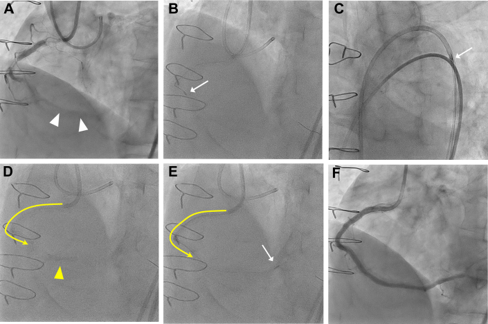
The delivery of the rotational atherectomy burr can sometimes be hindered in distal calcified lesions complicated by proximal vessel tortuosity or other obstacles. This problem may result in procedural failure or fatal complications, including coronary perforation, burr entrapment, or driveshaft fracture. To prevent these catastrophic outcomes and ensure successful burr delivery, we introduce the DELIVER (Deep Engagement of guide catheter or 5-F chiLd-guIde catheter for burr deliVEry and subsequent Rotational atherectomy) technique. This method involves deep catheter insertion beyond proximal vessel tortuosity or other obstacles, using strategies such as the distal balloon anchoring technique. Once the catheter is positioned, the rotational atherectomy burr is advanced through it to facilitate the atherectomy of the distal target lesion. This report presents 3 cases where the DELIVER technique was applied successfully to treat distal lesions. The technique enabled smooth and atraumatic burr delivery, even through tortuous arterial segments or other challenging anatomical structures.

Keywords: calcified lesion; deep engagement; distal lesion; rotational atherectomy; tortuous vessel.


Conflict of interest statement

Funding Support and Author Disclosures Drs Kaneko, Kashima, and Kuramitsu have served as proctors for Rotablator for Boston Scientific. All other authors have reported that they have no relationships relevant to the contents of this paper to disclose.

Figures

None
Graphical abstract
Visual Summary
Visual Summary
Summary of the DELIVER Technique (A) Calcified distal right coronary artery (RCA) stenosis and significant proximal vessel tortuosity. (B and C) A 6F guide catheter can be deeply inserted beyond a significantly tortuous segment via distal balloon anchoring. (D) Deeply inserted guide catheter enables safe delivery of a 1.5-mm burr distally and subsequent rotational atherectomy. (E) Baseline coronary angiogram revealing severe vessel tortuosity proximal to the calcified culprit lesion in the distal RCA. (F) Failure of distal delivery of the 1.5- or 1.25-mm burr owing to significant proximal tortuosity and calcification. (G) Deep insertion of a 5-F ST01 catheter via distal balloon anchoring. (H) Successful rotational atherectomy of the distal target lesion. (I) Baseline coronary angiography reveals a calcified mid-RCA lesion and a previously implanted stent in the ostial RCA. (J and K) Step-wise advancement of the guide catheter through the proximal stent via balloon-assisted tracking. (L) Successful delivery of a 1.75 mm burr and rotational atherectomy runs. Red arrowheads; proximal tortuous segment; yellow arrows, distal calcified lesion.
Figure 1
Figure 1
Possible Difficulties of Rotational Atherectomy for Distal Calcified Lesions in Tortuous Proximal Vessel Segments or Coronary Stents (A) A distal calcified lesion across a tortuous proximal vessel segment. (B) Deep cut at the lesser curvature. (C) Coronary perforation at the greater curvature. (D) Driveshaft fracture at the tortuous proximal vessel segment. (E) A distal calcified lesion across a proximal coronary stent. (F) Burr-passage failure or burr entrapment. Black arrowheads, distal calcified lesion; red arrowheads, proximal tortuous segment.
Figure 2
Figure 2
Deep Engagement of a 5F Child Guide Catheter Through an Extremely Long and Tortuous RCA and Subsequent Rotational Atherectomy (A) Baseline coronary angiography reveals the severe vessel tortuosity proximal to the occlusion and calcified culprit lesion (white arrowheads) in distal RCA. (B) Failure of the distal delivery of the 1.5- or 1.25-mm burr owing to significant proximal vessel tortuosity and calcification. (C) The 1.25-mm burr is unable to pass through a 7-F guide extension catheter at the entry port. (D) Deep insertion of the 5-F ST01 (Terumo) catheter (yellow line with arrow) via distal balloon anchoring (yellow arrowhead). (E) Smooth passage of a 1.25-mm burr (white arrow) through the 5-F ST01 to a point just proximal to the distal target lesion; subsequent rotational atherectomy is successful. (F) Final angiography demonstrates the excellent results. RCA = right coronary artery.
Figure 3
Figure 3
Deep Engagement of a 6-F Guide Catheter Through a Tortuous Saphenous Vein Graft and Rotational Atherectomy for Distal RCA Lesion (A and B) Baseline coronary angiography and computed tomography reveals calcified distal RCA stenosis and a significantly tortuous proximal saphenous vein graft (blue line) anastomosed to the mid-RCA. (C and D) A 6-F Judkins right 4.0 guide catheter (yellow line with arrow) is deeply inserted beyond the tortuous saphenous vein graft segment via distal balloon anchoring technique (yellow arrowhead). (E) Deeply inserted guide catheter enables safe delivery of the 1.5-mm burr (white arrow) distally and subsequent rotational atherectomy. (F) Final angiography demonstrates the excellent results. Abbreviation as in Figure 1.
Figure 4
Figure 4
Deep Engagement of a 6-F Guide Catheter Through a Stent in ostial RCA and subsequent RA for distal RCA lesion (A) Baseline coronary angiography reveals a calcified mid-RCA lesion (white arrowhead) and previously implanted stent in ostial RCA (white line). (B) Step-wise advancement of the guide catheter through the proximal stent by repeated 3.5-mm balloon dilatation and deflation (balloon-assisted tracking). (C) Further deep insertion of the guide catheter (yellow line with arrow) aided by distal balloon anchoring (yellow arrowhead). (D) Successful delivery of a 1.75-mm burr (white arrow) and rotational atherectomy. (E) Angiography after rotational atherectomy confirms improved coronary flow and enlarged lumen. (F) Final angiography demonstrates the excellent results. RA = right atrial; other abbreviation as in Figure 1.

References

    1. Shimony A., Zahger D., Straten M., et al. Incidence, risk factors, management and outcomes of coronary artery perforation during percutaneous coronary intervention. Am J Cardiol. 2009;104:1674–1677. - PubMed
    1. Généreux P., Madhavan M.V., Mintz G.S., et al. Ischemic outcomes after coronary intervention of calcified vessels in acute coronary syndromes. Pooled analysis from the HORIZONS-AMI (Harmonizing Outcomes With Revascularization and Stents in Acute Myocardial Infarction) and ACUITY (Acute Catheterization and Urgent Intervention Triage Strategy) trials. J Am Coll Cardiol. 2014;63:1845–1854. - PubMed
    1. Ayoub M., Tajti O., Ferenc M., et al. Feasibility and outcome of the Rotapro system in treating severely calcified coronary lesions: the Rotapro study. Cardiol J. 2023;30:526–533. doi: 10.5603/CJ.a2021.0128. - DOI - PMC - PubMed
    1. Bo Liang, Ning Gu. High-speed rotational atherectomy in coronary artery calcification: the randomized ROTAXUS and PREPARE-CALC trials. Catheter Cardiovasc Interv. 2022;100:61–71. - PubMed
    1. Sakakura K., Ito Y., Shibata Y., et al. Clinical expert consensus document on rotational atherectomy from the Japanese association of cardiovascular intervention and therapeutics: update 2023. Cardiovasc Interv Ther. 2023;38:141–162. - PMC - PubMed

Publication types

LinkOut - more resources