Review
. 2025 Mar 7;10(1):93.
doi: 10.1038/s41392-025-02124-y.

Tissue macrophages: origin, heterogenity, biological functions, diseases and therapeutic targets

Affiliations
Review

Tissue macrophages: origin, heterogenity, biological functions, diseases and therapeutic targets

Fan Guan et al. Signal Transduct Target Ther. .

Abstract

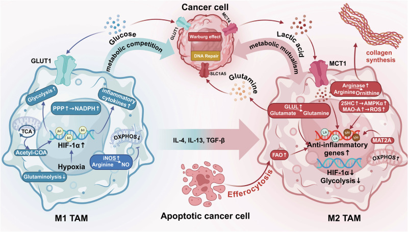
Macrophages are immune cells belonging to the mononuclear phagocyte system. They play crucial roles in immune defense, surveillance, and homeostasis. This review systematically discusses the types of hematopoietic progenitors that give rise to macrophages, including primitive hematopoietic progenitors, erythro-myeloid progenitors, and hematopoietic stem cells. These progenitors have distinct genetic backgrounds and developmental processes. Accordingly, macrophages exhibit complex and diverse functions in the body, including phagocytosis and clearance of cellular debris, antigen presentation, and immune response, regulation of inflammation and cytokine production, tissue remodeling and repair, and multi-level regulatory signaling pathways/crosstalk involved in homeostasis and physiology. Besides, tumor-associated macrophages are a key component of the TME, exhibiting both anti-tumor and pro-tumor properties. Furthermore, the functional status of macrophages is closely linked to the development of various diseases, including cancer, autoimmune disorders, cardiovascular disease, neurodegenerative diseases, metabolic conditions, and trauma. Targeting macrophages has emerged as a promising therapeutic strategy in these contexts. Clinical trials of macrophage-based targeted drugs, macrophage-based immunotherapies, and nanoparticle-based therapy were comprehensively summarized. Potential challenges and future directions in targeting macrophages have also been discussed. Overall, our review highlights the significance of this versatile immune cell in human health and disease, which is expected to inform future research and clinical practice.


Conflict of interest statement

Competing interests: The authors declare no competing interests. Consent for publication: All authors have agreed on the contents of the manuscript.

Figures

Fig. 1
Fig. 1
A timeline of research history and milestones of macrophages. NO Nitric Oxide, TAMs tumor-associated macrophages, CAR-M CAR-macrophage, CAR-iMac induced pluripotent stem cells (iPSCs)-derived CAR-expressing macrophage cells
Fig. 2
Fig. 2
CSF-1 could induce monocytes to differentiate into M0 macrophages. Then, M0 macrophages could further evolve into M1 or M2 macrophages stimulated by Th1-type or Th2-type cytokines. Due to their differences in activation patterns and other aspects, M2 macrophages could be further divided into four subtypes: M2a, M2b, M2c, and M2d. Various macrophage types express different molecular markers and secrete different materials, which play important roles in various physiological and pathological processes. CSF-1 colony stimulating factor-1, MHC-II major histocompatibility complex class II, iNOS inducible nitric oxide synthase, LPS Lipopolysaccharides IFN-γ interferon-gamma, TNF-ɑ tumor necrosis factor-alpha, IL-1β interleukin-1β, IL-8 interleukin-8, IL-12 interleukin-12
Fig. 3
Fig. 3
TRMs in inflammation and homeostasis. a The canonical pro-inflammatory response is initiated by either PRRs or opsonin receptors. PRRs can directly recognize DAMPs (usually cell-derived molecules, e.g., Biglycan, versican, F-actin) and PAMPs (usually microorganism-derived molecules, e.g., Foreign DNA, flagellin, mannose). The opsonin receptor-mediated recognition process involves binding foreign particles labeled by opsonins and opsonic receptors, including Fcγ receptors. The recognition activates actin polymerization, pro-inflammatory cytokines, and other responses. The phagosome fuses with the lysosome. In late endosomal MIICs, most newly synthesized MHC-II molecules are likely loaded in an HLA-DM-dependent mechanism. The antigens and MHC-II will form MHC-II peptide complexes and then be delivered to the plasma membrane to be available to stimulate antigen-specific CD4+ T lymphocytes. Pro-inflammatory cytokines (e.g., TNF-α, IL-1β, LPS). b Resolution of inflammation. Apoptotic cells can release “find me” signals (EG. ATP, Lys phosphatidylcholine, CX3CL1) to recruit TAMs. Phagocytosis is facilitated by receptors (e.g., BAI1, Mer, Axl, αvβ3-integrin) directly binding with eat-me signals (e.g., PtdSer, Calreticulin, LPC) on apoptotic cells or indirectly recognizing bridging molecules, such as MFGE8, C1q, protein S, etc., which bind to eat-me signals. The recognition activates actin polymerization, immune-resolution cytokines, and other responses. Pro-inflammation cytokines (TNF-α, IL-6, etc.) decrease, and immune-resolution cytokines (IL-10, TGF-β) increase. Some viruses display PtdSer on their surface and mimic apoptotic cells, which allows them to evade the immune system and facilitate entry into host cells. Tolerant responses have also been attributed to Dectin-1/2 and SIRPα. Altered glycosylation is a universal feature of cancer cells; some abnormal glycans (e.g., galectin 9) promote cancer growth and immune tolerance through Dectin 1/2 activation. Some tumor cells express CD47 that interacts with SIRPα, expressed on the surface of macrophages and dendritic cells, inhibiting phagocytosis and maintaining self-tolerance. c TRMs sense physical factors and cytokines in the microenvironment. SIRPα signal regulatory protein alpha, LPC lysophosphatidylcholine, PRRs pattern recognition receptors, PAMPs pathogen-associated molecular patterns, DAMPs damage-associated molecular patterns, MIIC MHC class II compartment, LPS lipopolysaccharides, LPC lysophosphatidylcholines, HLA human leukocyte antigen
Fig. 4
Fig. 4
Metabolic differences between M1 and M2 macrophages. In the tumor microenvironment, the metabolic differences between M1 and M2 macrophages are closely linked to the onset and progression of cancer. Typically, both M1 macrophages and cancer cells primarily undergo glycolysis, leading to metabolic competition. As the malignancy of the tumor progresses, M1 macrophages in the tumor microenvironment are gradually reprogrammed into M2 macrophages, shifting their metabolism to oxidative phosphorylation. Furthermore, the exchange of metabolic products between M2 macrophages and cancer cells further promotes the manifestation of the tumor’s biological characteristics. This process highlights the complexity of the tumor microenvironment and underscores the crucial role of macrophages in tumor development. ppp pentose phosphate pathway, NADPH nicotinamide adenine dinucleotide phosphate (reduced form), HIF-1α hypoxia-inducible factor-1 alpha, OXPHOS oxidative phosphorylation, GLUT1 glucose transporter type 1, MCT4 monocarboxylate transporter 4, MCT1 monocarboxylate transporter 1, SLC1A5 solute carrier family 1 member 5, IL-4 interleukin-4, IL-13 interleukin-13, TGF-β transforming growth factor-beta, FAO fatty acid oxidation, 25HC 25-hydroxycholesterol, AMPKα AMP-activated protein kinase alpha, MAO-A monoamine oxidase A, ROS reactive oxygen species, MAT2A methionine adenosyltransferase 2A
Fig. 5
Fig. 5
Role of Tissue Macrophages in Diseases. Macrophages play a pivotal role in developing a wide range of diseases, with distinct functions attributed to tissue-resident and monocyte-derived macrophages, as well as their M1/M2 polarization, which varies across different diseases. In autoimmune diseases such as rheumatoid arthritis and systemic sclerosis, macrophages damage tissue by releasing pro-inflammatory cytokines (e.g., IL-6 and TNF-α) and promote multi-tissue fibrosis through anti-inflammatory and fibrogenic factors. In cardiovascular diseases, including atherosclerosis and myocardial infarction, macrophage polarization facilitates phagocytosis of necrotic tissue, fibrotic repair, myocardial remodeling, and the formation and rupture of atherosclerotic plaques. AtoM arthritis-associated osteoclastogenic macrophages, IL-6 interleukin-6, TNF-α tumor necrosis factor-alpha, PANKL parathyroid hormone-related protein, CCL2 chemokine (C-C motif) ligand 2, ERK extracellular signal-regulated kinase, MAPK mitogen-activated protein kinase, FGF2 fibroblast growth factor 2, PGE2 prostaglandin E2, SPP1 secreted phosphoprotein 1, TGF-β transforming growth factor-beta, PDGF platelet-derived growth factor, CCL-18 chemokine (C-C motif) ligand 18, IL-34 interleukin-34, IL-7 interleukin-7, IL-1 interleukin-1, MCP-1 monocyte chemoattractant protein-1, NLRP3 NOD-like receptor pyrin domain-containing 3, KLF2 Krüppel-like factor 2, ADPN adiponectin, NF-κB nuclear factor kappa-light-chain-enhancer of activated B cells, HMGB1 high-mobility group box 1, IL-4 interleukin-4, IL-13 interleukin-13, OPN osteopontin, HIF-α hypoxia-inducible factor alpha
Fig. 6
Fig. 6
Role of Tissue Macrophages in Diseases. In neurodegenerative disorders such as Alzheimer’s and Parkinson’s diseases, macrophages are implicated in neuronal damage and neurotoxicity, involving the processing of amyloid-beta and alpha-synuclein. In metabolic disorders like obesity, which represents a state of systemic chronic inflammation, macrophages accelerate the development of insulin resistance and affect the efficacy of cell-based immunotherapies through tumor-associated macrophage interactions. Additionally, the diverse functions of macrophages are also evident in the progression of hepatic diseases, including hepatitis, liver cirrhosis, and hepatocellular carcinoma. p-Tau phosphorylated tau, APP amyloid precursor protein, SAPPβ soluble amyloid precursor protein beta, TREM2 triggering receptor expressed on myeloid cells 2, LRRK2 leucine-rich repeat kinase 2, NADPH nicotinamide adenine dinucleotide phosphate (reduced form), NOTCH NOTCH signaling pathway, mTOR1 mechanistic target of rapamycin complex 1, P53 tumor protein p53, CXCL10 C-X-C motif chemokine ligand 10, LGALS3 galectin-3 TNFsf12 tumor necrosis factor superfamily member 12

References

    1. Zhao, J., Andreev, I. & Silva, H. M. Resident tissue macrophages: Key coordinators of tissue homeostasis beyond immunity. Sci. Immunol.9, eadd1967 (2024). - PubMed
    1. Hume, D. A., Millard, S. M. & Pettit, A. R. Macrophage heterogeneity in the single-cell era: facts and artifacts. Blood142, 1339–1347 (2023). - PubMed
    1. Lazarov, T., Juarez-Carreño, S., Cox, N. & Geissmann, F. Physiology and diseases of tissue-resident macrophages. Nature618, 698–707 (2023). - PMC - PubMed
    1. Sakai, M. et al. Liver-derived signals sequentially reprogram myeloid enhancers to initiate and maintain kupffer cell identity. Immunity51, 655–670.e8 (2019). - PMC - PubMed
    1. Tober, J., McGrath, K. E. & Palis, J. Primitive erythropoiesis and megakaryopoiesis in the yolk sac are independent of c-myb. Blood111, 2636–2639 (2008). - PMC - PubMed

MeSH terms

LinkOut - more resources