. 2025 Mar 8;8(1):390.
doi: 10.1038/s42003-025-07838-x.

Extravascular coagulation regulates haemostasis independently of activated platelet surfaces in an in vivo mouse model

Affiliations

Extravascular coagulation regulates haemostasis independently of activated platelet surfaces in an in vivo mouse model

Asuka Sakata et al. Commun Biol. .

Abstract

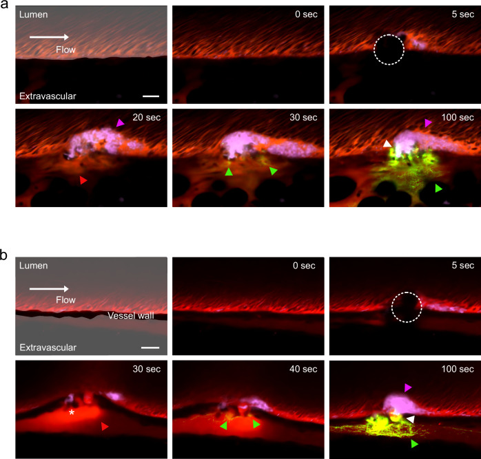
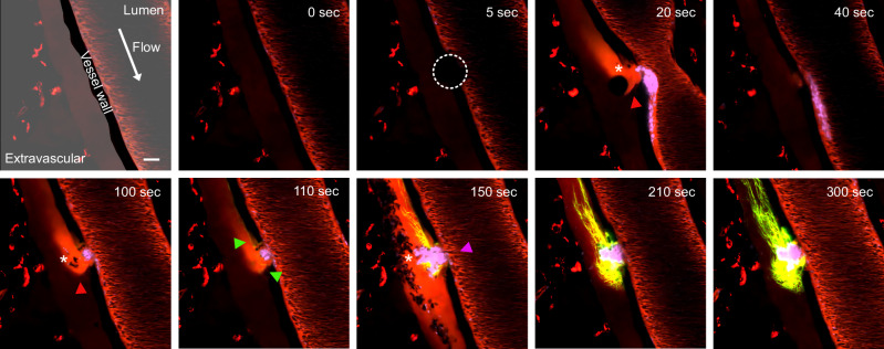
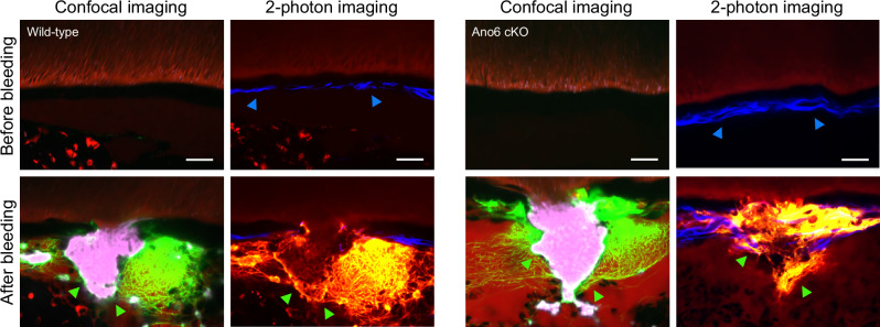
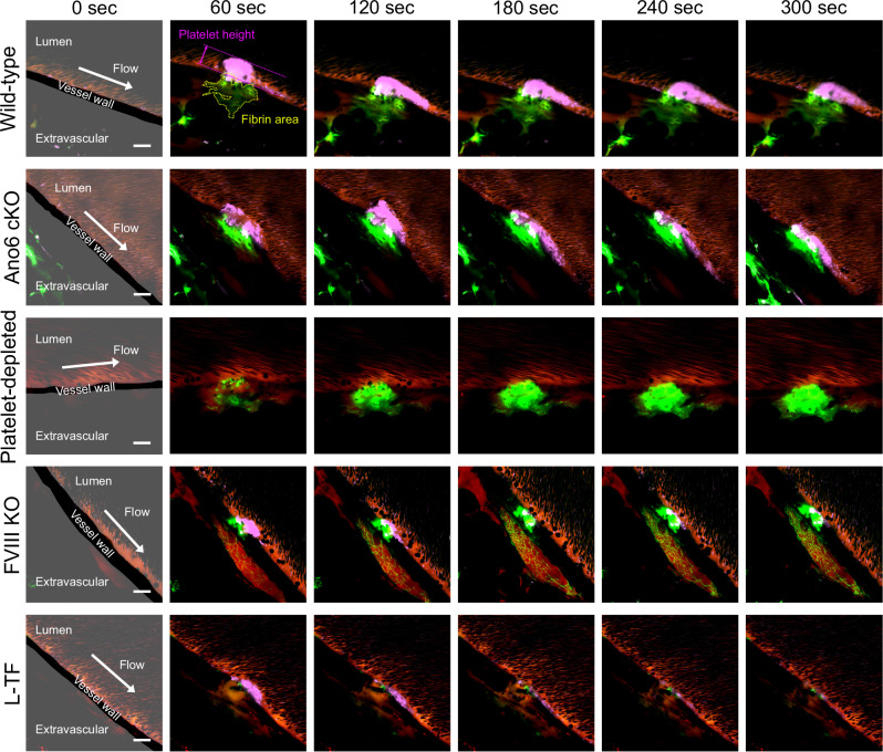
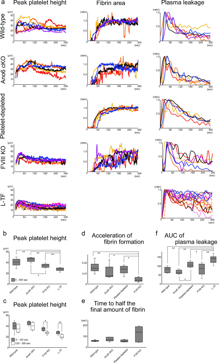
While the conventional understanding of haemostatic plug formation is that coagulation proceeds efficiently on the surface of activated platelets at the vascular injury site to form a robust haemostatic plug, this understanding does not explain the clinical reality that platelet dysfunction results in a mild bleeding phenotype, whereas coagulation disorders exhibit severe bleeding phenotypes, particularly in deep tissues. Here, we introduce an in vivo imaging method to observe internal bleeding and subsequent haemostatic plug formation in mice and report that haemostatic plug formation after internal bleeding, coagulation occurs primarily outside the blood vessel rather than on platelets. Experiments in mice with impaired platelet surface coagulation, depleted platelets, haemophilia A or reduced tissue factor expression suggest that this extravascular coagulation triggers and regulates haemostatic plug formation. Our discovery of the important role of extravascular coagulation in haemostasis may contribute to refining the treatment of haemostatic abnormalities and advancing antithrombotic therapy.


Conflict of interest statement

Competing interests: This study was funded by Chugai Pharmaceutical. The authors declare the following competing interests: A.S., K.T., Naoki M., S.H., R.K., Y.N., T.S., K.N., and M.S.: Members of the Medicinal Biology of Thrombosis and Haemostasis established by Nara Medical University and Chugai Pharmaceutical Co., Ltd. Naoki M., S.H., R.K., Y.N., and T.S.: Employees of Chugai Pharmaceutical Co., Ltd. Naoki M., S.H. and R.K.: Ownership of stock by Chugai Pharmaceutical Co., Ltd. M.S.: Patents for inventions related to products of Chugai Pharmaceutical Co., Ltd. K.T.: Grants or research support from the Japan Blood Products Organization, the Mother and Child Health Foundation and Novo Nordisk Pharma. M.S.: Takeda Pharmaceutical Co., Ltd., and CSL Behring; honoraria or consultation fees from Chugai Pharmaceutical Co., Ltd.; speaker bureau from Chugai Pharmaceutical Co., Ltd.; CSL Behring, Sanofi, Bayer, Novo Nordisk Pharma, Takeda Pharmaceutical Co., Ltd., Pfizer, and Fujimoto Seiyaku Corp. K.N.: Representative of Medicinal Biology of Thrombosis and Haemostasis collaborative research laboratory; research support from Chugai Pharmaceutical Co., Ltd.; grants or research support from Chugai Pharmaceutical Co., Ltd.; Takeda Pharmaceutical Co., Ltd.; KM Biologics Co., Ltd.; Sanofi Co., Ltd.; Novo Nordisk Pharma Co., Ltd.; Bayer Co., Ltd.; AbbVie GK LLC; Janssen Pharmaceutical K.K. Co., Ltd.; honouraria or consultation fees from Chugai Pharmaceutical Co., Ltd.; Sanofi Co., Ltd.; and CSL Behring.

Figures

Fig. 1
Fig. 1. Fibrin, a component of haemostatic plugs, is formed outside blood vessels.
Representative images of haemostatic plug formation after venous (a) and arterial (b) internal bleeding. The mice were intravenously injected with dextran-rhodamine B, Alexa Fluor 488-conjugated fibrinogen and the platelet imaging antibody X649 prior to observation. After two-photon excitation injury (circle of dots), platelet aggregate formation in the lumen (magenta triangle) and extravascular plasma leakage (red triangle) were observed. After arterial internal bleeding, red blood cell leakage was observed (white asterisk). Fibrin is formed in the extravascular area (green triangle). The haemostatic plug consisted of a platelet aggregate in the lumen, extravascular fibrin, and platelets and fibrin at the boundary of the lumen and extravascular space (white triangle). Scale bar = 20 μm. Green: fibrin/fibrinogen. Magenta: platelets. Red: plasma.
Fig. 2
Fig. 2. Haemostasis was achieved by the cessation of plasma leakage following the cessation of blood cell leakage.
Representative images of haemostatic plug formation after arterial internal bleeding from a large injury. The mice were intravenously injected with dextran-rhodamine B, Alexa Fluor 488-conjugated fibrinogen, and the platelet imaging antibody X649 prior to observation. After two-photon excitation injury (circle of dots), extravascular plasma leakage (red triangle) and red blood cell leakage were observed (white asterisk). Vasoconstriction temporarily reduced blood leakage, but it resumed when vasoconstriction was released. Fibrin begins to form in slow-flowing areas of the extravascular space (green triangle). Exacerbated blood cell leakage stopped when platelets clogged the site of vessel rupture (magenta triangle). Fibrin formation proceeded in the area of slowed flow caused by platelet blockage, resulting in the cessation of plasma leakage. Scale bar = 20 μm. Green: fibrin/fibrinogen. Magenta: platelets. Red: plasma.
Fig. 3
Fig. 3. Platelets are trapped by extravascular fibrin where collagen is damaged.
Representative images of haemostatic plugs after large arterial injury. The mice were intravenously injected with dextran-rhodamine B, Alexa Fluor 488-conjugated fibrinogen, and the platelet imaging antibody X649 prior to observation. Vessel observation before the arterial injury and after haemostatic plug formation via two-photon imaging revealed collagen (blue triangle) and platelets attached to the fibrin network (green triangle), where collagen ruptured. Similar findings were observed in Ano6 cKO mice with impaired platelet coagulation. Scale bar = 20 μm. In the confocal images, green indicates fibrin/fibrinogen, magenta indicates platelets, and red indicates plasma. In two-photon images, yellow-green indicates fibrin/fibrinogen, red indicates plasma, and blue indicates collagen.
Fig. 4
Fig. 4. Irreversible platelet activation does not occur in early haemostatic plugs.
Representative images of platelets that clog the liver vasculature after systemic thrombin injection (a) and haemostatic plugs that formed after large arterial injury (b). For arterial haemostatic plug observation, the mice were injected with Alexa Fluor 488-conjugated fibrinogen, PE-conjugated rat anti-mouse CD62P antibody, and platelet imaging antibody X649 prior to observation. For platelet observation after thrombin injection, the mice were injected with FITC-dextran, PE-conjugated rat anti-mouse CD62P antibody, and X649 antibody prior to observation. CD62P expression was detected on platelets that clog the liver vasculature (a) but was not detected in the in vivo internal bleeding/haemostasis model (b). Scale bar = 20 μm. Green: fibrin/fibrinogen. Magenta: platelets. Red: plasma.
Fig. 5
Fig. 5. Haemostatic plug formation after venous internal bleeding in wild-type, Ano6 cKO, platelet-depleted, FVIII KO, and L-TF mice.
Representative images of haemostatic plug formation after venous internal bleeding in wild-type, Ano6 cKO, platelet-depleted, FVIII KO, and L-TF mice. The mice were intravenously injected with dextran-rhodamine B, Alexa Fluor 488-conjugated fibrinogen, and the platelet imaging antibody X649 prior to observation. Scale bar = 20 μm. Green: fibrin/fibrinogen. Magenta: platelets. Red: plasma.
Fig. 6
Fig. 6. Haemostatic plug formation after arterial internal bleeding in wild-type, Ano6 cKO, platelet-depleted, FVIII KO, and L-TF mice.
Representative images of haemostatic plug formation after arterial internal bleeding in wild-type, Ano6 cKO, platelet-depleted, FVIII KO, and L-TF mice. The mice were intravenously injected with dextran-rhodamine B, Alexa Fluor 488-conjugated fibrinogen, and the platelet imaging antibody X649 prior to observation. Scale bar = 20 μm. Green: fibrin/fibrinogen. Magenta: platelets. Red: plasma.
Fig. 7
Fig. 7. Extravascular coagulation is important for platelet aggregate formation and the cessation of plasma leakage after venous internal bleeding.
Measurements of platelet aggregates, fibrin, and plasma leakage were taken from experimental mice (a) (wild-type, n = 6 lesions from 5 animals; Ano6 cKO, n = 5 lesions from 5 animals; platelet-depleted, n = 5 lesions from 5 animals; FVIII KO, n = 6 lesions from 4 animals; and L-TF mice, n = 16 lesions from 4 animals). Platelet aggregates were assessed by height from the vascular endothelium. Fibrin was quantified as the ratio of the fibrin area over time, normalized to the end of the observation period. Plasma leakage was expressed relative to the maximum signal outside the vessel. Box-and-whisker plot of the peak platelet aggregate height (b). Peak platelet aggregate height at 0–120 and 120–300 s (c). The increase in the fibrin area accelerated, as indicated by the ratio (d). Time required for the fibrin area to reach half the area at the end of observation (e). Area under the curve (AUC) of plasma leakage (f). *<0.05, **<0.01. c Wilcoxon signed-rank test; b, df Steel–Dwass test and Monte Carlo simulation.
Fig. 8
Fig. 8. Extravascular coagulation is important for the formation of platelet aggregates and the cessation of blood cell and plasma leakage after arterial internal bleeding.
Measurements of platelet aggregates, fibrin, and plasma leakage were taken from experimental mice (a) (wild-type, n = 6 lesions of 6 animals; Ano6 cKO, n = 7 lesions of 6 animals; platelet-depleted, n = 5 lesions of 5 animals; FVIII KO, n = 5 lesions of 5 animals; and L-TF mice, n = 6 lesions of 6 animals). Platelet aggregates were assessed by height from the vascular endothelium. Fibrin was quantified as the ratio of the fibrin area over time, normalized to the end of the observation period. Plasma leakage was expressed relative to the maximum signal outside the vessel. Box-and-whisker plot of the peak platelet aggregate height (b). Peak platelet aggregate height at 0–120 and 120–300 s (c). The increase in the fibrin area accelerated, as indicated by the ratio (d). Time required for the fibrin area to reach half the area at the end of observation (e). Area under the curve (AUC) of plasma leakage (f). The time from the start of bleeding to the cessation of blood cell leakage (g). The extent of platelet aggregate extrusion from the vessel was evaluated by measuring the distance from the vessel wall to the platelets, as shown in (h). †<0.1, *<0.05, **<0.01. c Wilcoxon signed-rank test; b, dh Steel–Dwass test and Monte Carlo simulation.
Fig. 9
Fig. 9. Extravascular coagulation triggers and regulates haemostasis.
When vascular continuity is lost, blood cells and plasma leak out of the vessels (a). Extravascular fibrin and platelets captured by fibrin inhibit blood cell and plasma leakage (b). Coagulation proceeds extravascularly, and the generated thrombin accelerates extravascular fibrin formation and activates platelets in the lumen (c). As extravascular fibrin grows, plasma leakage ceases, resulting in complete haemostasis (d).

References

    1. Hoffman, M. Remodeling the blood coagulation cascade. J. Thromb. Thrombolysis16, 17–20 (2003). - PubMed
    1. Dahlbäck, B. Blood coagulation. Lancet355, 1627–1632 (2000). - PubMed
    1. Kjalke, M. et al. Active site-inactivated factors VIIa, Xa, and IXa inhibit individual steps in a cell-based model of tissue factor-initiated coagulation. Thromb. Haemost.80, 578–584 (1998). - PubMed
    1. Oliver, J. A., Monroe, D. M., Roberts, H. R. & Hoffman, M. Thrombin activates factor XI on activated platelets in the absence of factor XII. Arterioscler. Thromb. Vasc. Biol.19, 170–177 (1999). - PubMed
    1. Bolton-Maggs, P. H. B. & Pasi, K. J. Haemophilias A and B. Lancet361, 1801–1809 (2003). - PubMed

LinkOut - more resources