Review
. 2025 Mar 7;25(1):83.
doi: 10.1186/s12935-025-03681-6.

JAK/STAT signaling as a key regulator of ferroptosis: mechanisms and therapeutic potentials in cancer and diseases

Affiliations
Review

JAK/STAT signaling as a key regulator of ferroptosis: mechanisms and therapeutic potentials in cancer and diseases

Yimeng Dai et al. Cancer Cell Int. .

Abstract

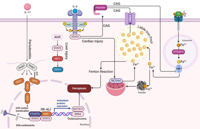
Ferroptosis is a distinct form of regulated cell death characterized by iron-dependent lipid peroxidation, playing a critical role in various diseases, including cancer, neurodegeneration, and tissue damage. This study reviews the intricate relationship between ferroptosis and the Janus kinase/signal transducer and activator of transcription (JAK/STAT) signaling pathway, highlighting its regulatory functions across multiple biological processes. Dysregulation of the JAK/STAT pathway is implicated in promoting or inhibiting ferroptosis, depending on the context. JAK2 promotes ferroptosis by activating STAT proteins, modulating the expression of key regulators like SLC7A11 and GPX4, and influencing iron homeostasis through pathways such as ferritinophagy and hepcidin regulation. STAT1 activation primarily enhances ferroptosis through the suppression of cystine-glutamate antiporter (System Xc-), leading to glutathione depletion and lipid peroxidation, contributing to cell death in conditions like Sjogren's syndrome and age-related macular degeneration. In contrast, STAT3 plays a protective role by upregulating SLC7A11 and GPX4, which inhibits ferroptosis and promotes cell survival, particularly in cancers such as hepatocellular carcinoma, prostate cancer, and renal cell carcinoma. This study also discusses STAT6's involvement in ferroptosis suppression in diseases like asthma and lung injury by regulating antioxidant defenses. Furthermore, the review explores potential therapeutic strategies targeting the JAK/STAT pathway to manipulate ferroptosis for disease treatment. In cancer therapy, modulating this pathway can enhance the effectiveness of ferroptosis inducers, offering promising avenues to overcome drug resistance. Additionally, the interplay between ferroptosis and JAK/STAT signaling in immune responses, oxidative stress, and lipid metabolism underscores its significance in disease progression and therapeutic intervention. By exploring these mechanisms, this study provides insights into the development of novel treatments targeting ferroptosis through JAK/STAT modulation, with implications for cancer, inflammatory diseases, and neurodegenerative conditions.

Keywords: Ferroptosis; JAK/STAT signaling; JAK2; STAT1; STAT3; STAT6; Therapy.


Conflict of interest statement

Declarations. Ethics approval and consent to participate: Not applicable. Informed consent to consent for publication: Not applicable. Generative AI and AI-assisted technologies in the writing process: During the preparation of this work, the authors used ChatGPT by OpenAI to improve paper readability. After using this tool/service, the authors reviewed and edited the content as needed and took full responsibility for the publication’s content. Competing interests: The authors declare no competing interests.

Figures

Fig. 1
Fig. 1
The molecular steps involved in the activation of the JAK-STAT signaling pathway, a critical mechanism for transmitting signals from extracellular cytokines to the nucleus, influencing gene expression. The process begins with cytokine-mediated receptor dimerization (step 1), where a cytokine binds to its specific receptor on the cell surface, leading to the dimerization (pairing) of the receptor subunits. This brings the JAKs in proximity. Once the receptors dimerize, JAK phosphorylates tyrosine residues (step 2) on the intracellular domain of the receptor, activating the JAK proteins themselves. This phosphorylation creates docking sites for STAT proteins. In step 3, these phosphorylated tyrosine residues recruit inactive STAT proteins to the receptor complex, where JAK then phosphorylates the STATs on specific tyrosine residues. This phosphorylation triggers dimerization of STATs (step 4), where two phosphorylated STAT molecules pair up. This activated STAT dimer is now able to translocate into the nucleus. In step 5, the dimerized STATs move into the nucleus, where they bind to specific sequences in the promoter region of target genes, known as STAT-binding sequences. This binding initiates transcription of cytokine-responsive genes, ultimately leading to cellular responses such as immune modulation, growth, or differentiation. Each step in this pathway is tightly regulated and essential for proper cellular function in response to external signals like cytokines [11]
Fig. 2
Fig. 2
This diagram illustrates the interconnected pathways involved in ferroptosis. First, the amino acid pathway is crucial for GSH production, a key antioxidant. Cystine enters the cell through the Xc- antiporter, is converted into cysteine, and combines with glutamate and glycine to form GSH. GPX4 then uses GSH to neutralize lipid peroxides, preventing ferroptosis. Second, the lipid pathway shows how PUFAs are processed by enzymes such as ACSL4, LPCAT3, and ALOX15, resulting in the formation of PUFA-OOH, which, if not neutralized by GPX4, triggers ferroptosis. Third, the iron pathway highlights how Fe2+ contributes to ferroptosis by generating ROS through the Fenton reaction. Iron enters cells via Tf, is reduced to Fe2+ by STEAP3, and either stored in ferritin or exported by FPN1. Excess Fe2+ leads to ROS production, enhancing lipid peroxidation and promoting ferroptosis. Lastly, the Nrf2 pathway serves as a protective mechanism against ferroptosis. Nrf2, typically inhibited by Keap1, is released during oxidative stress, translocates to the nucleus, and activates the expression of antioxidant genes like GPX4. This helps mitigate oxidative stress, regulate lipid peroxidation, and control iron metabolism, thereby preventing ferroptosis
Fig. 3
Fig. 3
The regulation of ferroptosis by JAK/STAT signaling pathways, focusing on the roles of different STAT proteins, including STAT1, STAT3, and STAT6, in modulating ferroptosis through pathways like SLC7A11 and GPX4 (Amino acid metabolism). STAT1 and ferroptosis: Upon activation by interferon-γ (IFN-γ), the JAK1/2-STAT1 pathway downregulates the cystine-glutamate antiporter (System Xc−), reducing cystine uptake and leading to GSH depletion. This suppression of the GSH/GPX4 axis results in increased lipid peroxidation and ferroptosis, contributing to cell death in diseases such as SS AMD. STAT3 and ferroptosis: The IL-6/JAK2/STAT3 pathway plays a crucial role in regulating ferroptos is resistance across various cancers, including RCC and HNSCC. IL-6 activates STAT3, promoting the transcription of SLC7A11, which enhances cystine uptake and GSH synthesis, thereby inhibiting ferroptosis. Additionally, the SHP-1/STAT3/SLC7A11 axis regulates the MCL1-BECN1 interaction, impacting ferroptosis in HCC and NSCLC. ARPC1A, transcriptionally regulated by STAT3, also inhibits ferroptosis in prostate cancer by reducing GPX4 and SLC7A11 expression, facilitating tumor progression. STAT6 and ferroptosis: In conditions such as asthma and ALI, IL-13 activates the JAK2/STAT6 pathway, which promotes the degradation of SLC7A11 via SOCS1, leading to increased ferroptosis in airway epithelial cells. Furthermore, STAT6 inhibits P53 acetylation, thereby enhancing SLC7A11 expression, which mitigates ferroptosis during lung injury
Fig. 4
Fig. 4
The regulation of ferroptosis through the interaction of iron metabolism and the STAT3/NRF2 pathway. In conditions like osteosarcoma and periodontitis, STAT3 activates NRF2, promoting antioxidant defense and osteogenic differentiation by upregulating key genes such as GPX4 and SLC7A11, which protect against ferroptosis. IL-6/STAT3 signaling increases hepcidin expression, inhibiting ferroportin and leading to iron accumulation and ferroptosis through the Fenton reaction. Additionally, STAT3 drives ferritin degradation via NCOA4, releasing iron and exacerbating ferroptosis in cardiac injury. In chemotherapy-resistant osteosarcoma, the STAT3/NRF2/GPX4 axis enhances resistance to ferroptosis, while targeting this pathway could sensitize cancer cells to ferroptosis inducers. The STAT3/HO-1 pathway in liver injury also contributes to ferroptosis by promoting iron dysregulation and oxidative stress. This integration highlights STAT3’s pivotal role in modulating ferroptosis through iron metabolism and antioxidant pathways across different diseases
Fig. 5
Fig. 5
The interaction of lipid metabolism pathways of ferroptosis with STAT proteins in human diseases. The figure illustrates how STAT proteins (STAT1, STAT3, and STAT6) regulate lipid metabolism and ferroptosis in various diseases. STAT1, activated by IL-10, promotes ferroptosis in diabetic nephropathy and intestinal injury by upregulating ACSL1/ACSL4 and enhancing lipid peroxidation. STAT3, induced by IL-9 and IL-6, modulates fatty acid oxidation and reduces ferroptosis in acute kidney injury and cancers by altering lipid metabolism. STAT6, activated by IL-4/IL-13, suppresses ferroptosis in cervical cancer via TAM-derived miRNA-660-5p, which downregulates ALOX15
Fig. 6
Fig. 6
Regulation of ferroptosis and STAT signaling pathways by various compounds in cancer models. The figure illustrates the role of key signaling molecules, such as JAK2, STAT3, STAT1, and STAT6, in mediating responses to natural and synthetic compounds targeting different cancer types. Inhibition or activation of these pathways leads to alterations in lipid peroxidation and ferroptosis

References

    1. Zhou Q, Meng Y, Li D, Yao L, Le J, Liu Y, et al. Ferroptosis in cancer: from molecular mechanisms to therapeutic strategies. Signal Transduct Target Ther. 2024;9:55. 10.1038/s41392-024-01769-5. - PMC - PubMed
    1. Dixon SJ, Lemberg KM, Lamprecht MR, Skouta R, Zaitsev EM, Gleason CE, et al. Ferroptosis: an iron-dependent form of nonapoptotic cell death. Cell. 2012;149:1060–72. - PMC - PubMed
    1. Berndt C, Alborzinia H, Amen VS, Ayton S, Barayeu U, Bartelt A, et al. Ferroptosis in health and disease. Redox Biol. 2024;75:103211. 10.1016/j.redox.2024.103211. - PMC - PubMed
    1. Luo Y, Alexander M, Gadina M, O’Shea JJ, Meylan F, Schwartz DM. JAK-STAT signaling in human disease: from genetic syndromes to clinical inhibition. J Allergy Clin Immunol. 2021;148:911–25. - PMC - PubMed
    1. Pang Q, Tang Z, Luo L. The crosstalk between oncogenic signaling and ferroptosis in cancer. Crit Rev Oncol Hematol. 2024;197:104349. - PubMed

LinkOut - more resources